. 2008 Nov;213(5):531-8.
doi: 10.1111/j.1469-7580.2008.00966.x.

Col2a1 lineage tracing reveals that the meniscus of the knee joint has a complex cellular origin

Affiliations

Col2a1 lineage tracing reveals that the meniscus of the knee joint has a complex cellular origin

Gareth Hyde et al. J Anat. 2008 Nov.

Abstract

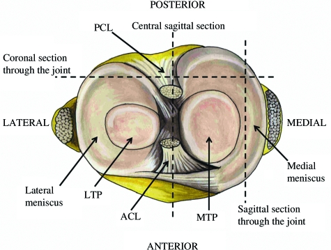
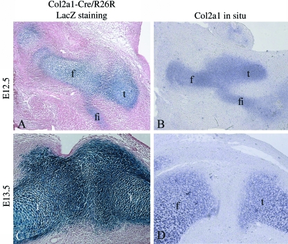
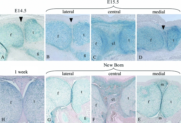
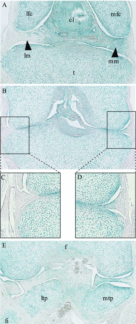
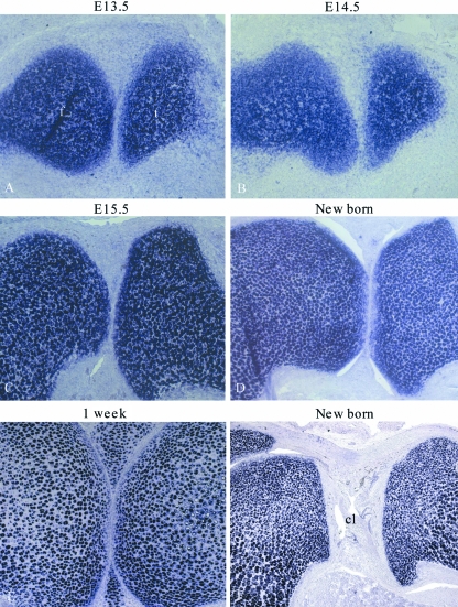
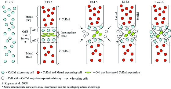
The knee joint consists of multiple interacting tissues that are prone to injury- and disease-related degeneration. Although much is known about the structure and function of the knee's constituent tissues, relatively little is known about their cellular origin and the mechanisms governing their segregation. To investigate the origin and segregation of knee tissues in vivo we performed lineage tracing using a Col2a1-Cre/R26R mouse model system and compared the data obtained with actual Col2a1 expression. These studies demonstrated that at E13.5 the interzone at the presumptive joint site forms when cells within the Col2a1-expressing anlagen cease expression of Col2a1 and not through cellular invasion into the anlagen. Later in development these interzone cells form the cruciate ligament and inner medial meniscus of the knee. At E14.5, after interzone formation, cells that had never expressed Col2a1 appeared in the joint and formed the lateral meniscus. Furthermore, cells with a Col2a1-positive expression history combined with the negative cells to form the medial meniscus. The invading cells started to express Col2a1 1 week after birth, resulting in all cells within the meniscus synthesizing collagen II. These findings support a model of knee development in which cells present in the original anlagen combine with invading cells in the formation of this complex joint.


Figures

Fig. 1
Fig. 1
A transverse view of the knee joint demonstrating how the meniscus forms a complete circle around the two condyles, joining with the cruciate ligament in the centre of the joint. The meniscus is divided into two halves, the lateral and medial menisci. The planes of sagittal and coronal sections are represented. Posterior cruciate ligament (PCL), anterior cruciate ligament (ACL), lateral tibial plateau (LTP), medial tibial plateau (MTP).
Fig. 2
Fig. 2
A comparison of Col2a1expression history with current Col2a1 expression. Sagittal sections through the knees of E12.5 and E13.5 Col2a1-Cre/R26R mouse embryos. The blue staining indicates β-galactosidase activity and occurs in cells that have expressed Col2a1(A + C). In situ hybridization detecting current Col2a1expression in comparable E12.5 and E13.5 murine knee sections (B + D). Femur (f), tibia (t), fibula (fi).
Fig. 3
Fig. 3
Analysis of Col2a1expression history in E14.5, E15.5, new born and 1-week murine knee joints. A sagittal section through the lateral meniscus of an E14.5 Col2a1-Cre/R26R knee (A). Sagittal sections through E15.5 (B–D) and new born Col2a1-Cre/R26R (E–G) murine knees: through the lateral meniscus (B + G), through the developing cruciate ligament (C + F) and through the medial meniscus (D + E). A section through the lateral meniscus of a 1-week Col2a1-Cre/R26R knee (H). Blue staining indicates β-galactosidase activity and occurs in cells that have expressed Col2a1. Arrowheads indicate the developing meniscus. Femur (f), tibia (t), fibula (fi), cruciate ligament (cl), meniscus (m).
Fig. 4
Fig. 4
Coronal sections through a new born Col2a1-Cre/R26R mouse knee. A section through the anterior of the knee (A). A section through the middle of the knee where the two femoral condyles are in contact with the tibial plateau (B), magnifications of the lateral (C) and medial (D) menisci. A section through the posterior of the knee (E). Blue staining indicates β-galactosidase activity and occurs in cells that have expressed Col2a1. Tibia (t), cruciate ligament (cl), lateral meniscus (lm), medial meniscus (mm), lateral femoral condyle (lfc), medial femoral condyle (mfc), femur (f), lateral tibial plateau (ltp) and medial tibial plateau (mtp), fibula (fi).
Fig. 5
Fig. 5
Col2a1 expression in the developing medial meniscus. In situhybridization against Col2a1on E13.5 (A), E14.5 (B), E15.5 (C), new born (D), and 1-week-old (E) sagittal knee sections through the medial meniscus. In situhybridization against Col2a1 on a new born sagittal knee section through the cruciate ligament (F). Femur (f), tibia (t) and cruciate ligament (cl).
Fig. 6
Fig. 6
A refined model of knee development in the mouse. Between E12.5 and E13.5 the Col2a1-positive anlagen is interrupted when the intermediate zone cells turn off expression of Col2a1. At the same time, epiphyseal chondrocytes begin to express Matn1, distinguishing them from articular chondrocytes and other skeletal tissues. By E14.5, cells with a Col2a1-negative expression history have invaded the joint and form the lateral and outer medial meniscus. The cells which have ceased Col2a1 expression form the cruciate ligament and the inner medial meniscus. By 1 week a mature joint is almost formed and the meniscal cells begin to express or re-express Col2a1. The cells of the cruciate ligament which previously turned off Col2a1 expression do not turn it back on. Articular chondrocytes (AC), epiphyseal chondrocytes (EC).

References

    1. Archer CW, Dowthwaite GP, Francis-West P. Development of synovial joints. Birth Defects Res C Embryo Today. 2003;69:144–155. - PubMed
    1. Craig FM, Bentley G, Archer CW. The spatial and temporal pattern of collagens I and II and keratan sulphate in the developing chick metatarsophalangeal joint. Development. 1987;99:383–391. - PubMed
    1. Duthon VB, Barea C, Abrassart S, Fasel JH, Fritschy D, Menetrey J. Anatomy of the anterior cruciate ligament. Knee Surg Sports Traumatol Arthrosc. 2006;14:204–213. - PubMed
    1. Francis-West PH, Abdelfattah A, Chen P, et al. Mechanisms of GDF-5 action during skeletal development. Development. 1999;126:1305–1315. - PubMed
    1. Frank CB. Ligament structure, physiology and function. J Musculoskelet Neuronal Interact. 2004;4:199–201. - PubMed

Publication types