. 2008 Nov 11:5:21.
doi: 10.1186/1476-9255-5-21.

Cellular and molecular mechanisms of cigarette smoke-induced lung damage and prevention by vitamin C

Affiliations

Cellular and molecular mechanisms of cigarette smoke-induced lung damage and prevention by vitamin C

Shuvojit Banerjee et al. J Inflamm (Lond). .

Abstract

Background: Cigarette smoke-induced cellular and molecular mechanisms of lung injury are not clear. Cigarette smoke is a complex mixture containing long-lived radicals, including p-benzosemiquinone that causes oxidative damage. Earlier we had reported that oxidative protein damage is an initial event in smoke-induced lung injury. Considering that p-benzosemiquinone may be a causative factor of lung injury, we have isolated p-benzosemiquinone and compared its pathophysiological effects with cigarette smoke. Since vitamin C is a strong antioxidant, we have also determined the modulatory effect of vitamin C for preventing the pathophysiological events.

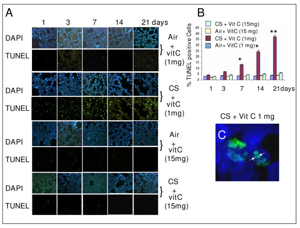
Methods: Vitamin C-restricted guinea pigs were exposed to cigarette smoke (5 cigarettes/day; 2 puffs/cigarette) for 21 days with and without supplementation of 15 mg vitamin C/guinea pig/day. Oxidative damage, apoptosis and lung injury were assessed in vitro, ex vivo in A549 cells as well as in vivo in guinea pigs. Inflammation was measured by neutrophilia in BALF. p-Benzosemiquinone was isolated from freshly prepared aqueous extract of cigarette smoke and characterized by various physico-chemical methods, including mass, NMR and ESR spectroscopy. p-Benzosemiquinone-induced lung damage was examined by intratracheal instillation in guinea pigs. Lung damage was measured by increased air spaces, as evidenced by histology and morphometric analysis. Oxidative protein damage, MMPs, VEGF and VEGFR2 were measured by western blot analysis, and formation of Michael adducts using MALDI-TOF-MS. Apoptosis was evidenced by TUNEL assay, activation of caspase 3, degradation of PARP and increased Bax/Bcl-2 ratio using immunoblot analysis and confocal microscopy.

Results: Exposure of guinea pigs to cigarette smoke resulted in progressive protein damage, inflammation, apoptosis and lung injury up to 21 days of the experimental period. Administration of 15 mg of vitamin C/guinea pig/day prevented all these pathophysiological effects. p-Benzosemiquinone mimicked cigarette smoke in causing protein modification and apoptosis in vitro and in A549 cells ex vivo as well as apoptosis and lung damage in vivo. All these pathophysiological events were also prevented by vitamin C.

Conclusion: p-Benzosemiquinone appears to be a major causative factor of cigarette smoke-induced oxidative protein damage that leads to apoptosis and lung injury. The pathophysiological events are prevented by a moderately large dose of vitamin C.


Figures

Figure 1
Figure 1
Histopathology of lung sections of guinea pigs exposed to CS showing progressive lung damage and prevention by moderately large dosage of vitamin C. A, Histology of lung sections of guinea pigs fed 1 mg or 15 mg vitamin C; Air, sham controls exposed to air; CS, exposed to CS; stained by hematoxylin and eosin. The number of guinea pigs used in each group was 4. Eight images were analyzed in 4 lung sections (2 images/section/animal) from each group (magnification ×10). B, C, Morphometric Measurements of the number of alveolar air space and surface density (S/V). Values are ± S.D. S/V has been calculated by the formula S/V (surface density) = π/4 × P/A, where P is the mean perimeter (contour length) of air space and A is the area of air space [6]. The number of air spaces and the S/V values calculated after 7, 14 and 21 days of smoke exposure are significantly less (p < 0.05)than that observed after 1 day exposure; * over the bars (Fig. 1 C) indicate p < 0.05 with respect to controls.
Figure 2
Figure 2
A, Detection of DNA strand breaks by TUNEL assay in lung cells of vitamin C-restricted (1 mg vitamin C/animal/day) or vitamin C-supplemented (15 mg/animal/day) guinea pigs exposed to air CS (as described under Methods). Following CS exposure, the guinea pigs were sacrificed after 1, 3, 7, 14 and 21 days. Lower Panel: the lung sections were stained with fluorescein labeled dUTP according to the protocols discussed under Methods. Green fluorescence indicates TUNEL positive cells. Upper Panel: Lung sections corresponding to the upper panel were stained with DAPI to identify the cell nuclei. B, Quantitative evaluation of TUNEL positive cells in lungs of guinea pigs exposed to air or CS. Eight images were analyzed in 4 lung sections (2 images/section/animal) from each group, respectively; the bars over the respective columns represent means ± SD; * indicates p < 0.05 and ** indicates p < 0.001 with respect to sham controls. C, Colocalization of TUNEL and DAPI staining in the nuclei of lungs of guinea pig fed 1 mg vitamin C/day and exposed to CS for 21 days; original magnification × 63; image captured by confocal microscopy (Zeiss LSM 510 META).
Figure 3
Figure 3
A, Immunoblots of caspase 3, PARP, Bax and Bcl-2 of the lung extracts of guinea pigs exposed to air or CS supplemented with 1 mg or 15 mg vitamin C/animal/day. Vit C means vitamin C. Air + vit C means exposed to air and fed 1 mg or 15 mg vitamin C/day; CS + vit C, means exposed to CS and fed 1 mg or 15 mg vitamin C/day. B, quantitative evaluation of Bax/Bcl-2 ratio. C, detection of apoptosis in the lung of guinea pig fed 1 mg vitamin C and exposed to CS for 21 days by immunofluorescence analysis using cleaved caspase 3 antibodies; lower panel I, stained with TRITC (red signal); II, stained with DAPI (blue); III, double staining with DAPI and TRITC; IV, enlarged version of III. D, colocalization of cleaved caspase 3 (CC3) in lungs of CS-exposed guinea pigs fed 1 mg vitamin C; red signal (TRITC) indicates cleaved caspase 3; blue signal, DAPI and green signal, TUNEL. Merged mens combination of TRITC, DAPI and TUNEL; image captured by laser scanning confocal microscope; original magnification × 63. E, Accumulation of Bax proteins in the lungs of CS-exposed guinea pigs fed 1 mg vitamin C/day; red signal, stained with TRITC and blue signal, stained with DAPI. Merged means combination of TRITC and DAPI.
Figure 4
Figure 4
A, Upper panel: Immunoblots of the DNP-derivatives of lung proteins of guinea pigs exposed to smoke from 1, 2 and 3 cigarettes. Twenty five μg proteins isolated from air-exposed or CS-exposed guinea pigs fed vitamin C-free diet for 7 days were converted to the DNP-derivative followed by immunoblotting as mentioned in the Methods. Lower panel: The membrane was reprobed with anti PARP antibodies which show that there was no PARP cleavage. B, Detection of DNA strand breaks (TUNEL assay) in lung cells of guinea pigs exposed to smoke from 1, 2 and 3 cigarettes. C, Quantitative evaluation of TUNEL assay and protein carbonyl formation in the lungs of guinea pigs exposed to smoke from 1, 2 and 3 cigarettes. Protein carbonyl was measured by densitometric scanning using Quantity One- 4.4 (Bio-Rad) Software. Bars over the respective columns represent means ± SD (n = 4). D, Western blots of mitochondrial and cytosolic cytochrome c of lungs of guinea pigs exposed to 1, 2 and 3 cigarettes; M represents mitochondria and C, cytosol. E, Immunoblots of VEGF in BALF isolated from guinea pigs with and without exposure to smoke from 3 cigaretes. Lane 1, sham control after exposure to air; lane 2, after exposure to 3 cigarettes; Similar results were obtained from 3 different animals. The upper panel represents the dimmer of VEGF (Mr 42 kDa) and lower panel, the monomer (Mr 21 kDa). Loading control (SDS-PAGE) is shown at the bottom. F, Immunoblots of VEGRF2 in lung tissue of guinea pigs with and without exposure to smoke from 3 cigarettes. Lane 1, sham control after exposure to air; lane 2, after exposure to 3 cigarettes; lower panel indicates loading control. Similar results were obtained from 3 different animals. G, western blots showing levels of MMP-9 and MMP-12 in lung tissue and BALF isolated from guinea pigs fed 1 mg vitamin C/day and exposed to air or CS for 14 days; In each of the blots under MMP-9 and MMP-12, the left blot indicates exposed to air and the right, exposed to CS.
Figure 5
Figure 5
Effect of N-acetyl cysteine (NAC) on CS-induced protein oxidation in vitro and apoptosis and lung damage in guinea pigs in vivo. A, CS-induced protein carbonyl formation in BSA in the presence and absence of NAC and vitamin C, as measured by immunoblot analysis. The incubation mixture contained BSA (1 mg) AECS (50 μl), NAC (100 μM) or vitamin C (200 μM) in a final volume of 200 μl of 50 mM potassium phosphate buffer, pH 7.4; incubated for 2 h at 37°C with shaking. After incubation, production of the DNP derivative was measured by immunoblot analysis, as describe before [6]. B, Detection of DNA strand breaks in lung cells of guinea pigs exposed to air or CS in the presence of NAC by TUNEL assay. After feeding vitamin C-free diet for 7 days, the guinea pigs were supplemented with 1 mg vitamin C/day and fed 15 mg NAC/animal/day and exposed to air or CS for 7 days, as described under Methods. C, Quantitative evaluation of TUNEL positive cells in lungs of guinea pigs exposed to CS in the presence or absence of NAC. Eight images were analyzed in 4 lung sections (2 images/section/animal) from each group, respectively; bars over the respective columns represent means ± SD. D, Histopathology profiles of lung sections of guinea pig after exposure to CS in the presence and absence of NAC (15 mg/animal/day). The number of guinea pigs used in each group was 4. Eight images were analyzed in 4 lung sections (2 images/section/animal) from each group (magnification ×10). E, F, Morphometric Measurements of alveolar air space, and surface density (S/V). Values are ± SD; number of images analyzed in each group:8.
Figure 6
Figure 6
p-BSQ mimics AECS causing oxidative modification and proteolysis in vitro: prevention by vitamin C. A, Dose-dependent AECS/p-BSQ/p-BQ-induced protein carbonyl formation in BSA in the presence or absence of vitamin C, as measured spectrophotometrically. The incubation mixture was same as in Fig.5A using AECS (50 μl), p-BSQ (90 nmoles) or p-BQ (45 nmoles). Small bars over the points in the graph represent mean ± SD. B, Conversion of p-BSQ to p-BQ by disproportionation reaction or by SOD/cytochrome c catalysed oxidation. p-BSQ (180 nmols) was incubated in 200 μl of 50 mM phosphate buffer, pH 7.4 at 37°C in air with or without the presence of Cu, Zn-SOD (30 μg) and cytochromec (180 nmoles), incubation was carried out in air. Aliquots were taken, diluted with mobile solvent and p-BQ estimated by HPLC. Details are given in Methods. ↓ Indicates addition of vitamin C (200 μM). C, Degradation of guinea pig lung microsomal proteins as a function of p-BSQ concentration. D, E, Degradation of guinea pig lung microsomal proteins by p-BSQ (180 nmoles) and p-BQ (90 nmoles) in the presence and absence of vitamin C (200 μM). Conditions are same as described previously [7]. Similar observations were made in three independent experiments.
Figure 7
Figure 7
AECS/p-BSQ/p-BQ-induced apoptosis in A549 lung epithelial cells ex vivo. A, Detection of DNA strand break using TUNEL assay. The cells were examined using a fluorescence microscope (Olympus B, magnification × 20). Digital images were captured with cool CCD camera (Olympus). Upper panel, TUNEL positive cells; lower panel, nuclei stained with DAPI. About 10 fields (150 cells/field) of three independent experiments were evaluated. A549 cells (2 × 10⁶) were treated with 50 μl of AECS, 180 nmoles p-BSQ or 90 nmoles p-BQ in culture medium at 37°C for 24 h. B, Quantitative evaluation of TUNEL positive cells treated with AECS/p-BSQ/p-BQ; PBS indicates control treated with phosphate buffered saline; small bars over the columns indicate ± SD. C, Activation of pro-caspase-3 analyzed by immunoblotting. D, Cleavage of poly ADP ribose polymerase (PARP) analysed by immunobloting using anti PARP antibody. For immunoblot assay of caspase 3 and PARP, cells were treated with AECS, p-BSQ or p-BQ for 12 h. Similar observations were made in three independent experiments.
Figure 8
Figure 8
Apoptosis and lung damage caused by intratracheal instillation of p-BSQ in guinea pigs and prevention by vitamin C. The guinea pigs were given a solution of 150 μg p-BSQ in 200 μl of normal saline/guinea pig/twice a day for 7 days by intratracheal instillation and then sacrificed. Details of intratracheal instillation are given in Materials and Methods. Control animals (PBS treated) received only saline (200 μl). Supplementation of vitamin C (15 mg/day) prevented apoptosis (about 80%) and lung damage (about 85%) in the p-BSQ treated group. In the saline-treated sham controls, administration of vitamin C (15 mg/day) did not produce any significant change in the morphology of alveolar cells or TUNEL assay (Fig. not shown). A, Upper panel shows TUNEL positive cells; lower panel, lung sections stained with DAPI. The number of guinea pigs used in each group was three. Six images were analyzed in three lung sections (2 images/section) from each group (magnification × 10). B, Histopathology (H and E staining) of lung sections after intratracheal instillation of PBS or p-BSQ. Marked enlargement of alveolar air spaces is found in the p-BSQ group (magnification × 20), which is markedly inhibited by vitamin C.
Figure 9
Figure 9
Mechanism of CS-induced lung damage and prevention by vitamin C.p-BSQ of CS induces all the pathological conditions via the formation of p-BQ. Vitamin C reduces and inactivates p-BQ and thereby prevents the lung injury

References

    1. Tuder RM, Petrache I, Elias JA, Voelkel NF, Henson PM. Apoptosis and emphysema: the Missing Link. Am J Respir Cell Mol Biol. 2003;28:551–554. - PubMed
    1. Demendts IK, Demoor T, Bracke KR, Joos GF, Brusselle GG. Role of apoptosis in the pathogenesis of COPD and pulmonary emphysema. Respir Res. 2006;7:53. - PMC - PubMed
    1. Stewart BW, Kleihues P. International Agency for Research on Cancer. Lyon, France; 2003. World Cancer Report; pp. 21–28.
    1. Chouchane S, Wooten JB, Tewes FJ, Wittig A, Muller BP, Veltel D, Diekmann J. Involvement of semiquinone radicals in the in vitro cytotoxicity of cigarette mainstream smoke. Chemical Research in Toxicology. 2006;19:1602–1610. - PubMed
    1. Pryor WA, Stone K, Zang L-Y, Bermudez E. Fractionation of Aqueous Cigarette Tar Extracts: Fractions That Contain the Tar Radical Cause DNA Damage. Chem Res Toxicol. 1998;11:441–448. - PubMed