. 2008 Nov 12:8:311.
doi: 10.1186/1471-2148-8-311.

Sox genes in the coral Acropora millepora: divergent expression patterns reflect differences in developmental mechanisms within the Anthozoa

Affiliations

Sox genes in the coral Acropora millepora: divergent expression patterns reflect differences in developmental mechanisms within the Anthozoa

Chuya Shinzato et al. BMC Evol Biol. .

Abstract

Background: Sox genes encode transcription factors that function in a wide range of developmental processes across the animal kingdom. To better understand both the evolution of the Sox family and the roles of these genes in cnidarians, we are studying the Sox gene complement of the coral, Acropora millepora (Class Anthozoa).

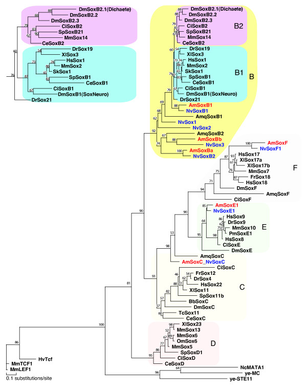
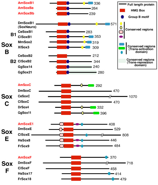
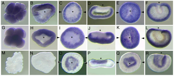
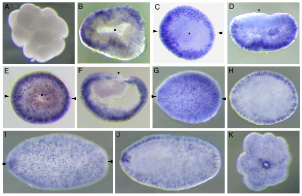
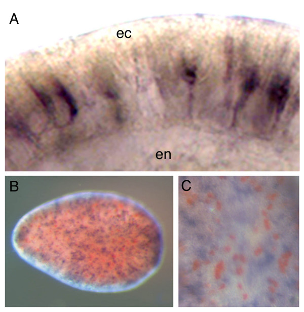
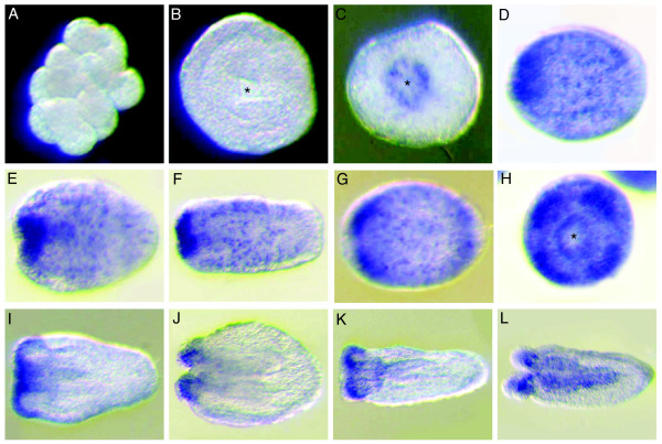
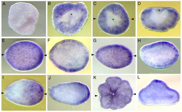
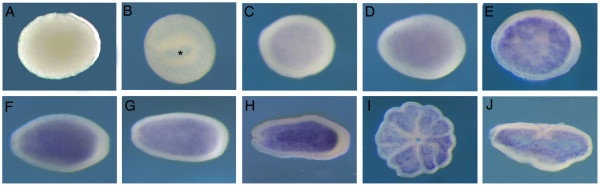
Results: Based on overall domain structures and HMG box sequences, the Acropora Sox genes considered here clearly fall into four of the five major Sox classes. AmSoxC is expressed in the ectoderm during development, in cells whose morphology is consistent with their assignment as sensory neurons. The expression pattern of the Nematostella ortholog of this gene is broadly similar to that of AmSoxC, but there are subtle differences--for example, expression begins significantly earlier in Acropora than in Nematostella. During gastrulation, AmSoxBb and AmSoxB1 transcripts are detected only in the presumptive ectoderm while AmSoxE1 transcription is restricted to the presumptive endoderm, suggesting that these Sox genes might play roles in germ layer specification. A third type B Sox gene, AmSoxBa, and a Sox F gene AmSoxF also have complex and specific expression patterns during early development. Each of these genes has a clear Nematostella ortholog, but in several cases the expression pattern observed in Acropora differs significantly from that reported in Nematostella.

Conclusion: These differences in expression patterns between Acropora and Nematostella largely reflect fundamental differences in developmental processes, underscoring the diversity of mechanisms within the anthozoan Sub-Class Hexacorallia (Zoantharia).


Figures

Figure 1
Figure 1
Maximum Likelihood phylogenetic analyses of Acropora Sox genes in MolPhy version 2.3[49] using the Dayhoff model of protein evolution and local rearrangement of the NJ trees (1,000 bootstraps). Acropora millepora Sox genes are shown in red, while Nematostella vectensis genes are shown in blue. The inset, showing the B1 and B2 families, has been stretched horizontally to clarify the relationship between the B1 and B2 genes for the reader. Species names are abbreviated as follows; Am, coral, Acropora millepora; Amq, demosponge, Amphimedon queenslandica; Bb, Japanese lancelet, Branchiostoma belcheri; Ce, nematode, Caenorhabditis elegans; Ci, ascidian, Ciona intestinalis; Dm, fruit-fly, Drosophila melanogaster; Dr, zebrafish, Danio rerio. Fr, Japanese pufferfish, Fugu rubripes; Gg, chicken, Gallus gallus; Hs, human, Homo sapiens; Hv, hydra, Hydra vulgaris; Mm, mouse, Mus musculus; Nc, red bread mold, Neurospora crassa; Nv, sea anemone, Nematostella vectensis; Om, rainbow trout, Oncorhynchus mykiss; Pm, sea lamprey, Petromyzon marinus; Sk, hemichordate, Saccoglossus Kowalevskii; Sp, sea urchin, Strongylocentrotus purpuratus; Tc, red flour beetle, Tribolium castaneum; Xl, frog, Xenopus laevis; ye-, yeast, Schizosaccharomyces pombe.
Figure 2
Figure 2
Schematic drawings, not to scale, comparing Acropora Sox proteins with those of various bilaterians, across the main Sox families (B-F). The conserved motifs compared are identified in the inset. Numbers of amino acids in the full length proteins are indicated on the right. Species names are abbreviated as follows; Am, coral, Acropora millepora; Ce, nematode, Caenorhabditis elegans; Ci, ascidian, Ciona intestinalis; Dm, fruit-fly, Drosophila melanogaster; Dr, zebrafish, Danio rerio. Fr, Japanese pufferfish, Fugu rubripes; Gg, chicken, Gallus gallus; Hs, human, Homo sapiens; Mm, mouse, Mus musculus; Xl, frog, Xenopus laevis.
Figure 3
Figure 3
Spatial expression patterns of AmSoxBb, AmSoxB1 and AmSoxE1 during early embryogenesis: A-F) AmSoxB1; (G-L) AmSoxBb; (M-R) AmSoxE1. (A, G, M) Early cleavage stage. (B, H, M) Blastula (prawnchip) stage. (C, I, O) Early donut stage, during gastrulation. (D, J, P) Transverse sections of C, I, O, respectively. (E, K, Q) Late donut stage, finishing gastrulation. (F, L, R) Transverse sections of E, K, Q respectively. Asterisks indicate the blastopore. Paired arrows on a panel of the figure indicate that the next panel is a section in the plane of the arrows (e.g. D is a transverse section of the embryo shown in C). The speed of embryonic development is temperature dependent, so we have not attempted to give ages of the embryonic stages in this and later figures. Typical ages for the various stages are available in Figure 2 of Ball et al. [38].
Figure 4
Figure 4
AmSoxC shows cell specific expression in the ectoderm from early embryogenesis through post-settlement: (A) Early cleavage stage. (B) Prawnchip stage. (C) Initiation of gastrulation at the early donut stage. (D) Transverse section of C. (E) Donut stage embryo, during gastrulation. (F) Longitudinal section of E. (G) Pear stage. (H) Longitudinal section of G showing that expression is restricted to the ectoderm. (I) Planula stage. (J) Longitudinal section of I. The cell specific expression in the ectoderm continues and oral pore staining is observed. (K) Post settlement polyp. Though the ectodermal cell specific expression becomes weaker, the oral pore staining is still maintained. Paired arrows on a panel of the figure indicate that the next panel is a section in the plane of the arrows (e.g. D is a transverse section of the embryo shown in C).
Figure 5
Figure 5
Cell morphology of AmSoxC expressing cells. (A) High magnification view of the ectoderm of in situ stained embryos (pear stage). (B) Double in situ hybridization of AmSoxC and Amlipase (planula stage). AmSoxC is stained black and Amlipase is red. (C) High magnification view of the surface of B. No overlapping staining is observed. Abbreviations: en = endoderm, ec = ectoderm.
Figure 6
Figure 6
Spatial expression pattern of NvSoxC during Nematostella development. Asterisks indicate the blastopore (B, C) or oral pore (H) when it faces out of the page, otherwise the oral pore is oriented to the left and aboral is to the right. (A) Early cleavage stage, (B) gastrula stage, (C) late gastrula stage, immediately after blastopore closure. (D-F) planula stage, (G) expression in pre-tentacles, (H) oral pore view of G, (I) early metamorphosing planula, (J) late metamorphosing planula stage with further retraction of pharynx towards aboral pole, (K) late metamorphosing planula which has finished elongating, (L) primary polyp.
Figure 7
Figure 7
Spatial expression pattern of AmSoxBa during development. (A) There is no expression in the early prawnchip stage, (B) Initiation of gastrulation in the late prawnchip stage. AmSoxB2 transcripts appear in the presumptive ectoderm with some cell specific staining. (C) At the donut stage the general ectodermal expression and some cell specific staining persist. (D) Transverse section of C. Expression is restricted to the presumptive ectoderm. (E) Pear stage (F) Longitudinal section of E shows ectodermal expression. (G) Late pear or early planula stage. (H) Longitudinal section of G. (I) Planula stage (96 h). Cell specific expression is restricted to the aboral half of the ectoderm. (J) Section image of I. (K) Post settlement polyp. (L) Transverse section of K. Expression is now missing from the aboral ectoderm. Asterisks indicate the position of the blastopore. (E-J) Oral pore is oriented to the left and aboral side to the right. Paired arrows on a panel of the figure indicate that the next panel is a section in the plane of the arrows (e.g. D is a transverse section of the embryo shown in C).
Figure 8
Figure 8
Spatial expression pattern of AmSoxF throughout development. An asterisk indicates the blastopore. (C-H) Oral pore is oriented to the left and aboral side to the right. (A) Unfertilized egg. (B) late donut stage (C-H) weak endodermal expression appears at the pre-pear stage, strengthening throughout the late pear and planula stages. (I-J) Endodermal expression continues post-settlement.

References

    1. Schepers GE, Teasdale RD, Koopman P. Twenty pairs of sox: extent, homology, and nomenclature of the mouse and human sox transcription factor gene families. Dev Cell. 2002;3(2):167–170. doi: 10.1016/S1534-5807(02)00223-X. - DOI - PubMed
    1. Bowles J, Schepers G, Koopman P. Phylogeny of the SOX family of developmental transcription factors based on sequence and structural indicators. Dev Biol. 2000;227(2):239–255. doi: 10.1006/dbio.2000.9883. - DOI - PubMed
    1. Cremazy F, Berta P, Girard F. Genome-wide analysis of Sox genes in Drosophila melanogaster. Mech Dev. 2001;109(2):371–375. doi: 10.1016/S0925-4773(01)00529-9. - DOI - PubMed
    1. Wegner M. From head to toes: the multiple facets of Sox proteins. Nucleic Acids Res. 1999;27(6):1409–1420. doi: 10.1093/nar/27.6.1409. - DOI - PMC - PubMed
    1. McKimmie C, Woerfel G, Russell S. Conserved genomic organisation of Group B Sox genes in insects. BMC Genet. 2005;6(1):26. doi: 10.1186/1471-2156-6-26. - DOI - PMC - PubMed

Publication types

Substances

LinkOut - more resources