. 2008 Nov 13:9:112.
doi: 10.1186/1471-2202-9-112.

The Kv2.1 K+ channel targets to the axon initial segment of hippocampal and cortical neurons in culture and in situ

Affiliations

The Kv2.1 K+ channel targets to the axon initial segment of hippocampal and cortical neurons in culture and in situ

Patrick D Sarmiere et al. BMC Neurosci. .

Abstract

Background: The Kv2.1 delayed-rectifier K+ channel regulates membrane excitability in hippocampal neurons where it targets to dynamic cell surface clusters on the soma and proximal dendrites. In the past, Kv2.1 has been assumed to be absent from the axon initial segment.

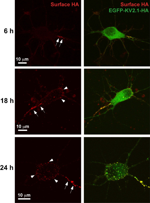
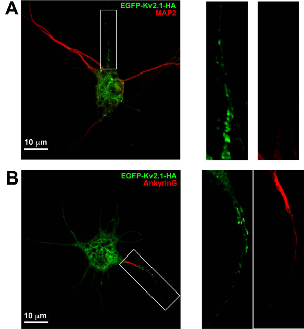
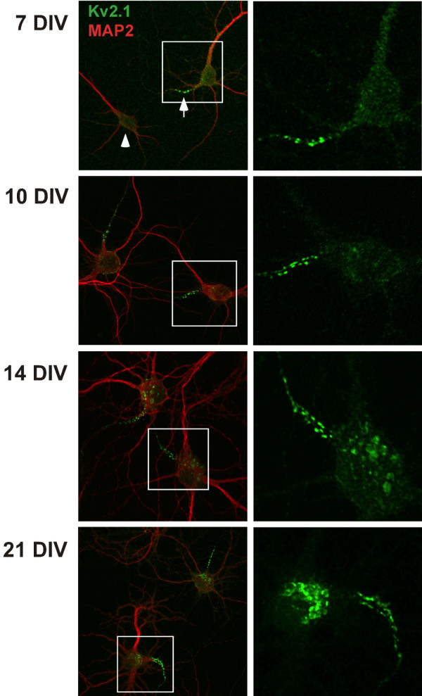
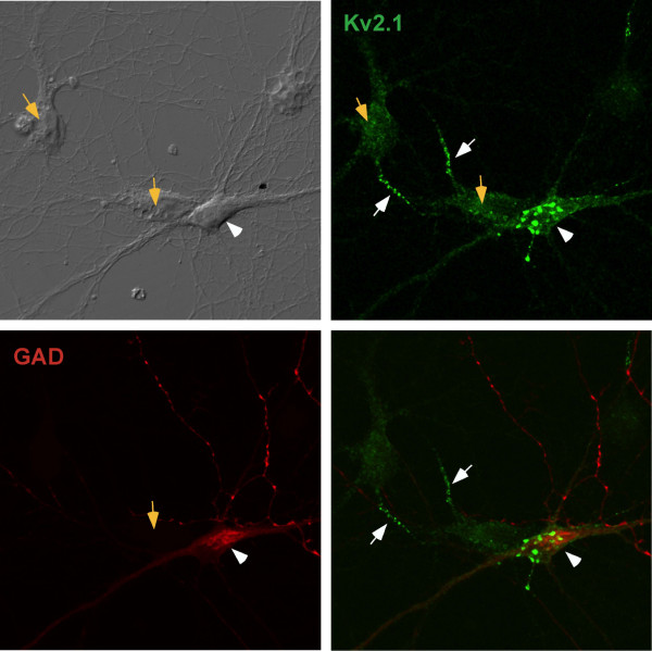
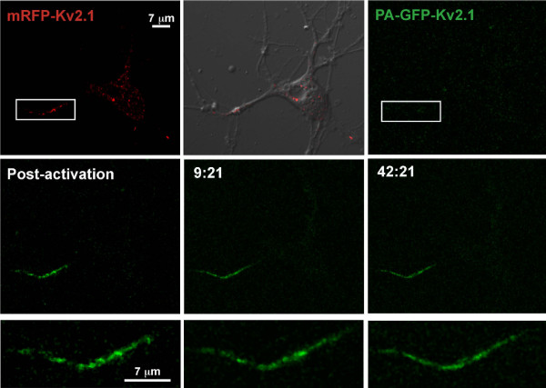
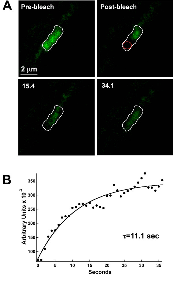
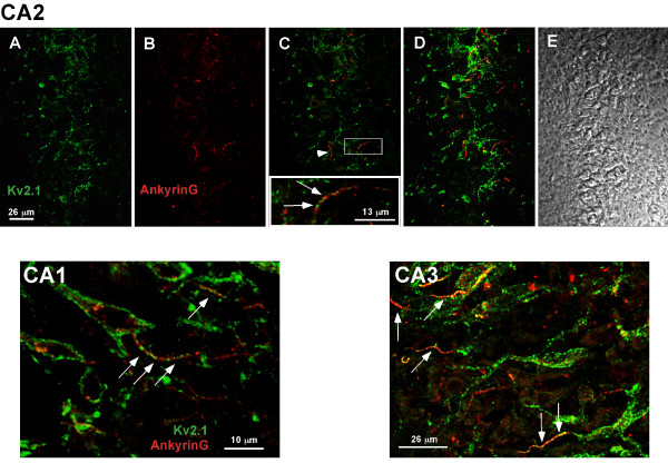
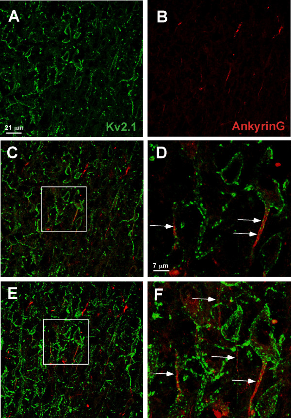
Results: Transfected and endogenous Kv2.1 is now demonstrated to preferentially accumulate within the axon initial segment (AIS) over other neurite processes; 87% of 14 DIV hippocampal neurons show endogenous channel concentrated at the AIS relative to the soma and proximal dendrites. In contrast to the localization observed in pyramidal cells, GAD positive inhibitory neurons within the hippocampal cultures did not show AIS targeting. Photoactivable-GFP-Kv2.1-containing clusters at the AIS were stable, moving <1 microm/hr with no channel turnover. Photobleach studies indicated individual channels within the cluster perimeter were highly mobile (FRAP tau=10.4+/-4.8 sec), supporting our model that Kv2.1 clusters are formed by the retention of mobile channels behind a diffusion-limiting perimeter. Demonstrating that the AIS targeting is not a tissue culture artifact, Kv2.1 was found in axon initial segments within both the adult rat hippocampal CA1, CA2, and CA3 layers and cortex.

Conclusion: In summary, Kv2.1 is associated with the axon initial segment both in vitro and in vivo where it may modulate action potential frequency and back propagation. Since transfected Kv2.1 initially localizes to the AIS before appearing on the soma, it is likely multiple mechanisms regulate Kv2.1 trafficking to the cell surface.


Figures

Figure 1
Figure 1
Surface accumulation of transfected Kv2.1 in a single neurite. Hippocampal neurons transfected with EGFP-Kv2.1-HA for 6, 18 and 24 h were labeled with Alexa 594-conjugated anti-HA monoclonal antibody 30 min prior to live cell imaging. Shown are representative maximum projection images comprised of multiple 0.3 μm optical sections for total EGFP-Kv2.1-HA (green, GFP signal) and surface (red, Alexa 594 anti-HA antibody binding). Kv2.1 surface clusters were observed exclusively in single a proximal neurite as early as 6 hours after transfection (arrows). At 18 and 24 hours post-transfection, the appearance of surface somato-dendritic clustering became apparent (arrowheads).
Figure 2
Figure 2
The single proximal neurite containing Kv2.1 corresponds to the axon initial segment. Hippocampal neurons grown for 10 days were transfected with EGFP-Kv2.1-HA and fixed 18 h later. Cultures were then labeled with either anti-MAP2 or anti-AnkG followed by Alexa 594-conjugated secondary antibodies. Shown in panel A is an example of Kv2.1 clustering in the proximal axon as indicated by the diminished MAP2 staining. Panel B illustrates intense AnkG staining in the Kv2.1 positive process. The right hand panels contain enlarged images of the neurites boxed in Panels A and B. Note that the diffuse GFP signal in the neurites represents intracellular GFP-Kv2.1.
Figure 3
Figure 3
Endogenous Kv2.1 preferentially accumulates in a MAP2-negative neurite. Hippocampal cultures grown for 7, 10, 14 and 21 days were fixed and immunostained for endogenous Kv2.1 (green) and the dendritic marker MAP2 (red). At 7 DIV, the strongest labeling for Kv2.1 in these cultures appeared in the proximal portion of a MAP2-negative process (arrow) although many neurons exhibited faint clusters evenly dispersed over the cell body and proximal neurites (arrowhead). Shown in the right hand panels are maximum projection images corresponding to Kv2.1 staining from the boxed regions. In the majority of neurons, the neurite with intense Kv2.1 labeling corresponded to the process with the least intense MAP2 staining, indicating the preferential accumulation of Kv2.1 at the proximal axon.
Figure 4
Figure 4
GAD-positive cells do not exhibit Kv2.1 expression in the AIS. DIV14 hippocampal neurons were fixed and stained for endogenous Kv2.1 and glutamic acid decarboxylase (GAD) to identify inhibitory neurons. The orange arrows indicate the GAD-negative neurons while the white arrowhead points to a GAD-positive cell. The white arrows indicate the AIS-localized Kv2.1 associated with the GAD-negative neurons. These results are consistent with Kv2.1 localization to the AIS only in excitatory, GAD-negative pyramidal neurons.
Figure 5
Figure 5
Kv2.1 clusters within the AIS are stable. Shown is a hippocampal neuron co-transfected with a 1:1 ratio of mRFP-Kv2.1 and PAGFP-Kv2.1-HA plasmid DNA. The proximal region of the single neurite expressing Kv2.1 (rectangle) was exposed to 405 nm laser light to photo-activate the PA-GFP. The top row illustrates the mRFP-Kv2.1 signal (red), the mRFP signal overlaid onto the corresponding DIC image and image acquisition under GFP optics. The middle row illustrates the PA-GFP signal immediately after photoactivation and at two representative time points (min:sec). The bottom row contains enlargements of the activated PA-GFP following photoactivation. Only small changes in cluster size and position occurred over the imaging period. There was no significant decrease in total AIS GFP fluorescence during the 42 minutes of acquisition, suggesting stable retention of Kv2.1 within the proximal axon. The imaging interval was 2 min.
Figure 6
Figure 6
FRAP analysis indicates Kv2.1 channels are mobile within the AIS cluster perimeter. Panel A shows FRAP of an AIS cluster. One half of an AIS cluster (white outline) was photo-bleached and FRAP then monitored every 1.1 sec. By 15 sec into the recovery, GFP fluorescence was readily observed diffusing from the unbleached half into the bleached region. Panel B shows the FRAP time course. FRAP was quantitated within a small region of interest drawn in the bleached region (red circle). The solid line represents a single exponential fit to the data; τ = 11.1 sec for the cluster illustrated. The mean FRAP time constant for AIS clusters was 10.4 ± 4.8 sec, n = 7.
Figure 7
Figure 7
Localization of Kv2.1 clusters to the AIS of hippocampal neurons in situ. Postnatal day 21 rat brains were formaldehyde-fixed, cryosectioned and immuno-stained with a polyclonal antibody against Kv2.1 and a monoclonal antibody against ankyrin G as described in Methods. The anti-Kv2.1 antibody was detected with Alexa 488-conjugated goat anti-rabbit secondary antibody (green) while the anti-AnkG monoclonal antibody was detected with Alexa 594-conjugated goat anti-mouse secondary (red). The top row of panels (A-E) illustrates the localization observed in the CA2 hippocampal layer. Single optical sections of either Kv2.1 or ankyrinG immuno-staining are shown in panels A and B, respectively. Panel C shows the overlay of these two images with the arrowhead pointing to an AIS domain with little or no Kv2.1 immuno-reactivity. However, as the insert containing a magnification of the boxed region indicates, Kv2.1 was most often found in the ankyrinG-positive AIS. Panel D shows a maximum projection image (Z-stack compression) and panel E the corresponding DIC image of the same field. The CA1 image represents a single optical section within the CA1 layer while the CA3 image is a maximum projection image of this hippocampal region. The arrows denote the expression of Kv2.1 within the AnkyrinG positive AIS domains.
Figure 8
Figure 8
Localization of Kv2.1 clusters to the AIS of layer IV cortical neurons in situ. Rat brain formaldehyde-fixed cyrosections obtained from post natal day 21 animals were immuno-stained with polyclonal antibody against Kv2.1 and monoclonal antibody against ankyrin G. The anti-Kv2.1 antibody was detected with Alexa 488-conjugated goat anti-rabbit secondary antibody (green) while the anti-AnkG monoclonal antibody was detected with Alexa 594-conjugated goat anti-mouse secondary antibody (red). Single optical sections of either Kv2.1 or ankyrinG immuno-staining are shown in panels A and B. Panel C shows the overlay of these two images and panel D contains a magnification of the boxed region in C. Panels E and F show a maximum projection image (Z-stack compression) corresponding to the areas shown in panels C and D. The arrows denote the expression of Kv2.1 within AnkG-positive AIS domains (arrows).

References

    1. Scannevin RH, Murakoshi H, Rhodes KJ, Trimmer JS. Identification of a cytoplasmic domain important in the polarized expression and clustering of the Kv2.1 K+ channel. J Cell Biol. 1996;135:1619–1632. doi: 10.1083/jcb.135.6.1619. - DOI - PMC - PubMed
    1. Antonucci DE, Lim ST, Vassanelli S, Trimmer JS. Dynamic localization and clustering of dendritic Kv2.1 voltage-dependent potassium channels in developing hippocampal neurons. Neuroscience. 2001;108:69–81. doi: 10.1016/S0306-4522(01)00476-6. - DOI - PubMed
    1. Du J, Tao-Cheng JH, Zerfas P, McBain CJ. The K+ channel, Kv2.1, is apposed to astrocytic processes and is associated with inhibitory postsynaptic membranes in hippocampal and cortical principal neurons and inhibitory interneurons. Neuroscience. 1998;84:37–48. doi: 10.1016/S0306-4522(97)00519-8. - DOI - PubMed
    1. Muennich EA, Fyffe RE. Focal aggregation of voltage-gated, Kv2.1 subunit-containing, potassium channels at synaptic sites in rat spinal motoneurones. J Physiol. 2004;554:673–685. doi: 10.1113/jphysiol.2003.056192. - DOI - PMC - PubMed
    1. Du J, Haak LL, Phillips-Tansey E, Russell JT, McBain CJ. Frequency-dependent regulation of rat hippocampal somato-dendritic excitability by the K+ channel subunit Kv2.1. J Physiol. 2000;522:19–31. doi: 10.1111/j.1469-7793.2000.t01-2-00019.xm. - DOI - PMC - PubMed

Publication types