. 2008 Nov 14:4:56.
doi: 10.1186/1744-8069-4-56.

Autonomic fiber sprouting in the skin in chronic inflammation

Affiliations

Autonomic fiber sprouting in the skin in chronic inflammation

Lina Almarestani et al. Mol Pain. .

Abstract

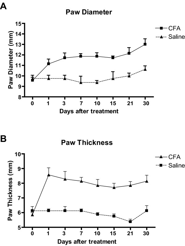
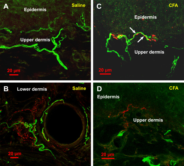
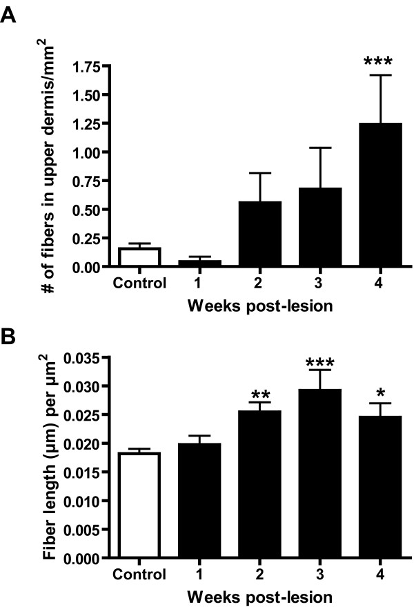
Pain is a major symptom associated with chronic inflammation. In previous work from our laboratory, we have shown that in animal models of neuropathic pain there is a sprouting of sympathetic fibers into the upper dermis, a territory normally devoid of them. However, it is not known whether such sympathetic sprouting, which is likely trophic factor mediated, also occurs in chronic inflammation and arthritis. In the present study, we used a rat model of chronic inflammation in which a small single dose of complete Freund's adjuvant (CFA) was injected subcutaneously, unilaterally, into the plantar surface of the hindpaw. This led to a localized long-term skin inflammation and arthritis in all joints of the hindpaw. Animals were perfused with histological fixatives at 1, 2, 3 or 4 weeks after the injection. Experimental animals treated with CFA were compared to saline-injected animals. We then investigated the changes in the pattern of peripheral innervation of the peptidergic nociceptors and sympathetic fibers in rat glabrous hindpaw skin. Antibodies directed towards calcitonin gene-related peptide (CGRP) and dopamine beta-hydroxylase (DBH) were used for the staining of peptidergic and sympathetic fibers, respectively. Immunofluorescence was then used to analyze the different nerve fiber populations of the upper dermis. At 4 weeks following CFA treatment, DBH-immunoreactive (IR) fibers were found to sprout into the upper dermis, in a pattern similar to the one we had observed in animals with a chronic constriction injury of the sciatic nerve in a previous publication. There was also a significant increase in the density of CGRP-IR fibers in the upper dermis in CFA treated animals at 2, 3 and 4 weeks post-injection. The increased peptidergic fiber innervation and the ectopic autonomic fibers found in the upper dermis may have a role in the pain-related behavior displayed by these animals.


Figures

Figure 1
Figure 1
Average maximum width ("diameter"), measured at the level of the digits, and dorso-ventral thickness of the hindpaw at several time before and after CFA or saline injection. Values are expressed as means ± SEM, n = 4. Note that in CFA-treated animals both parameters are already substantially increased and remain elevated at 30 days post-CFA, compared to saline.
Figure 2
Figure 2
Confocal images of the rat's hindpaw skin in saline-treated (A and B) and CFA-treated (C and D) rats, at 30 days post-injection. The material was processed for double immunofluorescence as described in the methods section, and examined with a Zeiss LSM 510 confocal microscope. CGRP immunoreactivity is shown in green and DBH immunoreactivity in red. Except for D that represents a single optical section, images represent z-stacks projected in the horizontal plane to show an extended focus. In A, note that in saline treated rats, the sympathetic fibers (DBH-IR), in red, are not observed in the upper dermis. In B, note that in the lower dermis of saline-treated rats sympathetic fibers can be observed in association with an arteriole, which is also innervated by CGRP-IR fibers. In contrast, in B and C, which were taken from CFA-treated rats, note the presence of sympathetic fibers in the upper dermis; although in both images they were observed travelling along the dermal-epidermal junction, most such fibers did not get as close to the epidermis. In C, note that the sympathetic fibers in the upper dermis often wrap around CGRP-IR fibers; arrows show zones of close apposition of the two fiber types (A), although they are detected in the lower dermis around blood vessels (B).
Figure 3
Figure 3
Quantitative analysis of sympathetic and sensory fibers in the upper dermis of saline-treated rats and in CFA-treated rats at 1, 2, 3 and 4 weeks post-injection. For sympathetic fibers (A), the number of fibers in the upper dermis was counted, whether for the sensory fibers the total length of fibers per mm2 was calculated. For details regarding quantification, see Material and Methods. In A, note that the average number of sympathetic fibers in the upper dermis was higher in the CFA group than in the saline group at 2, 3 and 4 weeks post-injection, although that increase reached significance levels at the 4 week time point only. One-way ANOVA followed by the Dunnett post hoc test. In B, note that there was a significant difference in the density of the sensory peptidergic (CGRP-IR) innervation when comparing saline and CFA-treated groups, starting at 2 weeks-post-CFA injection. Statistical analyses were performed by one-way ANOVA followed by the Dunnett post hoc test. Significance was established at P < 0.05.

References

    1. Philippe L, Gegout-Pottie P, Guingamp C, Bordji K, Terlain B, Netter P, et al. Relations between functional, inflammatory, and degenerative parameters during adjuvant arthritis in rats. Am J Physiol Regul Integr Comp Physiol. 1997;273:R1550–R1556. - PubMed
    1. Koufany M, Moulin D, Bianchi A, Muresan M, Sebillaud S, Netter P, et al. Anti-inflammatory effect of antidiabetic thiazolidinediones prevents bone resorption rather than cartilage changes in experimental polyarthritis. Arthritis Res Ther. 2008;10:R6. doi: 10.1186/ar2354. - DOI - PMC - PubMed
    1. Schoft LR, Anderson K, Jaffee BD. Rat models of arthritis: Similarities, differences, advantages, and disadvantages in the identification of novel therapeutics. In: Stevenson CS, Marshall LA, editor. In Vivo Models of Inflammation. Vol. 1. Basel: Birkhäuser Verlag; 2006. pp. 1–34.
    1. Bertorelli R, Corradini L, Rafiq K, Tupper J, Calò G, Ongini E. Nociceptin and the ORL-1 ligand [Phe1psi (CH2-NH)Gly2]nociceptin(1–13)NH2 exert anti-opioid effects in the Freund's adjuvant-induced arthritic rat model of chronic pain. British Journal of Pharmacology. 1999;128:1252–1258. doi: 10.1038/sj.bjp.0702884. - DOI - PMC - PubMed
    1. Stein C, Millan MJ, Herz A. Unilateral inflammation of the hindpaw in rats as a model of prolonged noxious stimulation: alterations in behavior and nociceptive thresholds. Pharmacol Biochem Behav. 1988;31:455–51. doi: 10.1016/0091-3057(88)90372-3. - DOI - PubMed

Publication types

MeSH terms