. 2008 Oct 13;1(1):5.
doi: 10.1186/1755-1536-1-5.

Redox mechanisms in hepatic chronic wound healing and fibrogenesis

Affiliations

Redox mechanisms in hepatic chronic wound healing and fibrogenesis

Erica Novo et al. Fibrogenesis Tissue Repair. .

Abstract

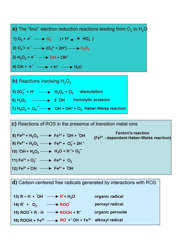
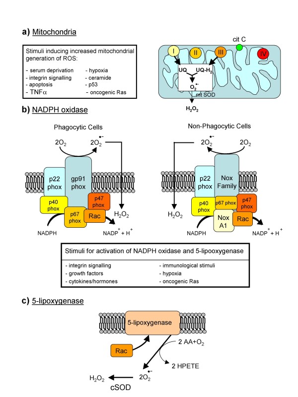
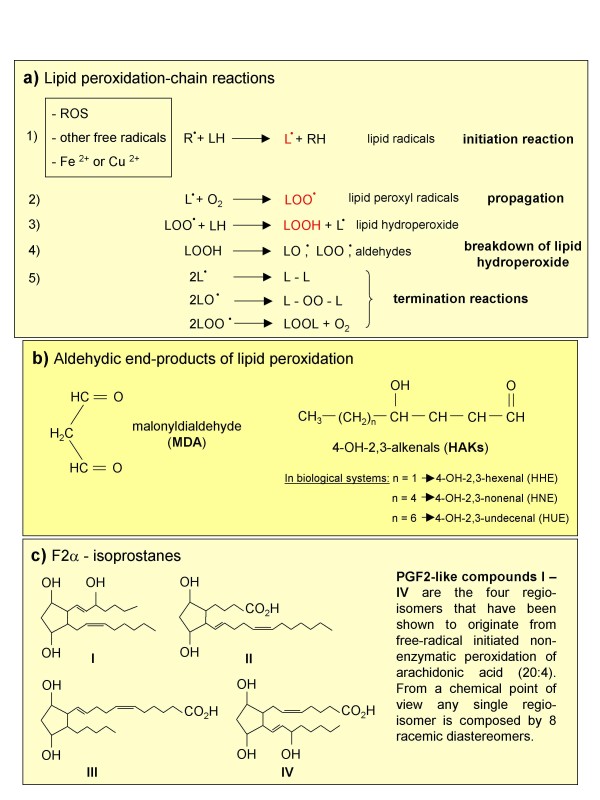
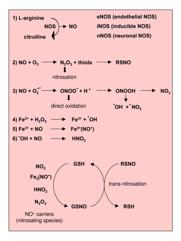
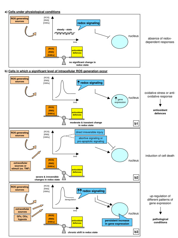
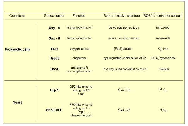
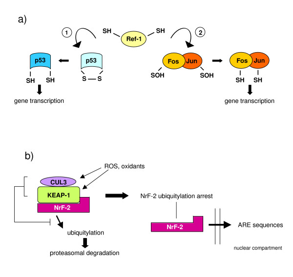
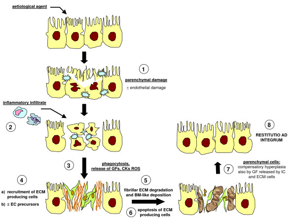
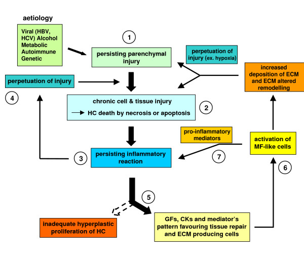
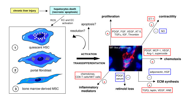
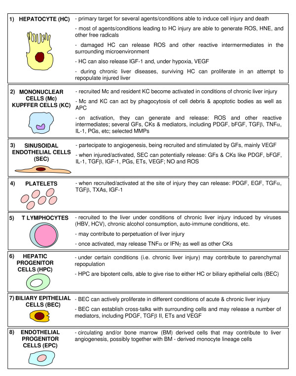
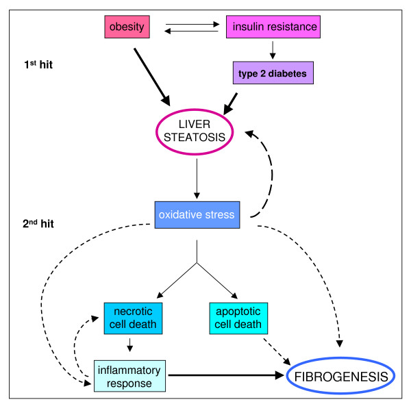
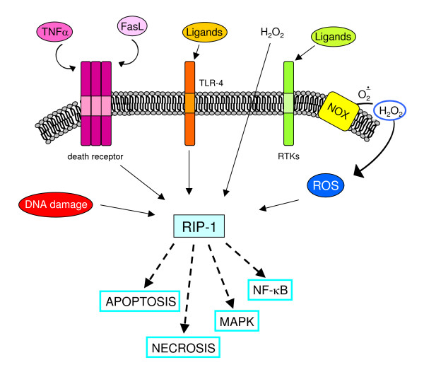
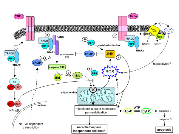
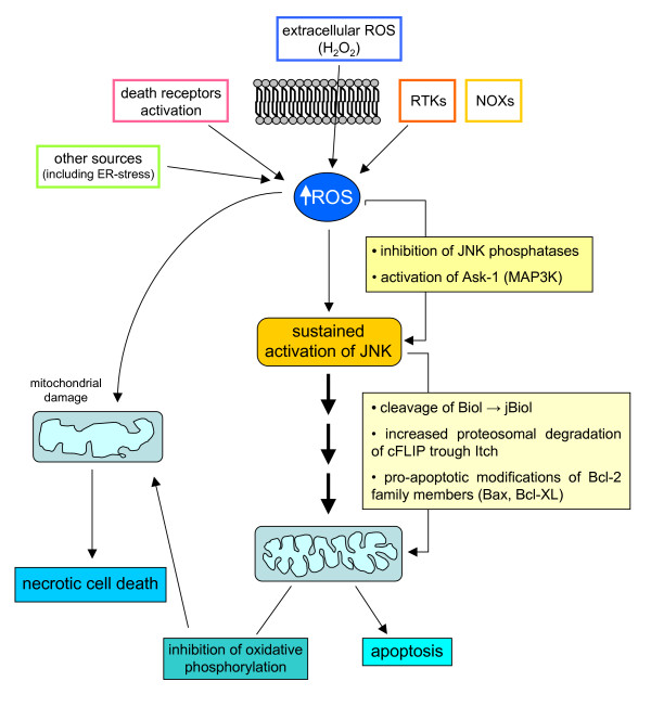
Reactive oxygen species (ROS) generated within cells or, more generally, in a tissue environment, may easily turn into a source of cell and tissue injury. Aerobic organisms have developed evolutionarily conserved mechanisms and strategies to carefully control the generation of ROS and other oxidative stress-related radical or non-radical reactive intermediates (that is, to maintain redox homeostasis), as well as to 'make use' of these molecules under physiological conditions as tools to modulate signal transduction, gene expression and cellular functional responses (that is, redox signalling). However, a derangement in redox homeostasis, resulting in sustained levels of oxidative stress and related mediators, can play a significant role in the pathogenesis of major human diseases characterized by chronic inflammation, chronic activation of wound healing and tissue fibrogenesis. This review has been designed to first offer a critical introduction to current knowledge in the field of redox research in order to introduce readers to the complexity of redox signalling and redox homeostasis. This will include ready-to-use key information and concepts on ROS, free radicals and oxidative stress-related reactive intermediates and reactions, sources of ROS in mammalian cells and tissues, antioxidant defences, redox sensors and, more generally, the major principles of redox signalling and redox-dependent transcriptional regulation of mammalian cells. This information will serve as a basis of knowledge to introduce the role of ROS and other oxidative stress-related intermediates in contributing to essential events, such as the induction of cell death, the perpetuation of chronic inflammatory responses, fibrogenesis and much more, with a major focus on hepatic chronic wound healing and liver fibrogenesis.


Figures

Figure 1
Figure 1
ROS are generated in biological systems through a number of interrelated reactions.
Figure 2
Figure 2
Major cellular sources of ROS in living cells.
Figure 3
Figure 3
Lipid peroxidation and the formation of non-radical intermediates.
Figure 4
Figure 4
Reactions leading to generation of NO and RNS.
Figure 5
Figure 5
Reactions of peroxynitrite leading to either apoptotic or necrotic cell death. NO and RNS may potentially prevent hepatocyte apoptosis as well as promote either necrotic or apoptotic cell death. The following mechanisms have been proposed. With regard to NO, RNS and prevention of apoptosis, the main molecular mechanisms resulting in an anti-apoptotic effect, related to S-nitrosating species, include [237-239]: stimulation of guanylate cyclase, leading to increased cyclic guanine monophosphate levels; the evolutionarily conserved inhibition of caspases by potentially reversible S-nitrosation of a critical cysteine residue at the caspase active site; activation of the Ras/Erk1/2 pro-survival pathway, which may result in activation of mitogen and stress activated kinase 1 (MSK1) and pp90 ribosomal S6 kinase (RSK), which in turn may inactivate the pro-apoptotic protein Bad or up-regulate anti-apoptotic proteins of the Bcl-2 family [237]; RNS also possibly acting by inhibiting leukocyte adhesion through S-nitrosation of critical -SH groups exposed by activated neutrophils and macrophages [240]. NO and RNS may prevent or promote cell death in relation to intracellular and intramitochondrial (because of mitochondrial NOS) levels of GSH and the concomitant cellular levels of transition metal ions. Moreover, NO may also lead to up-regulation of heme oxygenase 1 (HO-1) in hepatocytes and this may serve as a cytoprotective event [237,238]. The dark (that is, damaging) side of NO and RNS: in the presence of higher levels of ROS, the right NO/superoxide ratio or levels of molecular oxygen, NO may lead again to generation of highly reactive RNS, such as N2O3 or ONOO- at levels that are able to induce more aggressive oxidation, nitrosation/S-nitrosation and nitration of different biological macromolecules, potentially leading either to necrotic or apoptotic cell death. If NO-dependent pro-apoptotic mechanisms are concerned, the following have been shown to have a major role, with some again depending on S-nitrosating species: RNS and so called NO+ -carriers (nitrosating species) may result in activation of JNK, which, as previously reported for ROS, may sustain induction of apoptosis; NO, if generated at high levels in mitochondria, may result in ubiquinol auto-oxidation with concomitant production of superoxide, hydrogen peroxide and ONOO-, species that may be responsible for irreversible damage to complexes I and II of the respiratory chain, inhibition of ATP synthesis and eventually cytochrome c release and induction of caspase-dependent apoptosis. It should also be noted that, in the presence of significant redox stress, NO can potentiate damaging effects, resulting in a scenario of necrotic cell death rather than apoptosis. This is likely to occur particularly when the redox state is significantly affected, as in conditions resulting in depletion of GSH or significant alterations of the GSH/GSSG ratio.
Figure 6
Figure 6
Antioxidant (chain breaking) action of α-tocopherol and its recycling through ascorbate and GSH.
Figure 7
Figure 7
Overview of antioxidant enzymes.
Figure 8
Figure 8
Chemical structures of the most common chain-breaking antioxidants.
Figure 9
Figure 9
Alteration of redox homeostasis, redox signaling and cellular responses. Figure 9a: cells under physiological conditions in the absence of redox-dependent responses. Figure 9b: cells in which a significant level of intracellular ROS generation occurs as a moderate and transient change in redox state (b1), as a severe and irreversible change leading to cell death (b2) or as a chronic shift in redox state (b3).
Figure 10
Figure 10
Relevant examples of redox sensors in prokariotic cells and yeast. In the case of redox sensors described in yeast, the following can apply. The redox sensor Orp-1 of Saccharomyces cerevisiae (Oxidant receptor peroxidase-1, also known as Gpx3) is known to interact with hydrogen peroxide at Cys36, forming a -SOH group that, in turn, will lead to rearrangement (disulphide bonds) in the OxyR analogue Yap1 transcription factor and in the associated Ybp1 protein, leading ultimately to Yap1 nuclear translocation and Yap1-dependent gene activation [18,71]. Similar systems have also been described in Schizosaccharomyces pombe and a very similar mechanism has been described for PRX-Tpx1.
Figure 11
Figure 11
Redox sensors, redox signalling and control of redox sensitive transcription in higher eukaryotes. (a) Redox reactions involving transcription factors such as Ref1 (Redox-factor-1); Ref-1 is a ubiquitous reductase having cysteine residues (Cys65 and Cys94) that are believed to be critical for redox-dependent modification of several transcription factors, including AP-1 (activator protein-1), NF-κB (nuclear factor κB), p53, ATF/CREB (activating transcription factor/cAMP-response element-binding protein), and HIF-1α (hypoxia-inducible factor 1α). Ref-1 acts by reducing -SOH groups and/or oxidized cysteine residues or disulphide bonds present on transcription factors that, under these 'oxidized' conditions, have reduced or absent DNA-binding activity; the 'reduced' transcription factors then become able to bind their related sequences on DNA (shown here are the two examples Ref-1/p53 1 and Ref1/AP-1 2). (b) Nuclear translocation of transcriptional regulators that are maintained in an inactive form in another cellular compartment; a characteristic example is Nrf-2 (nuclear factor (erythroid-derived-2)-like-2), which is a transcriptional regulator able to bind to the so-called ARE (antioxidant responsive elements) regulatory sequences that are located on genes encoding a number of enzymes involved in detoxification, including those for glutathione S-transferases, NAD(P)H quinine oxidoreductase, the multidrug resistance-associated protein and cysteine-glutamate exchange transporter, thus up-regulating their transcription. In this case, Nrf-2 is usually bound to KEAP-1 (Kelch-like ECH associated protein-1) receptor or sensor, a protein rich in cysteine residues that usually forms a complex with cullin-3 and Nrf-2 to target the latter for proteasomal degradation. Exposure to oxidative stress (oxidation of Cys151, Cys273 and Cys288 combined with other reactions, including a Cys-zinc redox centre) results in modification of KEAP-1, leading to arrest of Nrf-2 ubiquitylation, allowing Nrf-2 to detach from KEAP-1 and translocate into the nucleus. (c) Modulation of transcription by alterations in the so-called 'redox buffer'; this concept indicates simply that several transcription factors as well as DNA modifying enzymes are sensitive to the most relevant reduced/oxidized molecular redox pairs, such as GSH/GSSG, NADPH/NADP and NADH/NAD. Examples of this way of coupling redox status to transcription factors or chromatin modifying enzymes include proteins that regulate circadian rhythms (Clock, NPAS2 and BMAL1), the protein for transcriptional silencing related to lifespan, SIRT1, and the transcriptional repressor C-terminal-binding protein (CtBP).
Figure 12
Figure 12
Acute liver injury: when healing is a coordinated and sequential process. A standard acute liver injury leading to irreversible parenchymal damage is followed by recruitment in the injured site of resident (Kupffer cells) or peripheral blood-derived activated monocyte/macrophages, resulting in phagocytosis, and the release of growth factors, cytokines, chemokines and ROS. Healing proceeds with recruitment of ECM-producing cells (HSCs and/or portal fibroblasts) and, likely, also of endothelial progenitor cells (EPCs): recruitment of these cells is essential to provide deposition of new ECM (basal membrane-like) and to form new sinusoids. Next, HSCs in excess will undergo apoptosis and the 'restitutio ad integrum' will require compensatory hyperplasia of hepatocytes that have survived the original injury as a response to a number of growth factors released by either inflammatory cells, endothelial cells or HSCs.
Figure 13
Figure 13
Schematic representation of events involved in fibrosclerotic development of CLDs. CLDs may involve different aetiological agents or conditions able to cause persisting parenchymal liver injury (1) and then hepatocyte (HC) cell death (either necrosis or apoptosis) (2). As a result, a persistent inflammatory reaction can occur (3), which may significantly affect the progression of the disease by either contributing to the perpetuation of injury (4) or 'creating' a growth factor, cytokine and mediator pattern favouring tissue repair and the activation of ECM-producing cells (5). This chronic scenario will lead to activation of myofibroblast-like cells that will contribute either to perpetuation of inflammation by releasing pro-inflammatory mediators (7) or to the wound healing response by excess and progressive accumulation of fibrillar (rich in collagen type I and III) extracellular matrix (ECM) components. If the aetiological agent or causal condition persists, the CLD can undergo a fibrosclerotic progression to cirrhosis and liver failure [74-81]. Cirrhosis in turn may be defined as an advanced end-stage of fibrosis, characterized by formation of regenerative nodules of parenchyma surrounded and separated by fibrotic septa, a scenario that is intrinsically associated with significant changes in hepatic angio-architecture [81-83].
Figure 14
Figure 14
Concepts and numbers. (a) The clinical impact of progressing fibrogenesis. (b) Predictors for fibrosis progression in CLDs. (c) Patterns of fibrosis progression in CLDs.
Figure 15
Figure 15
Origin of MF-like cells and their activation in the scenario of a CLD. Myofibroblast-like cells (MFs) may originate, under CLD conditions, from either quiescent HSCs, portal fibroblasts or bone marrow-derived MSCs able to engraft the chronically injured liver. Whatever the origin, MFs are believed to be characterised by the following properties and phenotypic responses: (a) high proliferative attitude; (b) increased ability to synthesise ECM components, particularly collagen type I and III; (c) altered ability to express matrix metallo-proteinases (MMPs) and related tissue inhibitors (TIMPs), resulting in an altered ability to remodel ECM in excess; (d) increased ability to migrate in response to different stimuli, including truly chemotactic ones; (e) increased synthesis of growth factors and pro-inflammatory cytokines and chemokines [76], including pro-angiogenic cytokines [81,83,92,93], that may act as paracrine as well as autocrine mediators; (f) contractility in response to vasoactive compounds like NO, endothelins and others; (g) the potential to undergo apoptosis in case of removal of the aetiological agent (that is, successful therapy, alcohol withdrawal, and so on) or causative conditions [80,81,91], although fibrosis regression has been mainly observed in experimental models. Here it should be mentioned that although there is no doubt that fibrosis is, at least in principle, a potentially reversible process, a complete reversion of cirrhosis (particularly for human cirrhotic livers) has never been convincingly documented [84] and human HSC/MFs have been shown to possess a peculiar survival attitude both in vitro as well as in vivo that may indeed favour progression over reversion [94].
Figure 16
Figure 16
The other cellular 'actors' in hepatic chronic wound healing: the roles of the different cell types, including those that may be related to redox state and signaling.
Figure 17
Figure 17
ROS may modulate receptor tyrosine kinase (RTK) signalling by regulating protein tyrosine phosphatases (PTPs) redox state. When a peptide ligand such as PDGF binds to its receptor RTK on the surface of a non-phagocytic cell (for example HSC/MFs), the signal can involve activation of PI3K and Rac, which in turn will result in activation of membrane NOX and generation of ROS. Within the cell ROS, such as H2O2, may act on a redox-sensitive cysteine residue in the active site of PTPs and transform the -SH group into the oxidized – SOH group (sulphenic acid), thus reversibly inactivating PTPs. Under physiological conditions and with low levels of ROS this change is rapidly reverted by reducing agents, with this transient redox inhibition of PTPs having a relevant role in RTK signalling. However, in conditions in which intracellular ROS are significantly increased, this may lead to more oxidation and then to irreversible changes, with formation at the level of the sensitive cysteine residues of sulphinic and sulphonic acid. These oxidized forms of PTPs are inactive and this will result in long-lasting blocking of PTP-dependent receptor dephosphorylation, allowing a positive reinforcement of RTK downstream signal transduction. The intracellular thiol/disulfide balance potentially plays a relevant role here: cellular levels of GSH or other reducing agents, for example, may operate to revert the sulphenic acid group in the active site of PTP to the thiolate anion, converting PTP back to the active state.
Figure 18
Figure 18
Two examples of ROS involvement in cytokine-dependent NF-κB activation. (a) NF-κB activation by IL-1β. In some cells IL-1β induces MyD88-dependent endocytosis of IL-R1; during endocytosis Rac1 recruits NOX2 in the endosomal compartment. NOX2 activation generates superoxide that spontaneously dismutates into H2O2, which then diffuses in the cytoplasm and triggers TRAF6 association with the ligand-receptor complex on the endosome, leading finally to NF-κB activation. (b) NF-κB activation by LPS. NF-κB activation by LPS through TLR4 activation involves Myd88 recruitment, which links TLR-4 activation to IRAK and TRAF6, mediating NF-κB activation. The involvement of ROS is consequent to direct interaction, followed by the Rac1-mediated activation of TLR-4 by NOX4 (or another NOX isoform, depending on the target cells). At present it is uncertain whether H2O2 operates (as for IL-1) by triggering activation of TRAF6.
Figure 19
Figure 19
Oversimplified scheme of responses induced by increased intracellular levels of ROS.
Figure 20
Figure 20
Increased levels of iron can contribute to increased generation of ROS and other radical or non-radical intermediates, resulting in a potentiation of cytotoxic, pro-inflammatory or pro-fibrogenic consequences. The role of iron in CLD progression. Hereditary hemochromatosis (HH) is a long-lasting disease in which hepatic iron levels increase progressively over a long period during which no or relatively modest inflammation and injury can be detected; when hepatic levels of iron increase to over 60 mmol/g dry weight, HSCs become activated and fibrogenesis becomes significant [113], although this transition (from non-fibrotic to fibrotic and then later cirrhotic) is not yet completely clear and other risk factors (ethanol consumption and ALD, chronic infection by HCV, concomitant metabolic conditions leading to NAFLD) are likely to be involved. However, with regard to patients with chronic HCV infection, it has been proposed that mutations in the hereditary hemochromatosis HFE gene may be responsible not only for derangement of iron homeostasis and HH, but may also worsen or accelerate the course of CLD by eliciting a turn-over of redox active iron in both the liver and plasma; in other words, the hypothesis is that HFE mutations may additionally result in increased intracellular production of ROS and free radicals taking place in hepatocytes or, also on the basis of recent knowledge on the role of hepcidin (see the section 'ROS-dependent sustained activation of JNK: a common step in oxidative stress-dependent cell death') and the iron transporter ferroportin, may affect the ability of Kupffer cells to handle and retain iron [112,114,115]. Chronic HCV infection. In addition to what has been reported for HH, it should be recalled that non-hereditary (that is, secondary) increased hepatic iron levels have been shown to represent a significant determinant for both the severity and progression rate of CLD associated with chronic HCV infection. Along these lines, different laboratories have shown a correlation between liver iron levels and HSC activation as well as fibrosis progression [114,115], which can be significantly prevented by phlebotomy. It should be noted (reviewed in [113]), however, that other researchers did not find evidence for such a correlation. NAFLD and NASH. With regard to NAFLD, current evidence suggests that metabolic disturbances leading to steatosis (as associated with obese or overweight patients and, usually, with the so-called metabolic syndrome, often also including diabetes and insulin resistance) are likely to represent the 'first hit'. In order for NAFLD to progress to non-alcoholic steatohepatitis or NASH a 'second hit' (see later) is believed to be necessary and usually identified as occurrence of oxidative stress. Along these lines, iron is a rather obvious candidate because of its well known role as an ideal metal catalyst for the generation of ROS and other free-radical or non-radical intermediates. Alcoholic liver disease (ALD). Homologous considerations (that is, the role of hepatic iron levels) may be advanced for ALD and may help to explain, at least in part, why only approximately 30% of patients with high levels of chronic alcohol consumption are likely to develop cirrhosis over time. As recently reviewed [113], there are several reasons to believe that iron is a serious and, likely, independent candidate factor able to contribute to progression of ALD to cirrhosis. For example, in the pre-cirrhotic stage, approximately 30% of ALD patients show an elevated hepatic iron index, but when the ALD progresses to cirrhosis the percentage of ALD patients having iron overload rises up to 60%. Interestingly, it was recently suggested that ethanol consumption is able to alter IL-6-dependent expression of hepcidin, a condition resulting in enhanced absorption of iron and hepatic siderosis [118]. Other mechanisms that may enhance iron hepatic levels have been recently reviewed by Brittenham [119].
Figure 21
Figure 21
The two hit theory of NAFLD progression. Current literature indicates that the crucial derangement in NAFLD is represented by insulin resistance (IR), which is a key feature of the metabolic syndrome, a clinical entity that also includes type 2 diabetes mellitus, hypertrigliceridemia, hypertension, a decreased level of high-density lipoprotein and obesity. Indeed, as suggested by several authors, steatosis may simply represent the hepatic manifestation of the metabolic syndrome ([143,144] and references therein). The first hit is a metabolic one dominated by IR, with NAFLD being associated with both hepatic and adipose tissue IR as well as reduced insulin sensitivity of the whole human body [143,144,146]. This can lead from one side to 1) a significant reduction of glucose disposal and to a lack of suppression of hepatic glucose production as well as 2) a defect of disposal of free fatty acids (FFAs) at the adipocyte level (and skeletal muscle) that, in turn, 3) will open the way to high circulating levels of FFAs (and hypertrigliceridemia) coming from either subcutaneous and visceral fat, which will cause a persistent excess delivery of FFAs to the liver, the ultimate cause of liver steatosis [142-146]. Molecular details on how the excess load of FFAs to hepatocytes can lead to steatosis can be found in more specialized reviews [142,143], with increased de novo synthesis of fatty acids and triglycerides as well as both impaired oxidation (mitochondrial plus peroxisomal) and impaired efflux of fatty acids (by reduced synthesis of Apo-B100 or transport of VLDL particles) being the candidate responsible processes.
Figure 22
Figure 22
Receptor interacting protein (RIP) kinase 1 as a crucial cellular crossroads affecting whether target cells survive or die. ROS may increase in the cells also as a consequence of increased release by mithocondria, as in the case of TNFα and FasL-related responses. Activation of death receptor (DR), Toll-like receptors (TLRs) as well as signalling pathways initiated upon detection of intracellular stress (including oxidative stress itself and/or DNA damage) all have been reported to converge on RIP, particularly RIP1; the cellular context will then drive the RIP-related response of target cells towards survival by preferentially inducing activation of NF-κB and/or MAPK, or to cell death by inducing either true apoptosis or a form of caspase-independent cell death [193], although this is an oversimplified scheme (for example, sustained JNK activation is a well known event leading to cell death).
Figure 23
Figure 23
TNF-induced and ROS-dependent events in target cells: the role of JNK activation in mediating cell death. TNF interaction with its type I receptor will generate a sequence of events (depicted in Figure 23) that is not designed to lead uniquely to cell death [197-200] and in which ROS may play a crucial role. 1) The interaction between TNF and TNFRI is first followed by association with the intracytoplasmic receptor tail of the adapter TRADD (TNF-receptor associated death domain), the protein kinase RIP1 and TRAF-2 and TRAF-5 (TNF-receptor associated factors 2 and 5); this works as a signalling complex that activates NF-κB (through Inhibitor of NF-κB kinase (IKK)) and MAPK cascades that regulate the AP-1 transcription factor. Activation of NF-κB-binding activity and transcription of dependent genes is potentially pro-survival by up-regulating the synthesis of cFLIP (c FLICE inhibitory protein), which can inhibit activation of caspase 8/10, the starting point for TNF-mediated induction of classic apoptosis. 2) At this point the TRADD/RIP-1/TRAF-2 complex can dissociates from the receptor by a still unknown mechanism and bind to FADD (Fas-ligand associated death domain) to form another complex leading to recruitment of several molecules of pro-caspase 8/10 and to their autocatalytic cleavage, resulting in activation; however, this event occurs only in cells having low levels of cFLIP, suggesting that apoptosis as well as caspase-independent or necrotic cell death (see later) may occur when the preventive pro-survival function of NF-κB fails. When activation of caspase 8/10 ocurs, the scenario can still prefigure two alternatives. 3) Activated caspase 8/10 may cleave the pro-apoptotic protein Bid (to tBid), which then translocates to mitochondria, causing permeabilization of mitochondrial outer membrane, cytochrome c release, apoptosome assembly and activation of executioner caspases, leading to classic apoptosis. This pro-apoptotic scenario may be amplified by the recruitment to TNFRI of FAN (Factor-associated neutral sphingomyelinase), which is able to produce sphingosine that, in turn, permeabilizes lysosomal membranes, leading to the release of cathepsins. These lysosomal proteases, which can be activated following engagement of different death receptors, act as pro-apoptotic proteases able to cleave Bid as well as to induce oligomerization of Bax or interact directly with mitochondrial outer membrane (note that this pathway, for the sake of clarity, has been omitted from Figure 23). It should be noted that in hepatocytes it has been reported that generation of ROS following activation of death receptors (by Fas or TNF) is strictly dependent on Bid cleavage to tBid and its subsequent translocation to mitochondria [201]. 4) The second alternative is the one more strictly related to ROS and involves the crossroads kinase RIP1. RIP1 has multiple crucial roles; it is involved in the activation of NF-κB, but may also contribute to apoptosis when cleaved by caspase-8 to cRIP1 (4a), an event preventing or reducing activation of NF-κB and sustaining enhanced interaction between TRADD, FADD and procaspases 8/10. However, it has also been described (4b) that the kinase domain of RIP1 is essential to activate JNK, which in turn (although this is just one of the JNK-dependent actions) will favour TNFRI-related apoptosis and mitochondrial outer membrane permeabilization by eliciting cFLIP phosphorylation and degradation. Concerning the involvement of ROS, however, RIP1 (reviewed in [107,193,194]) has been shown (5) to be able also to translocate to mitochondria (prevented by NF-κB) following TNF stimulation of cells: this event induces mitochondrial outer membrane permeabilization and increases release of ROS in the cytoplasm without leading to cytochrome c release and apoptosome complex formation. This increased TNF-related mitochondrial generation of ROS is crucial because it can lead target cells to apoptosis or, more likely, to a necrotic or caspase-independent form of cell death: in this latter scenario a significant role is attributed to ROS-mediated sustained activation of JNK (6), surely operating in hepatocytes [186,187], which, as we will see (Figure 24), is a crucial crossroads for ROS-related irreversible injury of parenchymal cells as elicited by the different aetiologies leading to CLDs. TNF (7) may lead to ROS-dependent and JNK-mediated cell death (necrotic type) also by involving activation of Nox1: however, this mechanism has been described in fibroblasts and, at present, we do not know whether it may apply to hepatocytes or other liver cell populations [195,202].
Figure 24
Figure 24
ROS-dependent sustained activation of JNK isoforms as a crucial event in inducing cell death. ROS-mediated sustained activation of JNK isoforms is likely to rely on inhibition of JNK phosphatases and/or activation of the upstream kinase ASK-1, finally resulting in mitochondrial outer membrane permeabilization. To explain this later, crucial event, the following hypotheses have been proposed: a) JNK may, in a caspase-independent way that has still not been characterized, promote the cleavage of the BH3 domain of Bid, resulting in the production of jBid, which should operate in a pro-apoptotic way similarly to tBid [209]; b) JNK may favour apoptosis by increasing proteasomal degradation of cFLIP (the inhibitor of pro-caspase 8/10 activation) by activating the ubiquitin ligase Itch [199]; c) by pro-apoptotic modifications of proteins belonging to the Bcl-2 family, such as Bax or Bcl-XL [187,194].
Figure 25
Figure 25
ER stress and ROS in NAFLD/NASH. This figure describes the most relevant features of ER stress following excess accumulation of FFAs in hepatocytes and also involving oxidative stress and ROS (the latter may originate in NAFLD from deranged mitochondria, CYP2E1 and CYP4A isoforms, or from peroxisomes). On the basis of what is described in the text (sections 'ROS and oxidative stress-related reactive intermediates in NAFLD and NASH: their generation and the role in causing steatosis' and 'Free fatty acids, endoplasmic reticulum stress, oxidative stress and cell death: hepatocyte injury in NAFLD but not only'), the following major features can be offered. a) When the UPR response fails to solve the problem of protein folding caused by different conditions able to induce ER stress (see text for details), including increases in FFA levels, this is followed by an induction of apoptotic cell death that can use both mitochondrial pathways as well as other independent pathways. b) ROS and oxidative stress are able to disrupt ER functions, a major cause seemingly being ROS-dependent increased release of calcium from ER stores: excess calcium has been reported to induce mitochondrial outer membrane permeabilization and, in turn, increased mitochondrial ROS release as a further contribution to increased intracellular levels by other sources and causes operating in NAFLD-related hepatocytes. c) In such a complex scenario, ER stress can result in apoptosis by a number of mechanisms, including: damage to mitochondria leading to cytochrome release, apoptosome formation and related sequential activation of executioner caspase 9 and 3; IRE-1 recruitment of TRAF-2 in order to activate either ASK-1 and then JNK, a potentially pro-apoptotic pathway that can be further sustained by ROS, or (at least in mice) caspase 12, which, in turn, can activate caspases 9 and 3; and activation of PERK and ATF6 (p90), which can lead through nuclear translocation of ATF4 and ATF6(p50), respectively, to transcriptional up-regulation of CHOP, a factor that promotes apoptosis by either inhibiting expression of Bcl-XL or up-regulating expression of pro-apoptotic proteins such as Gadd34, Trb3 and Dr5.
Figure 26
Figure 26
The central role of mitochondrial damage in ethanol-dependent and ROS-mediated hepatocyte injury.
Figure 27
Figure 27
ROS and related mediators as pro-fibrogenic stimuli: a 'stellate centric' view.
Figure 28
Figure 28
Intracellular generation of ROS in human HSC/MFs exposed to PDGF-BB or to hydrogen peroxide. Detection of intracellular generation of ROS was performed by using the conversion of 2',7'-dichlorodihydrofluorescein diacetate (DCFH-DA) probe in human HSC/MFs. Experimental conditions included control cells and cells treated with PDGF-BB (10 ng/ml) or H2O2 (50 μM, positive control) for 15 minutes. Cells were observed and photographed under a Zeiss fluorescence microscope equipped with phase contrast objectives. Images of the same fields were collected and images in the right column offer, for all conditions, the overlay of fluorescence and phase contrast images (E Novo et al, unpublished data).
Figure 29
Figure 29
Redox-dependent development of autoantibodies against oxidative stress-modified epitopes.

Similar articles

Cited by

References

    1. Commoner B, Townsend J, Pake GE. Free radicals in biological materials. Nature. 1954;174:689–691. doi: 10.1038/174689a0. - DOI - PubMed
    1. Harman D. Aging: a theory based on free radical and radiation chemistry. J Gerontol. 1956;11:298–300. - PubMed
    1. Slater TF. Necrogenic action of carbon tetrachloride in the rat: a speculative mechanism based on activation. Nature. 1966;209:36–40. doi: 10.1038/209036a0. - DOI - PubMed
    1. McCord JM, Fridovich I. Superoxide dismutase: an enzymic function for erythrocuprein (hemocuprein) J Biol Chem. 1969;244:6049–6055. - PubMed
    1. Slater TF. Free Radical Mechanisms in Tissue Injury. London, UK: Pion Ltd; 1972. - PMC - PubMed

LinkOut - more resources