. 2008 Nov 3;1(1):2.
doi: 10.1186/1755-8417-1-2.

Smad4 haploinsufficiency: a matter of dosage

Affiliations

Smad4 haploinsufficiency: a matter of dosage

Paola Alberici et al. Pathogenetics. .

Abstract

Background: The inactivation of tumor suppressor genes follows Alfred Knudson's 'two-hit' model: both alleles need to be inactivated by independent mutation events to trigger tumor formation. However, in a minority of tumor suppressor genes a single hit is sufficient to initiate tumorigenesis notwithstanding the presence of the wild-type allele, a condition known as haploinsufficiency. The SMAD4 gene is an intracellular mediator of the TGF-beta and BMP signal transduction pathways and a tumor suppressor involved in pancreatic and colorectal tumorigenesis. In Smad4-mutant mouse models, haploinsufficiency characterizes the development of gastrointestinal polyps with initial retention of the wild-type allele and protein expression within the nascent tumors and in their direct microenvironment. Similarly, germline SMAD4 mutations are responsible for a subset of patients affected by juvenile polyposis syndrome, an autosomal dominant intestinal cancer syndrome. To date, the molecular and cellular consequences of SMAD4 haploinsufficiency on TGF-beta and BMP signaling and on genome-wide gene expression have not been investigated.

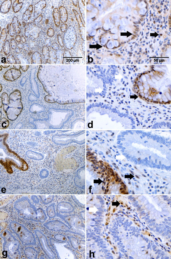
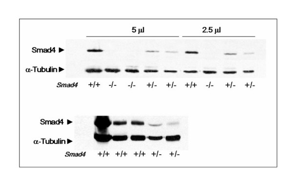
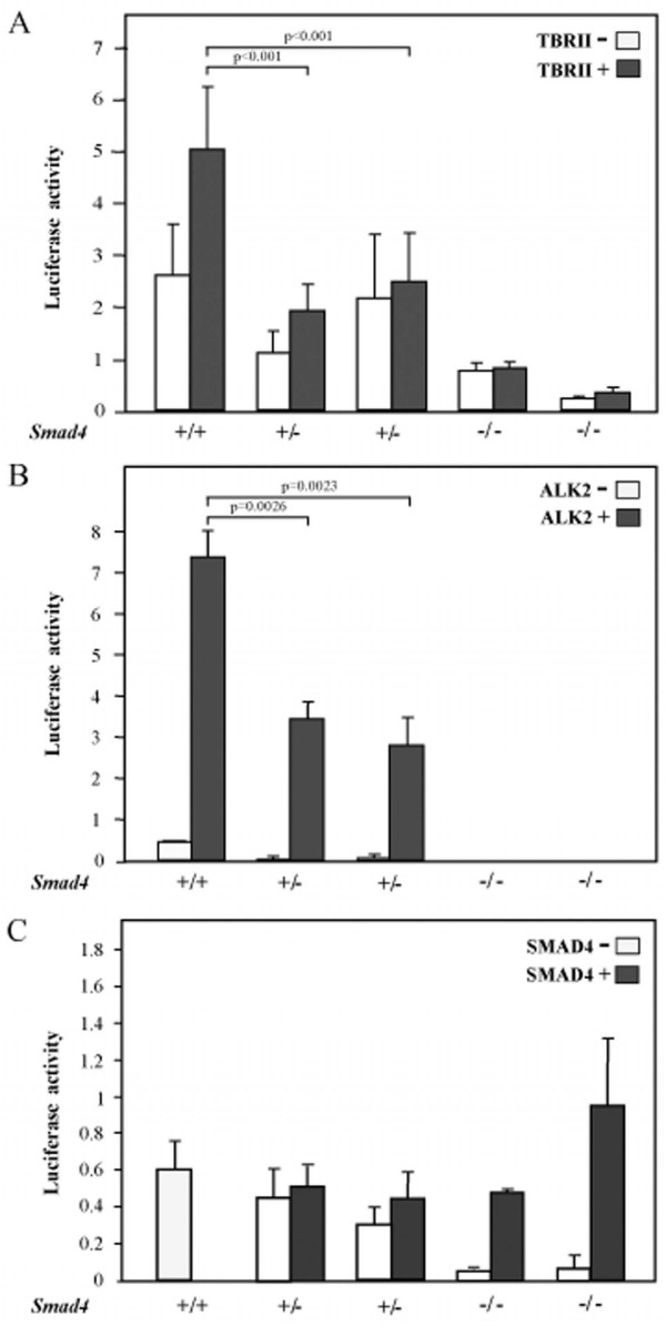
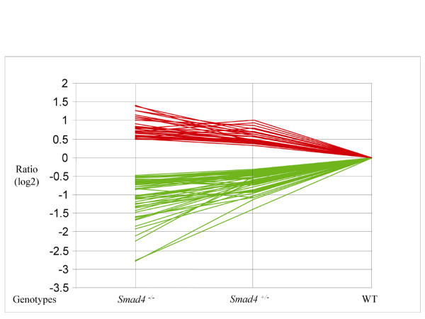
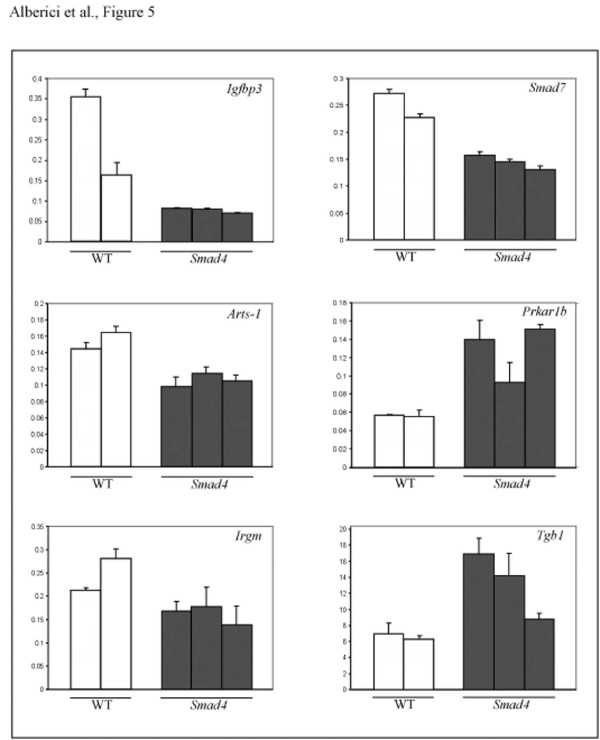
Results: Here we show that, similar to previous observations in Smad4-mutant mouse models, haploinsufficiency characterizes a substantial fraction of the juvenile polyps arising in patients with germline SMAD4 mutations. Also, mouse embryonic and intestinal cells heterozygous for a targeted Smad4 null mutation are characterized by a corresponding 50% reduction of the Smad4 protein levels. Reporter assays revealed that mouse Smad4+/- cells exert intermediate inhibitory effects on both TGF-beta and BMP signaling. Genome-wide expression profiling analysis of Smad4+/- and Smad4-/- cells pinpointed a subset of dosage-dependent transcriptional target genes encompassing, among others, members of the TGF-beta and Wnt signaling pathways. These SMAD4 dosage-dependent transcriptional changes were confirmed and validated in a subset of target genes in intestinal tissues from juvenile polyposis syndrome patients.

Conclusion: Smad4 haploinsufficiency is sufficient to significantly inhibit both TGF-beta and BMP signal transduction and results in the differential expression of a broad subset of target genes likely to underlie tumor formation both from the mesenchymal and epithelial compartments. The results of our study, performed in normal rather than tumor cells where additional (epi-) genetic alterations may confound the analysis, are relevant for our understanding and elucidation of the initial steps underlying SMAD4-driven intestinal tumorigenesis.


Figures

Figure 1
Figure 1
Immunohistochemical analysis of SMAD4 protein expression in juvenile polyps from patients carrying SMAD4 germline mutations. SMAD4 immunohistochemical analysis of hamartomatous polyps obtained from unrelated Juvenile Polyposis syndrome patients with established SMAD4 germline mutations (see Table 1). Images were taken at 10× (a, c, e, g,) and 40× (b, d, f, h). Filled (black) arrows indicate cells scored as positive, whereas white arrows point to negatives. a-b. Example of a polyp positive for SMAD4 nuclear staining in both the epithelial and stromal compartment (scored as double positive in Table 1). c-d. Polyp with heterogeneous SMAD4 expression pattern with patches of positively and negatively staining epithelial glands. Most of the stromal cells appear negative with few exceptions. This tumor was scored as +/- (epithelial) and – (stromal) in Table 1. e-f. Example of a juvenile polyp with heterogeneous SMAD4 expression in the epithelial compartment but with a more pronounced positive staining of the stromal tumor microenvironment. This tumor was scored as +/- (epithelial) and + (stromal) in Table 1. g-h. Example of a juvenile polyp characterized by negative SMAD4 staining throughout the epithelial cells. In the stromal compartment however, several positively staining cells are present. This tumor was scored as – (epithelial) and + (stromal) in Table 1.
Figure 2
Figure 2
Western analysis of Smad4-mutant embryonic stem cell lines. SMAD4 western blot analysis demonstrates haploinsufficiency in embryonic stem (ES) and adult intestinal cells from Smad4+/E6sad mice. Upper panel: ES cell lysates loaded at two different protein amounts; lower panel: normal intestinal tissue lysates from wild-type and Smad4+/E6sad mice.
Figure 3
Figure 3
TGF-β and BMP reporter assay analysis of Smad4-mutant embryonic stem cell lines. TGF-β and BMP reporter assay analysis of Smad4-mutant embryonic stem (ES) cell lines. A. TGF-β (CAGA12-MLP-luciferase) and B. BMP (BRE-luciferase) reporter assays were carried out in wild-type (+/+), Smad4+/E6sad(+/-), and Smad4E6sad/E6sad (-/-) ES cell lines. Normalized CAGA-luc and BRE-luc levels are indicated for each cell line. For each genotype reporter activities are shown for cells transfected (TBRII+ and ALK2+) and non transfected (TBRII- and ALK2-) with the corresponding receptor-expressing vector. In C. the TGF-β reporter assays analysis was carried out after transfection with a SMAD4-expressing vector. Each bar represents the average of three independent experiments, and the error bars represent the standard deviation. For the CAGA12-MLP-luciferase assay, three independent ES wild-type clones have been employed. The WT bar represents the average of the luciferase activities measured in two independent experiments. p values were calculated by the statistical two samples t-test (two tails).
Figure 4
Figure 4
Trend analysis of Smad4 dosage-dependent transcriptional targets. Trend analysis of the 79 genes found to be differentially expressed in both Smad4+/E6sad and Smad4E6sad/E6sad embryonic stem cells. The fold changes are represented as (log2) ratio values and plotted as red and green lines for up- and down-regulated genes, respectively.
Figure 5
Figure 5
qPCR validation of the expression profiling results. Quantitative PCR analysis of a selection of six genes differentially regulated in Smad4+/E6sad cells. Gene expression was quantified in normal intestinal tissues from two wild-type (WT) and three Smad4+/E6sad (Smad4) animals and is plotted as ratio over the reference gene (see Materials and Methods). Each bar represents the average of three independent experiments.

References

    1. Payne SR, Kemp CJ. Tumor suppressor genetics. Carcinogenesis. 2005;26:2031–2045. - PubMed
    1. Knudson AG., Jr Mutation and cancer: statistical study of retinoblastoma. Proc Natl Acad Sci USA. 1971;68:820–823. - PMC - PubMed
    1. Fodde R, Smits R. Cancer biology. A matter of dosage. Science. 2002;298:761–763. - PubMed
    1. Howe JR, Roth S, Ringold JC, Summers RW, Jarvinen HJ, Sistonen P, Tomlinson IP, Houlston RS, Bevan S, Mitros FA, Stone EM, Aaltonen LA. Mutations in the SMAD4/DPC4 gene in juvenile polyposis. Science. 1998;280:1086–1088. - PubMed
    1. Xu X, Brodie SG, Yang X, Im YH, Parks WT, Chen L, Zhou YX, Weinstein M, Kim SJ, Deng CX. Haploid loss of the tumor suppressor Smad4/Dpc4 initiates gastric polyposis and cancer in mice. Oncogene. 2000;19:1868–1874. - PubMed

LinkOut - more resources