. 2008 Nov 17:2:98.
doi: 10.1186/1752-0509-2-98.

Interlinked nonlinear subnetworks underlie the formation of robust cellular patterns in Arabidopsis epidermis: a dynamic spatial model

Affiliations

Interlinked nonlinear subnetworks underlie the formation of robust cellular patterns in Arabidopsis epidermis: a dynamic spatial model

Mariana Benítez et al. BMC Syst Biol. .

Abstract

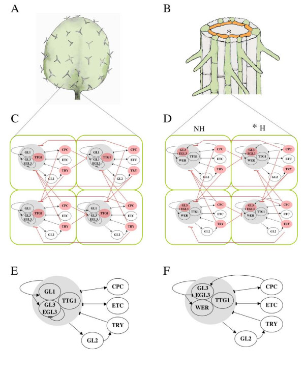
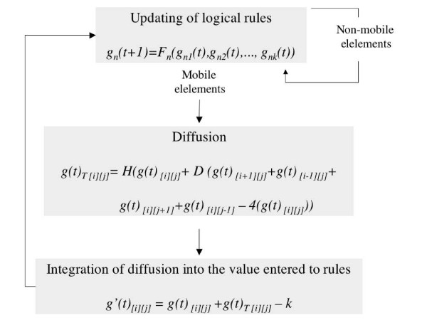
Background: Dynamical models are instrumental for exploring the way information required to generate robust developmental patterns arises from complex interactions among genetic and non-genetic factors. We address this fundamental issue of developmental biology studying the leaf and root epidermis of Arabidopsis. We propose an experimentally-grounded model of gene regulatory networks (GRNs) that are coupled by protein diffusion and comprise a meta-GRN implemented on cellularised domains.

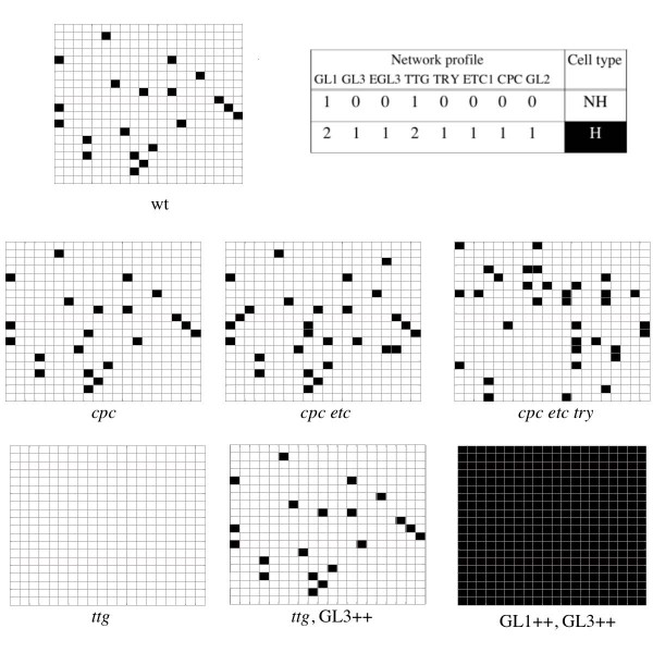
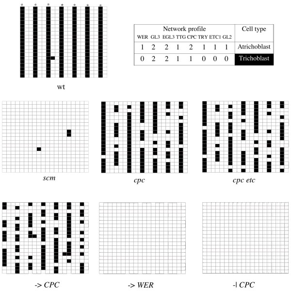
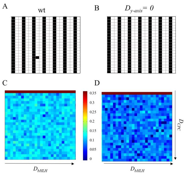
Results: Steady states of the meta-GRN model correspond to gene expression profiles typical of hair and non-hair epidermal cells. The simulations also render spatial patterns that match the cellular arrangements observed in root and leaf epidermis. As in actual plants, such patterns are robust in the face of diverse perturbations. We validated the model by checking that it also reproduced the patterns of reported mutants. The meta-GRN model shows that interlinked sub-networks contribute redundantly to the formation of robust hair patterns and permits to advance novel and testable predictions regarding the effect of cell shape, signalling pathways and additional gene interactions affecting spatial cell-patterning.

Conclusion: The spatial meta-GRN model integrates available experimental data and contributes to further understanding of the Arabidopsis epidermal system. It also provides a systems biology framework to explore the interplay among sub-networks of a GRN, cell-to-cell communication, cell shape and domain traits, which could help understanding of general aspects of patterning processes. For instance, our model suggests that the information needed for cell fate determination emerges from dynamic processes that depend upon molecular components inside and outside differentiating cells, suggesting that the classical distinction of lineage versus positional cell differentiation may be instrumental but rather artificial. It also suggests that interlinkage of nonlinear and redundant sub-networks in larger networks is important for pattern robustness. Pursuing dynamic analyses of larger (genomic) coupled networks is still not possible. A repertoire of well-characterised regulatory modules, like the one presented here, will, however, help to uncover general principles of the patterning-associated networks, as well as the peculiarities that originate diversity.


Figures

Figure 1
Figure 1
Cellular patterns and meta-gene regulatory network models for leaf and root Arabidopsis epidermis. Spaced-out pattern of trichome distribution in the leaf of Arabidopsis thaliana (A). Root-hairs (green) are arranged in bands that overlie the junction of two cortex cells (yellow) (B). Coupled gene regulatory network (GRN) model for cell type determination in leaf epidermis (C). GRN underlying cell-fate determination in root epidermis (D). (E) and (F) represent the GRN for leaf and root epidermis, respectively. In both networks, nodes correspond to genes, arrows stand for positive regulatory interactions and flat-end edges stand for negative ones. Red nodes represent elements that are able to move among cells and couple the GRN into meta-GRN. Red lines stand for intercellular interactions established by mobile elements. Asterisks in (B) and (D) indicate the H position where the cortex-related signal is acting.
Figure 2
Figure 2
Diagrammatic representation of the model structure. In every time-step, the nodes' states are updated according to logical rules [see Additional file 1], then the mobile elements are allowed to diffuse and, finally, diffusion is considered to recalculate the states of mobile elements. These new values are entered into the logical rules in the next iteration. The state of non-mobile elements is only determined by the logical rules applied every time-step.
Figure 3
Figure 3
The model renders cellular patterns similar to those observed in the leaf epidermis. The simulated cellular patterns for wild-type (wt) and mutant networks are consistent with the patterns reported in the literature. Black squares correspond to trichomes and white ones to pavement cells (non-hair cells). Captions under the matrixes indicate the simulated genotype that gave rise to each of them (++ stands for overexpression, while lower case italics indicate loss of function). The table shows that the network profiles typical of hair and non-hair cells are recovered by the meta-GRN model. These simulations were all performed in 20 × 20 matrices with parameter values DCPC = 0.05, DTRR = 0.05, DTTG = 0.03.
Figure 4
Figure 4
The model renders cellular patterns similar to those observed in the root epidermis. Cellular patterns obtained from simulations of wild-type (wt) and mutant networks are consistent with the patterns reported in the literature. Black squares correspond to trichoblasts and white ones to atrichoblasts. Captions under the matrixes indicate the simulated genotype that gave rise to each of them (lower case italics indicate loss of function, -> indicates the simulation of a positive upstream signal, while -| stands for a negative one). Asterisks indicate the hair (H) position. The table shows that the network profiles characteristic of hair and non-hair cells are recovered by the coupled GRN model (B). These simulations were all performed in 20 × 20 matrices with the following parameter values: DCPC = 0.01, DGL3 = 0.01, DEGL3 = 0.01.
Figure 5
Figure 5
Spatial root-like pattern is stabilised by differential diffusion in the x and y axes. The pattern generated by the coupled GRN model gives rise to a striped pattern with some 'errors' that would correspond to ectopic hairs (A). A similar pattern but with fewer or no errors is obtained when diffusion rate in the x axis is larger than that in the y axis (Dy-axis = 0) and the same random seed is taken (B). The parameter spaces for each case are presented below their typical cell arrangements (C), (D). The colour scale indicates the logarithm of the average number of ectopic cell-types for every combination of parameters. Note that, overall, the parameter space obtained for differential diffusion exhibits fewer ectopic trichoblasts.

Similar articles

Cited by

References

    1. Dover G. Dear Mr Darwin: Letters on the Evolution of Life and Human Nature. University of California Press; 2006.
    1. Scheres B. Plant cell identity. The role of position and lineage. Plant Physiol. 2001;125:112–114. doi: 10.1104/pp.125.1.112. - DOI - PMC - PubMed
    1. Dinneny JR, Benfey PH. Plant stem cell niches: standing the test of time. Cell. 2008;132:553–557. doi: 10.1016/j.cell.2008.02.001. - DOI - PubMed
    1. von Dassow G, Meir E, Munro E, Odell G. The segment polarity network is a robust developmental module. Nature. 2000;406:188–193. doi: 10.1038/35018085. - DOI - PubMed
    1. Albert R, Othmer H. The topology of the regulatory interactions predicts the expression pattern of the segment polarity genes in Drosophila melanogaster. J Theor Biol. 2003;223:1–18. doi: 10.1016/S0022-5193(03)00035-3. - DOI - PMC - PubMed

Publication types

MeSH terms