. 2009 Mar 15;177(2):348-54.
doi: 10.1016/j.jneumeth.2008.10.023. Epub 2008 Oct 30.

In-tube transfection improves the efficiency of gene transfer in primary neuronal cultures

Affiliations

In-tube transfection improves the efficiency of gene transfer in primary neuronal cultures

Marc W Halterman et al. J Neurosci Methods. .

Abstract

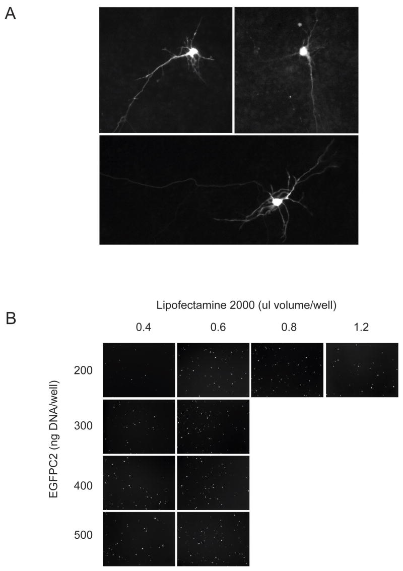
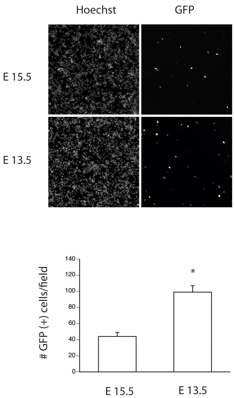
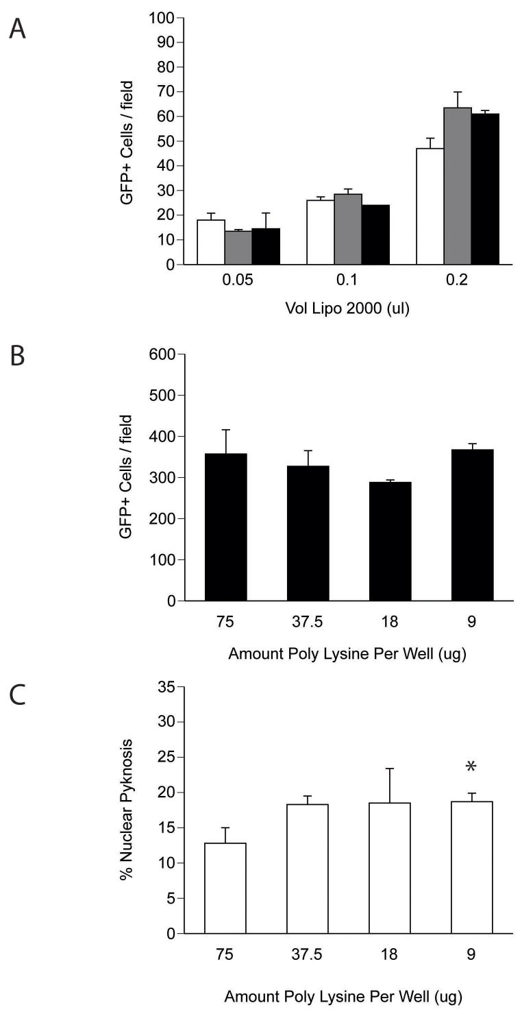
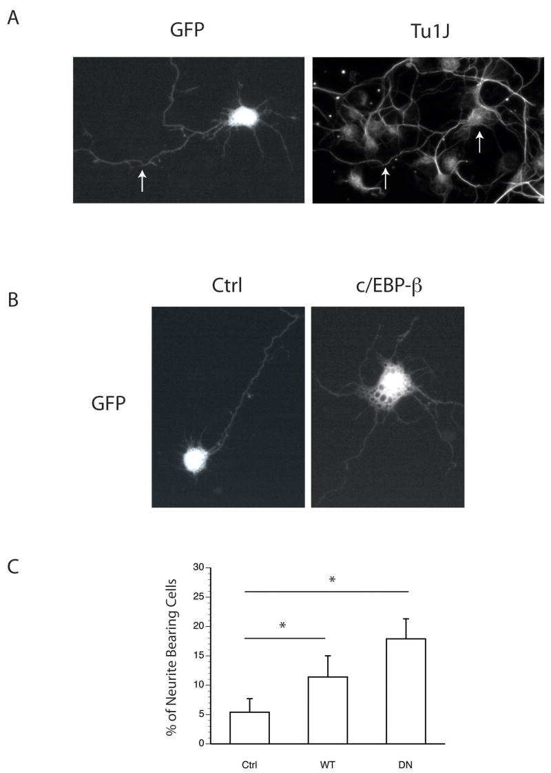
To facilitate genetic studies in primary neurons, we analyzed the efficiency of cationic lipid-mediated plasmid DNA transfection using adherent and acutely dissociated neuronal suspensions derived from embryonic mouse cortical tissue. Compared to transfections using adherent cultures, the in-tube procedure enhanced the delivery of a GFP reporter plasmid between four- to eightfold depending on the age of the harvested embryo. The procedure required relatively brief complex incubation times, and supported the transfection of cells expressing the neuronal markers NeuN and TuJ1 with improved uniformity in transfection events across the well surface. To demonstrate the utility of this approach in studying the genetic mechanisms controlling neuron development, we provide data regarding the role of the bZIP transcription factor c/EBP-beta in regulating neurite outgrowth. It is anticipated that this in vitro protocol will facilitate the identification of novel genes involved in both developmental and disease-relevant signaling pathways.


Figures

Figure 1
Figure 1. Transfection of primary cortical neuronal cultures using standard approaches
(A) Transfection results obtained using adherent DIV 4 mouse cortical cultures exposed to lipid:DNA complexes. Fluorescence microscopic images were obtained 24 h after transfection (40× power). (B) Optimization of the in-well transfection technique using acutely dissociated cortical neurons. After the DNA:lipid were allowed to complex in the poly-L-lysine treated microtiter plate, acutely dissociated neuron suspensions were added, and imaged after 24 h later for transfection efficiency. The images shown (20× power) represent those fields containing the highest density of transfection events.
Figure 2
Figure 2. In-tube transfection of E13.5 cortical cultures improves transfection rates
Low power micrograph (4×) of transfection rates obtained using the in-tube transfection technique. Acutely dissociated primary cultures from E15.5 and E13.5 mice were transfected using the in-tube method and analyzed for the number GFP+ cells 24 h post-transfection. Hoechst 33342 staining was used to calculate the total cells per field counts. The data represent the average number of GFP+ cells obtained under 40× power from eight non-overlapping fields and significance was determined by Student’s t- testing (avg ± stdv; * = p < 0.02).
Figure 3
Figure 3. Optimization of transfection parameters using dissociated suspension cortical neurons
(A) Effect of incubation time on in-tube transfection rates. Cell-lipid-DNA complexes were incubated in 1.5 ml microtubes at RT for the times incubated before plating (10 min - open bars, 20 min - grey bars, 40 min - black bars). (b) Poly-L-lysine concentration does not influence transfection rates using the in-tube technique. The number of GFP+ cells per 20× field was analyzed across the range of poly-L-lysine doses used during the coating procedure. (C) Effect of poly-L-lysine on survival. DIV 1 transfected cultures were fixed and stained with Hoechst stain and the fraction of pyknotic to total nuclei was assessed as an indicator of cell viability. The data represent the percent of pyknotic nuclei observed under high power (63×) from five non-overlapping fields consisting of between 200–400 cells. Significance was determined by Student’s t-testing (avg ± stdv; * = p < 0.02, n=3 wells).
Figure 4
Figure 4. In-tube transfection facilitates genetic analyses in primary neuronal cultures
(A) In-tube transfection targets cultured cells expressing the neuronal marker TuJ1. The arrows highlight the overlap between GFP signal and TuJ1 immunoreactivity in the cell soma and axon. Acute cultures were in-tube transfected and fixed at DIV 1 before immunostaining. (B–C) E15.5 cortical neurons were transfected with control, and c/EBP test plasmids as shown, and analyzed for the percent of cells bearing neurites > 1× the somal diameter. Data are presented as the average ± stdv and significance was determined by Student’s t-testing. (* = p < 0.05; n=4).

References

    1. Alvarez-Buylla A, Lois C. Neuronal stem cells in the brain of adult vertebrates. Stem Cells. 1995;13:263–272. - PubMed
    1. Ango F, Albani-Torregrossa S, Joly C, Robbe D, Michel JM, Pin JP, Bockaert J, Fagni L. A simple method to transfer plasmid DNA into neuronal primary cultures: functional expression of the mGlu5 receptor in cerebellar granule cells. Neuropharmacology. 1999;38:793–803. - PubMed
    1. Bailey SN, Ali SM, Carpenter AE, Higgins CO, Sabatini DM. Microarrays of lentiviruses for gene function screens in immortalized and primary cells. Nat Methods. 2006;3:117–122. - PubMed
    1. Biewenga JE, Destree OH, Schrama LH. Plasmid-mediated gene transfer in neurons using the biolistics technique. J Neurosci Methods. 1997;71:67–75. - PubMed
    1. Blencowe BJ. Splicing on the brain. Nat Genet. 2005;37:796–797. - PubMed

Publication types

MeSH terms

LinkOut - more resources