. 2009 Jan;37(1):96-110.
doi: 10.1093/nar/gkn913. Epub 2008 Nov 16.

Contributions of the two accessory subunits, RNASEH2B and RNASEH2C, to the activity and properties of the human RNase H2 complex

Affiliations

Contributions of the two accessory subunits, RNASEH2B and RNASEH2C, to the activity and properties of the human RNase H2 complex

Hyongi Chon et al. Nucleic Acids Res. 2009 Jan.

Abstract

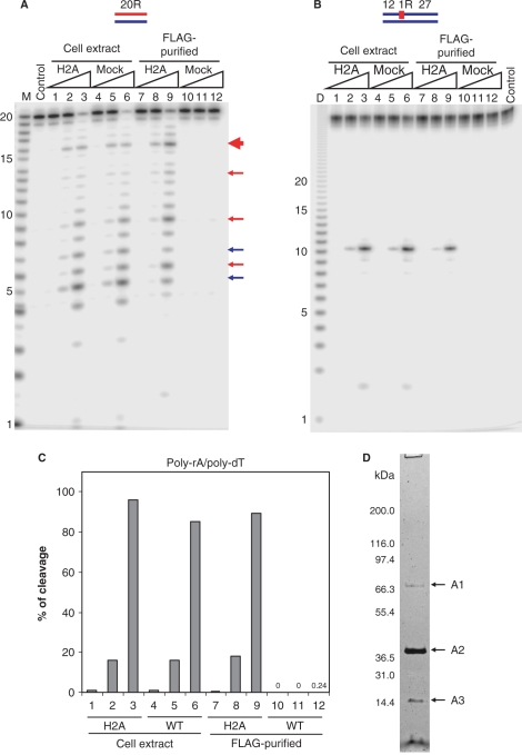
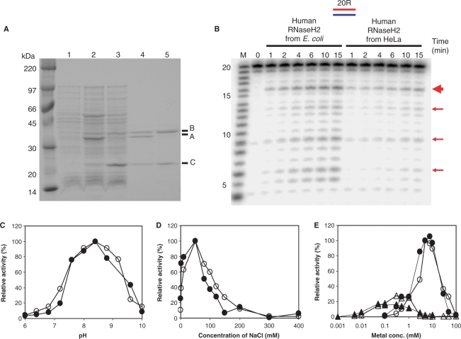
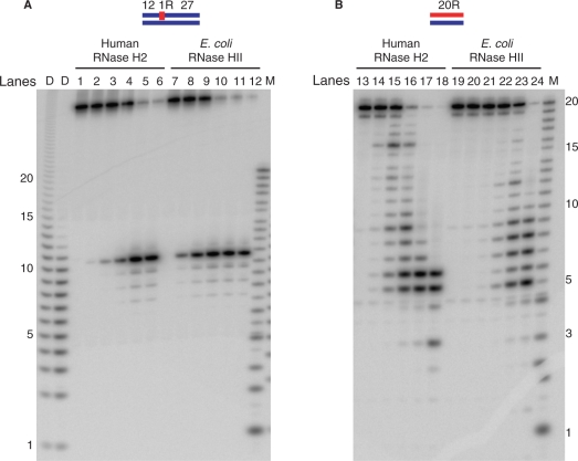
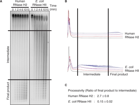
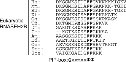
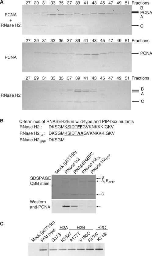
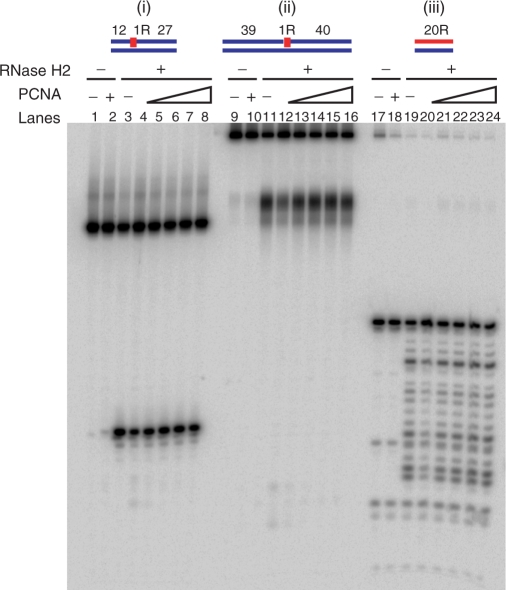
Eukaryotic RNase H2 is a heterotrimeric enzyme. Here, we show that the biochemical composition and stoichiometry of the human RNase H2 complex is consistent with the properties previously deduced from genetic studies. The catalytic subunit of eukaryotic RNase H2, RNASEH2A, is well conserved and similar to the monomeric prokaryotic RNase HII. In contrast, the RNASEH2B and RNASEH2C subunits from human and Saccharomyces cerevisiae share very little homology, although they both form soluble B/C complexes that may serve as a nucleation site for the addition of RNASEH2A to form an active RNase H2, or for interactions with other proteins to support different functions. The RNASEH2B subunit has a PIP-box and confers PCNA binding to human RNase H2. Unlike Escherichia coli RNase HII, eukaryotic RNase H2 acts processively and hydrolyzes a variety of RNA/DNA hybrids with similar efficiencies, suggesting multiple cellular substrates. Moreover, of five analyzed mutations in human RNASEH2B and RNASEH2C linked to Aicardi-Goutières Syndrome (AGS), only one, R69W in the RNASEH2C protein, exhibits a significant reduction in specific activity, revealing a role for the C subunit in enzymatic activity. Near-normal activity of four AGS-related mutant enzymes was unexpected in light of their predicted impairment causing the AGS phenotype.


Figures

Figure 1.
Figure 1.
RNase H activity in cell extracts and FLAG-tag purified fractions from RNASEH2A expressing and untransduced HeLa cell lines. The 5′-end 32P-labeled 20-bp RNA/DNA hybrid (A) and DNA12–RNA1–DNA27/DNA40 hybrid (B) were cleaved with increasing amounts of the total cell extracts and FLAG-tag purified fractions from RNASEH2A expressing cells (H2A) and untransduced cells (mock) at 37°C for 15 min. The 500 μl cell extracts from 107 cells yielded 200 μl of FLAG-purified samples as described in Materials and methods section. Ten picomoles of substrates were treated with 1 μl of the samples in 10 μl of reaction mixtures. Protein samples were diluted in Dilution Buffer. Lanes 1, 4, 7 and 10 contained 0.002 μl equivalents of the undiluted sample, lanes 2, 5, 8 and 11 contained 0.02 μl equivalents and lanes 3, 6, 9 and 12 contained 0.2 μl equivalents. After digestion the reactions were electrophoresed in a 20% TBE-urea PAGE and the gel analyzed on a phosphoimager. Note (B) the mobilities of the DNA12 product of DNA12–RNA1–DNA27/DNA40 migrates faster than the RNA size markers due to inherent differences in migration in the gels between RNA and DNA. In (A), major cleavage sites of 20-bp RNA/DNA hybrid with RNase H1 and RNase H2 are indicated with blue and red arrows, respectively. The main cleavage product of RNase H2 is indicated by a thick red arrow. Molecular size markers are indicated as M (products of digestion of 32P-labeled 20-mer RNA by Phosphodiesterase I) (measuring the sites of cleavage from the 5′- label of the 20-mer RNA) and D (products of digestion of 32P uniformly labeled poly-rA/poly-dT by mouse RNase H1) (measuring the sizes of products that have uniform sequences). (C) Uniformly 32P-labeled poly-rA/poly-dT (1 μM) was cleaved with increasing amount of the total cell extracts and FLAG-tag purified fractions. Amounts of samples in lanes 1–12 are equivalent to those of (A). The ratios of cleavage products were determined by measuring the acid-soluble radioactivity. (D) SDS–PAGE of the purified RNase H2 from HeLa RNASEH2A cells with the two-step affinity immunopurification. HeLa RNASEH2A cells were extracted and subjected to anti-FLAG and anti-HA two-step purification. The purified sample was analyzed by SDS–PAGE stained with silver staining. The fragment indicated with A1, A2 and A3 were identified by mass spectrometry, as described in text.
Figure 2.
Figure 2.
Comparison of enzymatic characteristics of human RNase H2 purified from HeLa cells and E. coli. (A) SDS–PAGE of human RNase H2 expressed in E. coli. Whole-cell extracts of overnight culture of E. coli MIC1066 transformed with pET15b (lane 1), pET-hH2ABC (lane 2) and pET-hH2BC2 (lane 3), and purified human RNase H2 A/B/C complex (lane 4) and B/C complex (lane 5) were analyzed by 10–20% gradient SDS–PAGE (Bio-Rad) and the gel was stained with CBB. The molecular weight marker is Rainbow Marker from Amersham Bioscience. The proteins corresponding to RNASEH2A, RNASEH2B and RNASEH2C are indicated as A, B and C, respectively. (B) Cleavage of 20-bp substrate with human RNase H2 purified from HeLa cells and E. coli. The 5′-end 32P-labeled 20-bp RNA/DNA hybrid (10 pmol) was hydrolyzed with human RNase H2 purified from HeLa cells (as in lane 8 of Figure 1A) and E. coli (12 fmol) at 37°C for the time (min) indicated above each lane in 10 μl reaction mixtures. Molecular size marker is indicated with M (products of digestion of 32P-labeled 20-mer RNA by Phosphodiesterase I). The digested products were analyzed by 20% TBE-urea PAGE. Prominent cleavage sites were indicated with red arrows. The thick red arrow indicates the main cleavage site. Dependence of pH (C), salt concentration (D) and Mg2+ and Mn2+ ions concentration (E) of human RNase H2 purified from HeLa cells (filled) and E. coli (open) were analyzed using uniformly 32P-labeled poly-rA/poly-dT substrate. MgCl2 and MnSO4 concentrations were indicated by circle and triangle in (E), respectively.
Figure 3.
Figure 3.
Comparison of cleavage pattern of short substrates with human RNase H2 and E. coli RNase HII. The 5′-end 32P-labeled DNA12–RNA1–DNA27/DNA40 hybrid (A) and 20-bp RNA/DNA hybrid (B) were cleaved with human RNase H2 and E. coli RNase HII at 37°C for 15 min. The reaction volume was 10 μl and the substrate concentration was 1 μM. Amounts of proteins for human RNase H2 were 1.2 fmol (lanes 2 and 14), 12 fmol (lanes 3 and 15), 120 fmol (lanes 4 and 16), 1.2 pmol (lanes 5 and 17), 12 pmol (lanes 6 and 18). The amounts of protein for E. coli RNase HII were 7.6 fmol (lanes 8 and 20), 76 fmol (lanes 9 and 21), 760 fmol (lanes 10 and 22), 7.6 pmol (lanes 11 and 23), 76 pmol (lanes 12 and 24). Lanes 1, 7, 13 and 19 contained no enzymes. The digested products were analyzed by 20% TBE-urea PAGE. Molecular size markers are indicated as M (products of digestion of 32P-labeled 20-mer RNA by Phosphodiesterase I) and D (products of digestion of 32P-labeled poly-rA/poly-dT by mouse RNase H1).
Figure 4.
Figure 4.
Cleavage of poly-rA/poly-dT substrate with human RNase H2 and E. coli RNase HII. (A) The uniformly 32P-labeled poly-rA/poly-dT (1 μM) was digested with human RNase H2 (0.25 fmol) and E. coli RNase HII (76 fmol) at 37°C for the times indicated for each lane. Samples were analyzed by 20% TBE-urea PAGE as described in Materials and methods section. (B) Graphical representations of degradation of the substrate at the time points 0 (black), 2 (red), 6 (purple) and 15 min (blue) are shown. (C) Processivity values are from at least three independent experiments.
Figure 5.
Figure 5.
Relative specific activity and processivity. (A) Schematic representation of the recombinant human RNase H2 three subunits. Numbers represent the position of the amino acid residues relative to the N-terminal methionine after the pET15b-derived His-tag. AGS-related mutations examined in this study are shown above each subunit. Conserved catalytic residues (D34, E35, D141 and D169) are shown below RNASEH2A. The region shown in black (K294 to F301) in RNASEH2B represents the PIP-box. (B, C) A bar graph of relative specific activity and processivity: (B) Relative specific activity (red) and processivity (green) of human RNase H2 wild type and AGS-related mutants and E. coli RNase HII (EcHII) was analyzed using uniformly 32P-labeled poly-rA/poly-dT as a substrate. (C) Relative specific activity of human RNase H2 wild-type and AGS-related mutants was analyzed by 32P-labeled DNA12–RNA1–DNA27/DNA40 hybrid. The relative values were normalized to wild type RNase H2 (100%). The error bars represent the standard deviation of at least three independent measurements.
Figure 6.
Figure 6.
PCNA binding motif at the C-terminus of eukaryotic RNASEH2B. Amino acid sequences of C-terminus of archaeal RNase HII and eukaryotic RNASEH2B and the consensus PCNA interacting protein box (PIP-box) are aligned. m is aliphatic hydrophobic, Φ is aromatic and x is any residue (32). Conserved residues are in boldface. Hs, Homo sapiens; Mm, Mus musculus; Rn, Rattus norvegicus; Xt, Xenopus tropicalis; Gg, Gallus gallus; Tn, Tetraodon nigroviridis; Dm, Drosophila melanogaster; Ce, Caenorhabditis elegans; Am, Apis mellifera; Dd, Dictyostelium discoideum; Os, Oryza sativa; Spo, Schizosaccharomyces pombe; Sce, Saccharomyces cerevisiae.
Figure 7.
Figure 7.
Physical interaction of RNase H2 with PCNA. (A) Physical interaction between RNase H2 and PCNA was analyzed using gel filtration column chromatography. Mixture of RNase H2 and PCNA (top), PCNA alone (middle) and RNase H2 alone (bottom) were analyzed on the gel filtration column. The indicated fractions were subjected to 10–20% gradient SDS–PAGE and the proteins were visualized by CBB. A representative result is shown in this figure. A, B, C, and PCNA mark the migration of RNASEH2A, RNASEH2B, RNASEH2C and PCNA, respectively. (B) PCNA interacts with C-terminal tail of RNASEH2B: Bacterial lysate containing untagged PCNA was incubated with bacterial lysates containing His-tagged wild-type RNase H2 A/B/C complex, B/C complex, mutant A/B/C complex with two F to A mutations in PIP-box (RNase H2FA) and mutant A/B/C complex with PIP-box deleted (RNase H2ΔPIP). Pulled-down samples were analyzed by SDS–PAGE with CBB stain and western blotting with anti-PCNA antibody. (C) AGS-related mutations do not affect physical interaction between RNase H2 and PCNA: Bacterial lysate containing untagged PCNA was incubated with bacterial lysates containing His-tagged wild-type RNase H2 and mutant RNase H2 with AGS-related mutations. Pulled-down samples were analyzed by western blotting with anti-PCNA antibody.
Figure 8.
Figure 8.
Cleavage of oligomeric substrates with RNase H2 in the presence of PCNA. The 5′-end 32P-labeled DNA12–RNA1–DNA27/DNA40 hybrid (i), DNA39–RNA1–DNA40/DNA80 hybrid (ii) and 20-bp RNA/DNA hybrid (ii) were cleaved with human RNase H2 (3 fmol for DNA12–RNA1–DNA27/DNA40 and DNA39–RNA1–DNA40/DNA80 hybrids, 1.2 fmol for 20-bp RNA/DNA hybird) at 37°C for 15 min. The reaction volume was 10 μl. Substrates amounts were 150 fmol. PCNA quantities were 300 fmol (lanes 4, 12 and20), 1 pmol (lanes 5, 13 and 21), 3 pmol (lanes 6, 14 and 22), 10 pmol (lanes 7, 15 and 23), 30 pmol (lanes 2, 8, 10, 15, 18 and 24). Lanes 1, 9 and 17 contained no enzyme. Reactions were analyzed by 20% TBE-urea PAGE.

References

    1. Crouch RJ, Toulme JJ, editors. Ribonucleases H. Paris: INSERM; 1998.
    1. Jeong HS, Backlund PS, Chen HC, Karavanov AA, Crouch RJ. RNase H2 of Saccharomyces cerevisiae is a complex of three proteins. Nucleic Acids Res. 2004;32:407–414. - PMC - PubMed
    1. Crow YJ, Leitch A, Hayward BE, Garner A, Parmar R, Griffith E, Ali M, Semple C, Aicardi J, Babul-Hirji R, et al. Mutations in genes encoding ribonuclease H2 subunits cause Aicardi-Goutières syndrome and mimic congenital viral brain infection. Nat. Genet. 2006;38:910–916. - PubMed
    1. Rice G, Patrick T, Parmar R, Taylor CF, Aeby A, Aicardi J, Artuch R, Montalto SA, Bacino CA, Barroso B, et al. Clinical and molecular phenotype of Aicardi-Goutières syndrome. Am. J. Hum. Genet. 2007;81:713–725. - PMC - PubMed
    1. Crow YJ, Hayward BE, Parmar R, Robins P, Leitch A, Ali M, Black DN, van Bokhoven H, Brunner HG, Hamel BC, et al. Mutations in the gene encoding the 3′-5′ DNA exonuclease TREX1 cause Aicardi-Goutières syndrome at the AGS1 locus. Nat. Genet. 2006;38:917–920. - PubMed

Publication types

MeSH terms