. 2009 Jan;29(2):503-14.
doi: 10.1128/MCB.01354-08. Epub 2008 Nov 17.

Artemis and nonhomologous end joining-independent influence of DNA-dependent protein kinase catalytic subunit on chromosome stability

Affiliations

Artemis and nonhomologous end joining-independent influence of DNA-dependent protein kinase catalytic subunit on chromosome stability

Travis H Stracker et al. Mol Cell Biol. 2009 Jan.

Abstract

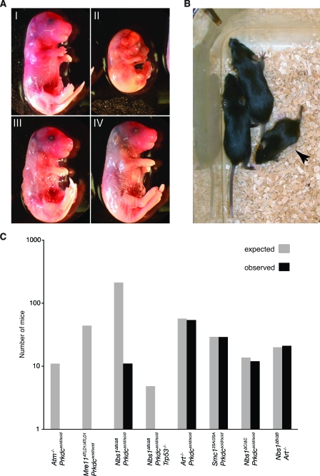
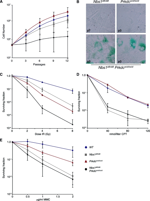
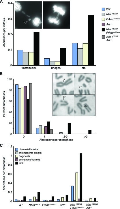
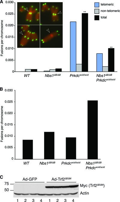
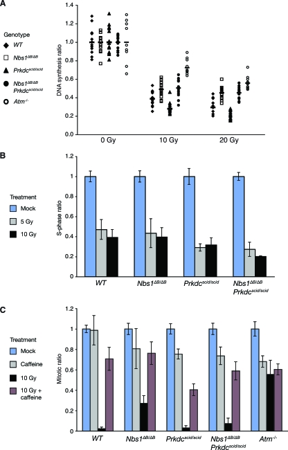
Deficiency in both ATM and the DNA-dependent protein kinase catalytic subunit (DNA-PKcs) is synthetically lethal in developing mouse embryos. Using mice that phenocopy diverse aspects of Atm deficiency, we have analyzed the genetic requirements for embryonic lethality in the absence of functional DNA-PKcs. Similar to the loss of ATM, hypomorphic mutations of Mre11 (Mre11(ATLD1)) led to synthetic lethality when juxtaposed with DNA-PKcs deficiency (Prkdc(scid)). In contrast, the more moderate DNA double-strand break response defects associated with the Nbs1(DeltaB) allele permitted viability of some Nbs1(DeltaB/DeltaB) Prkdc(scid/scid) embryos. Cell cultures from Nbs1(DeltaB/DeltaB) Prkdc(scid/scid) embryos displayed severe defects, including premature senescence, mitotic aberrations, sensitivity to ionizing radiation, altered checkpoint responses, and increased chromosome instability. The known functions of DNA-PKcs in the regulation of Artemis nuclease activity or nonhomologous end joining-mediated repair do not appear to underlie the severe genetic interaction. Our results reveal a role for DNA-PKcs in the maintenance of S/G(2)-phase chromosome stability and in the induction of cell cycle checkpoint responses.


Figures

FIG. 1.
FIG. 1.
Genetic interactions with the Prkdcscid allele. (A) Embryos isolated from Nbs1+/ΔB Prkdcscid/scid breedings at E15.5. (I) Nbs1+/+ Prkdcscid/scid embryo. (II) Nbs1ΔB/ΔB Prkdcscid/scid embryo. (III and IV) Nbs1+/ΔB Prkdcscid/scid embryos. (B) Female Nbs1ΔB/ΔB Prkdcscid/scid pup (arrow) with Nbs1+/ΔB Prkdcscid/scid littermates at 1 month of age. All double mutants were runted, and 10 of 11 had a short, rigid, kinked tail. (C) Synthetic lethality was observed in Atm/ Prkdcscid/scid and Mre11ATLD1/ATLD1 Prkdcscid/scid embryos. Partial synthetic lethality of the Nbs1ΔB and Prkdcscid alleles was observed, while Nbs1ΔB/ΔB Art/, Smc12SA/2SA Prkdcscid/scid, Nbs1ΔC/ΔC Prkdcscid/scid, and Art/ Prkdcscid/scid double mutants were born at expected ratios. The loss of p53 did not eliminate the synthetic lethality of the Nbs1ΔB/ΔB Prkdcscid/scid genotype.
FIG. 2.
FIG. 2.
Analysis of cell growth and survival. (A) The cell growth of primary MEFs of the indicated genotypes was analyzed using a modified 3T3 protocol. Nbs1ΔB/ΔB Prkdcscid/scid cell cultures showed little proliferation compared to WT, Prkdcscid/scid, or Nbs1ΔB/ΔB cell cultures. (B) Early-passage MEFs were stained for SA-β-Gal activity. p3 Nbs1ΔB/ΔB Prkdcscid/scid cultures showed increased SA-β-Gal activity compared to p3 Prkdcscid/scid or p7 Nbs1ΔB/ΔB MEFs, where little activity is detectable. (C) SV40-transformed EFs of the indicated genotypes were exposed to IR and assessed for colony formation. Nbs1ΔB/ΔB Prkdcscid/scid EFs were acutely sensitive to IR-induced damage, and this sensitivity was also observed in MEF cultures (data not shown). Enhanced sensitivity in Nbs1ΔB/ΔB Art/ cell cultures was not observed (see Fig. S3 in the supplemental material). (D and E) SV40-transformed MEFs were exposed to the indicated doses of CPT for 24 h (D) or to the DNA cross-linker MMC for 2 h (E), and survival was assessed by monitoring colony formation. Double-mutant Nbs1ΔB/ΔB Prkdcscid/scid MEFs did not display increased sensitivity to either agent compared to that of single-mutant Nbs1ΔB/ΔB MEFs.
FIG. 3.
FIG. 3.
Genomic instability in fibroblast cultures. (A) Increased mitotic aberrations in DAPI-stained Nbs1ΔB/ΔB Prkdcscid/scid MEFs were observed. Examples of the aberrations scored, the micronucleus (left; arrowhead) and chromosome bridges (right), are shown. (B) Metaphase aberrations in early-passage (p3 to p5) EFs of the indicated genotypes were scored. Nbs1ΔB/ΔB Prkdcscid/scid double mutants showed increased spontaneous aberrations compared to other mutants, which were indistinguishable from the WT. Examples of a chromosome fusion (top left), a chromosome fragment (top right), and two chromatid breaks (bottom left and right) are shown and are indicated by arrowheads. (C) Aberrations in Nbs1ΔB/ΔB Prkdcscid/scid cultures were primarily chromatid breaks and fragments. Similar results were obtained in analyses of MEF cultures (see Fig. S1 in the supplemental material).
FIG. 4.
FIG. 4.
(A) Analysis of spontaneous chromosome fusions in SV40-transformed cultures of the indicated genotypes by telomere-specific FISH. Examples of chromosome fusions with telomeric sequence (top left and bottom left), a chromatid fusion involving telomeric sequence (top right), and a chromosome fusion without telomeric sequence (bottom right) are shown and are indicated by arrowheads. The total fusions per chromosome are shown and are further classified as telomeric or nontelomeric. Increased spontaneous fusions involving telomere sequence were observed in transformed Prkdcscid/scid MEFs. Increased fusions in Nbs1ΔB/ΔB Prkdcscid/scid cultures were also observed, but their frequency was significantly reduced compared to that in Prkdcscid/scid single mutants (**, P = 2.5e−9; Wilcoxon rank sum test). DAPI banding data indicating that fusions were nonclonal are presented in Table S3 in the supplemental material. (B) Telomeric fusions induced by Trf2ΔBΔM expression. Similar numbers of fusions were induced after Trf2ΔBΔM expression regardless of the genotype. Fusion numbers were normalized to those in cultures infected with Ad-GFP to account for spontaneous fusion levels. (C) Western blot analysis of the expression of Myc-tagged Trf2ΔBΔM in Ad-GFP- or Trf2ΔBΔM-expressing adenovirus (Ad-Trf2ΔBΔM)-infected MEFs. Lanes: 1, WT; 2, Nbs1ΔB/ΔB; 3, Prkdcscid/scid; and 4, Nbs1ΔB/ΔB Prkdcscid/scid. Actin was included as a control for protein loading.
FIG. 5.
FIG. 5.
Influence of DNA-PKcs on checkpoint induction. (A) The intra-S-phase checkpoint defect of Nbs1ΔB/ΔB mutants was indistinguishable from that of Nbs1ΔB/ΔB Prkdcscid/scid double mutants. Prkdcscid/scid MEFs showed significantly stronger arrest than WT MEFs (Wilcoxon rank sum test; at 10 Gy, P = 3.18e−2 [two sided], and at 20 Gy, P = 2.98e−5 [two sided]). Atm/ cultures are shown for comparison. (B) G1/S checkpoint responses in early-passage MEFs. Cultures were irradiated with 5 or 10 Gy, and DNA synthesis levels were normalized to those in untreated cultures. Both Prkdcscid/scid and Nbs1ΔB/ΔB Prkdcscid/scid MEFs showed stronger G1/S checkpoint arrest than WT MEFs. Representative flow cytometry data are included in Fig. S5 in the supplemental material. (C) Deficient G2/M checkpoint arrest in Nbs1ΔB/ΔB mutants was rescued by the Prkdcscid/scid mutation. MEFs of the indicated genotypes were mock treated, incubated with 10 mM caffeine, exposed to 10 Gy of IR, or exposed to 10 Gy IR and treated with 10 mM caffeine, and their mitotic indices were determined 1 h posttreatment. Treatment with caffeine abolished the checkpoint response in all backgrounds. Representative flow cytometry data are included in Fig. S6 in the supplemental material.

Similar articles

Cited by

References

    1. Bailey, S. M., M. N. Cornforth, A. Kurimasa, D. J. Chen, and E. H. Goodwin. 2001. Strand-specific postreplicative processing of mammalian telomeres. Science 2932462-2465. - PubMed
    1. Bailey, S. M., J. Meyne, D. J. Chen, A. Kurimasa, G. C. Li, B. E. Lehnert, and E. H. Goodwin. 1999. DNA double-strand break repair proteins are required to cap the ends of mammalian chromosomes. Proc. Natl. Acad. Sci. USA 9614899-14904. - PMC - PubMed
    1. Bressan, D. A., B. K. Baxter, and J. H. Petrini. 1999. The Mre11-Rad50-Xrs2 protein complex facilitates homologous recombination-based double-strand break repair in Saccharomyces cerevisiae. Mol. Cell. Biol. 197681-7687. - PMC - PubMed
    1. Buscemi, G., C. Savio, L. Zannini, F. Micciche, D. Masnada, M. Nakanishi, H. Tauchi, K. Komatsu, S. Mizutani, K. Khanna, P. Chen, P. Concannon, L. Chessa, and D. Delia. 2001. Chk2 activation dependence on Nbs1 after DNA damage. Mol. Cell. Biol. 215214-5222. - PMC - PubMed
    1. Carney, J. P., R. S. Maser, H. Olivares, E. M. Davis, M. Le Beau, J. R. Yates III, L. Hays, W. F. Morgan, and J. H. Petrini. 1998. The hMre11/hRad50 protein complex and Nijmegen breakage syndrome: linkage of double-strand break repair to the cellular DNA damage response. Cell 93477-486. - PubMed

Publication types

MeSH terms

LinkOut - more resources