. 2009 Jan;29(2):547-58.
doi: 10.1128/MCB.00329-08. Epub 2008 Nov 17.

USP19 deubiquitinating enzyme supports cell proliferation by stabilizing KPC1, a ubiquitin ligase for p27Kip1

Affiliations

USP19 deubiquitinating enzyme supports cell proliferation by stabilizing KPC1, a ubiquitin ligase for p27Kip1

Yu Lu et al. Mol Cell Biol. 2009 Jan.

Erratum in

  • Mol Cell Biol. 2009 Jun;29(11):3241

Abstract

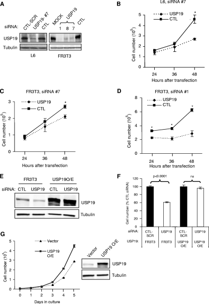
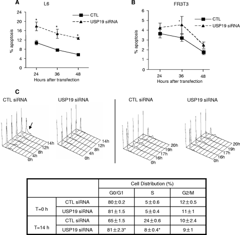
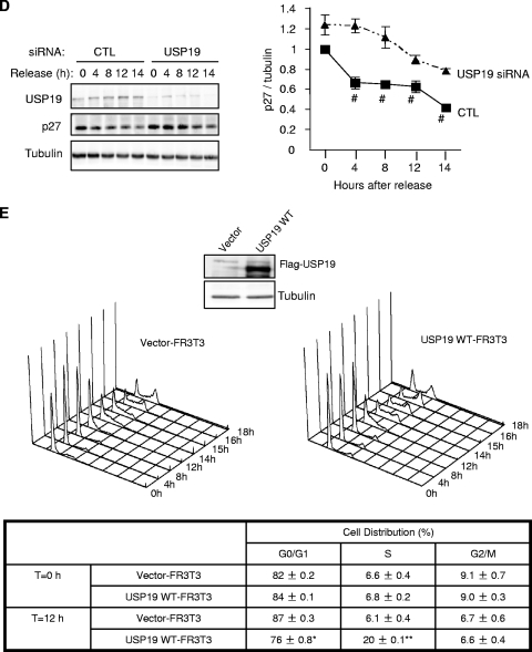
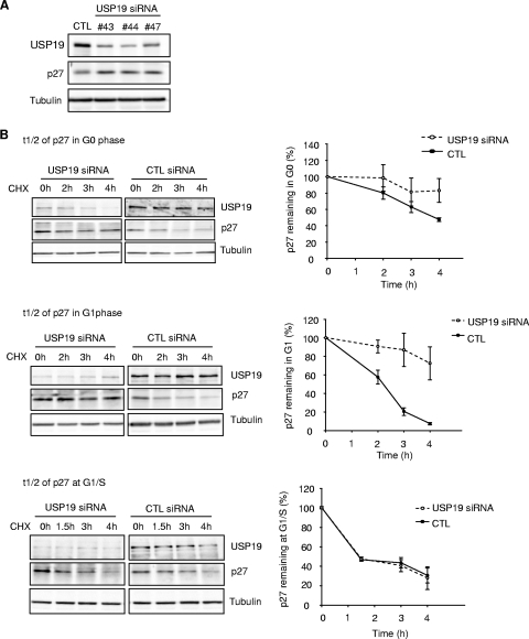
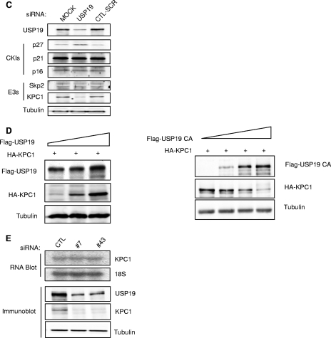
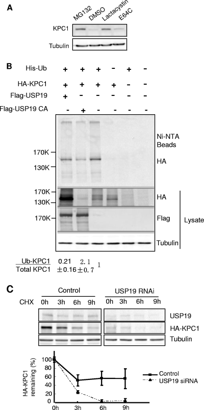
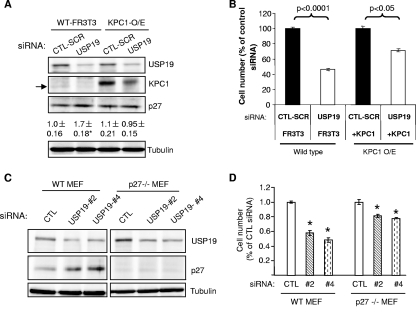
p27(Kip1) is a cyclin-dependent kinase inhibitor that regulates the G(1)/S transition. Increased degradation of p27(Kip1) is associated with cellular transformation. Previous work demonstrated that the ubiquitin ligases KPC1/KPC2 and SCF(Skp2) ubiquitinate p27(Kip1) in G(1) and early S, respectively. The regulation of these ligases remains unclear. We report here that the USP19 deubiquitinating enzyme interacts with and stabilizes KPC1, thereby modulating p27(Kip1) levels and cell proliferation. Cells depleted of USP19 by RNA interference exhibited an inhibition of cell proliferation, progressing more slowly from G(0)/G1 to S phase, and accumulated p27(Kip1). This increase in p27(Kip1) was associated with normal levels of Skp2 but reduced levels of KPC1. The overexpression of KPC1 or the use of p27(-/-) cells inhibited significantly the growth defect observed upon USP19 depletion. KPC1 was ubiquitinated in vivo and stabilized by proteasome inhibitors and by overexpression of USP19, and it also coimmunoprecipitated with USP19. Our results identify USP19 as the first deubiquitinating enzyme that regulates the stability of a cyclin-dependent kinase inhibitor and demonstrate that progression through G(1) to S phase is, like the metaphase-anaphase transition, controlled in a hierarchical, multilayered fashion.


Figures

FIG. 1.
FIG. 1.
Depletion of USP19 leads to reduced cell proliferation. (A) Effectiveness of USP19 siRNA oligonucleotides in depleting the enzyme. L6 myoblasts and FR3T3 fibroblasts were transfected with USP19 or scrambled (CTL-SCR) or nonspecific control siRNA oligonucleotides (CTL) or transfected without siRNA oligonucleotides (MOCK). Forty-eight hours after transfection, cells were harvested and the lysate subjected to immunoblot analysis with antibody against either USP19 or γ-tubulin. #7, 1, 8, and 7, USP19 siRNA oligonucleotides. (B to D) Cells depleted of USP19 show reduced proliferation. L6 myoblasts (B) or FR3T3 fibroblasts (C, D) were transfected with USP19 siRNA oligonucleotide no. 7 (#7) (B and C) or no. 1 (#1) (D) or nonspecific or scrambled control (CTL) siRNA oligonucleotides. Cell numbers were determined at the indicated times after transfection. Shown are means ± standard deviations of the results. *, significantly different from results for control oligonucleotide transfection (P < 0.001). (E) Normal FR3T3 cells or those stably overexpressing (O/E) USP19 bearing mutations that do not change the coding sequence but render the mRNA resistant to siRNA-mediated degradation were transfected with USP19 siRNA or scrambled siRNA (CTL). After 48 h, cells were harvested and lysates prepared and analyzed by immunoblotting. (F) Normal proliferation in cells expressing USP19 resistant to siRNA. Cells were treated as described for panel E and counted at 48 h after transfection with the indicated siRNA oligonucleotides. Shown are means ± standard errors of the results. (G) Expression of exogenous USP19 increases the rate of cell proliferation. FR3T3 cells stably overexpressing Flag-tagged USP19 were cultured for the indicated number of days. Vector-transfected FR3T3 cells were used as control. Shown are means ± standard errors of the results.
FIG. 2.
FIG. 2.
Cells depleted of USP19 show only a small increase in apoptosis but impaired progression from G0 to S phase. (A, B) Apoptosis is mildly increased in USP19-depleted L6 myoblasts or FR3T3 fibroblasts. Cells were treated with the indicated siRNA oligonucleotides. At the indicated times, cells were harvested and analyzed by FACS to quantify apoptotic cells (identified as annexin V positive, propidium iodide negative). *, significantly different from results for control at the indicated time points (P < 0.001). (C) FR3T3 cells depleted of USP19 showed delayed entry into S phase. Cells were serum starved for 36 h. They were then transfected with scrambled control (CTL) or USP19 siRNA oligonucleotides. Following another 36 h of serum starvation, cells were stimulated to reenter cell cycle with DMEM containing 20% FBS. Cells were harvested at the indicated times following serum stimulation and subjected to FACS analysis. Arrow indicates entry into S phase seen in CTL cells at 14 h (left) and at ∼17 h in cells transfected with USP19 siRNA oligonucleotides (right). At 17 h, CTL cells were starting to reenter G1. Table shows percentages of cells in different phases of the cell cycle before and 14 h after release from G0. *, significantly different (P < 0.001) from results for CTL. (D) p27Kip1 accumulates in USP19-depleted cells. Equal amounts of protein from lysates (from experiment described in panel C) were analyzed by immunoblotting with the indicated antibodies. Shown are a representative blot and quantitation of results for multiple samples. Values for USP19 siRNA-treated cells are significantly different from those for CTL-treated cells. #, P < 0.001; two-way analysis of variance. (E) FR3T3 cells overexpressing USP19 enter S phase earlier. Cells stably transfected with plasmid overexpressing USP19 or an empty vector (control) were starved of serum for 72 h and then stimulated to reenter cell cycle with DMEM containing 20% FBS. Cells were harvested at the indicated times following serum stimulation and subjected to FACS analysis as described for panel C above. Means ± standard errors of the results are shown. Asterisks indicate means that are significantly different from those for control cells (*, P < 0.01; **, P < 0.001). WT, wild type.
FIG. 2.
FIG. 2.
Cells depleted of USP19 show only a small increase in apoptosis but impaired progression from G0 to S phase. (A, B) Apoptosis is mildly increased in USP19-depleted L6 myoblasts or FR3T3 fibroblasts. Cells were treated with the indicated siRNA oligonucleotides. At the indicated times, cells were harvested and analyzed by FACS to quantify apoptotic cells (identified as annexin V positive, propidium iodide negative). *, significantly different from results for control at the indicated time points (P < 0.001). (C) FR3T3 cells depleted of USP19 showed delayed entry into S phase. Cells were serum starved for 36 h. They were then transfected with scrambled control (CTL) or USP19 siRNA oligonucleotides. Following another 36 h of serum starvation, cells were stimulated to reenter cell cycle with DMEM containing 20% FBS. Cells were harvested at the indicated times following serum stimulation and subjected to FACS analysis. Arrow indicates entry into S phase seen in CTL cells at 14 h (left) and at ∼17 h in cells transfected with USP19 siRNA oligonucleotides (right). At 17 h, CTL cells were starting to reenter G1. Table shows percentages of cells in different phases of the cell cycle before and 14 h after release from G0. *, significantly different (P < 0.001) from results for CTL. (D) p27Kip1 accumulates in USP19-depleted cells. Equal amounts of protein from lysates (from experiment described in panel C) were analyzed by immunoblotting with the indicated antibodies. Shown are a representative blot and quantitation of results for multiple samples. Values for USP19 siRNA-treated cells are significantly different from those for CTL-treated cells. #, P < 0.001; two-way analysis of variance. (E) FR3T3 cells overexpressing USP19 enter S phase earlier. Cells stably transfected with plasmid overexpressing USP19 or an empty vector (control) were starved of serum for 72 h and then stimulated to reenter cell cycle with DMEM containing 20% FBS. Cells were harvested at the indicated times following serum stimulation and subjected to FACS analysis as described for panel C above. Means ± standard errors of the results are shown. Asterisks indicate means that are significantly different from those for control cells (*, P < 0.01; **, P < 0.001). WT, wild type.
FIG. 3.
FIG. 3.
Levels of the cyclin-dependent kinase inhibitor (CKI) p27Kip1 and the ubiquitin protein ligase (E3) KPC1 are modulated by USP19. (A) Multiple independent USP19 siRNA oligonucleotides (#43, no.43; #44, no.44; #47, no. 47) increase p27Kip1 levels in asynchronous FR3T3 cells. (B) Depletion of USP19 decreases the rate of degradation of p27Kip1 in G0 and G1 phases but not at the G1/S transition. FR3T3 cells transfected with USP19 no. 7 siRNA or scrambled control oligonucleotides were synchronized in G0 phase as described for Fig. 2. Cells were stimulated to reenter the cell cycle with DMEM medium containing 10% FBS and 2 mM thymidine (to accumulate cells at G1/S). At 7 h (G1) or 12.5 h (G1/S, 15 h for USP19 siRNA-transfected cells which progressed more slowly) of stimulation (Fig. 2C) (aliquots of cells were analyzed by FACS to confirm positions in cell cycle), the cells were incubated with cycloheximide to block protein synthesis and measure the rate of degradation of p27Kip1. At the indicated times, cell protein was analyzed by immunoblotting with the indicated antibodies. Shown are representative immunoblots and quantitation of p27Kip1 levels from triplicate samples. Rates of degradation of p27Kip1 were significantly inhibited in USP19 siRNA-treated cells in G0 (P < 0.05) and G1 phases (P < 0.001) but not in S phase (not significant) (all analyses were by two-way analysis of variance). t1/2, half-life; CHX, cycloheximide. (C) Levels of KPC1 but not Skp2 are decreased in USP19-depleted cells. Proliferating FR3T3 cells were treated with scrambled control or USP19 siRNA oligonucleotides. Cells were harvested 48 h later, and lysates subjected to immunoblot analysis with the indicated antibodies. Levels of p27Kip1 but not p21 or p16 are increased upon USP19 depletion. Levels of KPC1 but not Skp2 are decreased upon USP19 depletion. (D) USP19 modulates levels of KPC1. FR3T3 cells were transfected with plasmid expressing HA-KPC1 (+) and increasing amounts of a plasmid expressing Flag-USP19 (left) or catalytically inactive Flag-USP19 C545A mutant and His-tagged ubiquitin (right). Proteins from cell extracts were analyzed by immunoblotting with anti-Flag (USP19), anti-HA (KPC1), and anti-γ-tubulin antibodies. (E) Silencing of USP19 does not lower KPC1 levels by lowering mRNA levels. FR3T3 cells were transfected with USP19 siRNA oligonucleotides no. 7 (#7) or no. 43 (#43) or control oligonucleotides (CTL). Two days later, RNA or protein was isolated and analyzed by RNA blot analysis (top) or by immunoblotting (below), respectively.
FIG. 3.
FIG. 3.
Levels of the cyclin-dependent kinase inhibitor (CKI) p27Kip1 and the ubiquitin protein ligase (E3) KPC1 are modulated by USP19. (A) Multiple independent USP19 siRNA oligonucleotides (#43, no.43; #44, no.44; #47, no. 47) increase p27Kip1 levels in asynchronous FR3T3 cells. (B) Depletion of USP19 decreases the rate of degradation of p27Kip1 in G0 and G1 phases but not at the G1/S transition. FR3T3 cells transfected with USP19 no. 7 siRNA or scrambled control oligonucleotides were synchronized in G0 phase as described for Fig. 2. Cells were stimulated to reenter the cell cycle with DMEM medium containing 10% FBS and 2 mM thymidine (to accumulate cells at G1/S). At 7 h (G1) or 12.5 h (G1/S, 15 h for USP19 siRNA-transfected cells which progressed more slowly) of stimulation (Fig. 2C) (aliquots of cells were analyzed by FACS to confirm positions in cell cycle), the cells were incubated with cycloheximide to block protein synthesis and measure the rate of degradation of p27Kip1. At the indicated times, cell protein was analyzed by immunoblotting with the indicated antibodies. Shown are representative immunoblots and quantitation of p27Kip1 levels from triplicate samples. Rates of degradation of p27Kip1 were significantly inhibited in USP19 siRNA-treated cells in G0 (P < 0.05) and G1 phases (P < 0.001) but not in S phase (not significant) (all analyses were by two-way analysis of variance). t1/2, half-life; CHX, cycloheximide. (C) Levels of KPC1 but not Skp2 are decreased in USP19-depleted cells. Proliferating FR3T3 cells were treated with scrambled control or USP19 siRNA oligonucleotides. Cells were harvested 48 h later, and lysates subjected to immunoblot analysis with the indicated antibodies. Levels of p27Kip1 but not p21 or p16 are increased upon USP19 depletion. Levels of KPC1 but not Skp2 are decreased upon USP19 depletion. (D) USP19 modulates levels of KPC1. FR3T3 cells were transfected with plasmid expressing HA-KPC1 (+) and increasing amounts of a plasmid expressing Flag-USP19 (left) or catalytically inactive Flag-USP19 C545A mutant and His-tagged ubiquitin (right). Proteins from cell extracts were analyzed by immunoblotting with anti-Flag (USP19), anti-HA (KPC1), and anti-γ-tubulin antibodies. (E) Silencing of USP19 does not lower KPC1 levels by lowering mRNA levels. FR3T3 cells were transfected with USP19 siRNA oligonucleotides no. 7 (#7) or no. 43 (#43) or control oligonucleotides (CTL). Two days later, RNA or protein was isolated and analyzed by RNA blot analysis (top) or by immunoblotting (below), respectively.
FIG. 4.
FIG. 4.
KPC1 is a substrate of the ubiquitin-proteasome pathway and is stabilized by USP19. (A) KPC1 is stabilized by proteasome inhibitors. FR3T3 cells were treated with a proteasome inhibitor, either MG132 (10 μΜ) or lactacystin (5 μΜ), or with cysteine protease inhibitor E64C (10 μΜ) or vehicle (DMSO [dimethyl sulfoxide]) for 6 h. Cells were then harvested and blotted with anti-KPC1 antibody. (B) KPC1 is polyubiquitinated in vivo, and the ubiquitination is modulated by USP19. FR3T3 cells were transfected with the indicated plasmids. Cells were treated with MG132 for 6 h before lysis to accumulate ubiquitinated proteins. Lysate was either analyzed by immunoblotting with the indicated antibodies or incubated with Ni+-agarose beads. Bound ubiquitinated proteins were washed extensively and then eluted with Laemmli buffer and analyzed by immunoblotting with anti-HA antibody. Quantitation and ratios of the levels of ubiquitinated KPC1 (anti-HA blot of proteins bound to Ni+-NTA beads) to total KPC1 (anti-HA blot of lysate) (means ± standard errors) are indicated below. Means for cells overexpressing wild-type USP19 or the USP19CA mutant are significantly different (P value of <0.025 by one-way analysis of variance) from that for non-USP19-overexpressing cells, which was set at 1. Ub, ubiquitin; +, present; −, absent. (C) Silencing of USP19 destabilizes KPC1. FR3T3 cells stably overexpressing KPC1 were transfected with USP19 siRNA or control oligonucleotides. Forty-eight hours later, the cells were incubated with cycloheximide. At the indicated times, cell protein was analyzed by immunoblotting with the indicated antibodies. Shown are representative immunoblots and quantitation of KPC1 levels from triplicate samples. Error bars show standard errors. CHX, cycloheximide; RNAi, RNA interference.
FIG. 5.
FIG. 5.
USP19 interacts with KPC1 and, like KPC1, is localized to the cytosol. (A) Interaction between endogenous KPC1 and USP19. FR3T3 cell lysates were subjected to IP using anti-KPC1 antibody, followed by immunoblot analysis with anti-USP19 or anti-KPC1 antibodies. As a control (CTL) for specificity of the antibodies, the experiment was also conducted in cells in which USP19 had been depleted by RNA interference. IgG, immunoglobulin G. (B) FR3T3 cells were transfected with the indicated combinations of plasmids expressing Flag-USP19 and HA-KPC1. Extracts of the transfected cells were subjected to IP using anti-Flag or anti-HA antibody. The immunoprecipitates were blotted with the indicated antibodies. +, present; −, absent. (C) Subcellular fractions of lysate from FR3T3 cells were subjected to immunoblotting with anti-USP19, anti-KPC1, anti-histone H2A (nuclear marker), and anti-α-tubulin (cytoplasmic marker) antibodies.
FIG. 6.
FIG. 6.
USP19 modulates cell growth through KPC1/p27Kip1-dependent and -independent pathways. (A, B) KPC1 overexpression normalizes p27Kip1 levels and partially rescues growth defects of USP19-depleted cells. Wild-type (WT) or FR3T3 cells stably overexpressing (O/E) KPC1 were transfected with USP19 siRNA or scrambled control oligonucleotide (CTL-SCR). After 48 h, the cells were counted or harvested in a denaturing buffer. (A) Proteins from cells exposed to the indicated siRNA oligonucleotides were analyzed by immunoblotting with the indicated antibodies (arrow indicates endogenous KPC1, which migrates faster than overexpressed epitope-tagged KPC1). Shown below the p27Kip1 blot is quantitation of p27Kip1 protein as means ± standard errors. *, P value of <0.025 compared to result for CTL-SCR. (B) Cell numbers in the different treatment groups. Shown are means ± standard errors. (C, D) Ability of USP19 depletion to inhibit cell growth is partially blocked in MEF lacking p27Kip1. Wild-type or p27Kip1−/− MEF were transfected with USP19 siRNA (#2, no. 2; #4, no. 4) or nonspecific control oligonucleotide (CTL). After 48 h, the cells were counted or harvested in a denaturing buffer. (C) Proteins from cells exposed to the indicated siRNA oligonucleotides were analyzed by immunoblotting with the indicated antibodies. (D) Cell numbers in the different treatment groups. Shown are means ± standard errors. *, P value of <0.001 compared to result for CTL.

References

    1. Bloom, J., and M. Pagano. 2003. Deregulated degradation of the cdk inhibitor p27 and malignant transformation. Semin. Cancer Biol. 1341-47. - PubMed
    1. Burrows, J. F., M. J. McGrattan, A. Rascle, M. Humbert, K. H. Baek, and J. A. Johnston. 2004. DUB-3, a cytokine-inducible deubiquitinating enzyme that blocks proliferation. J. Biol. Chem. 27913993-14000. - PubMed
    1. Carrano, A. C., E. Eytan, A. Hershko, and M. Pagano. 1999. SKP2 is required for ubiquitin-mediated degradation of the CDK inhibitor p27. Nat. Cell Biol. 1193-199. - PubMed
    1. Crosas, B., J. Hanna, D. S. Kirkpatrick, D. P. Zhang, Y. Tone, N. A. Hathaway, C. Buecker, D. S. Leggett, M. Schmidt, R. W. King, S. P. Gygi, and D. Finley. 2006. Ubiquitin chains are remodeled at the proteasome by opposing ubiquitin ligase and deubiquitinating activities. Cell 1271401-1413. - PubMed
    1. Deng, C., P. Zhang, J. W. Harper, S. J. Elledge, and P. Leder. 1995. Mice lacking p21CIP1/WAF1 undergo normal development, but are defective in G1 checkpoint control. Cell 82675-684. - PubMed

Publication types

MeSH terms