. 2009 Feb;77(2):770-82.
doi: 10.1128/IAI.00994-08. Epub 2008 Nov 17.

Dendritic cell and NK cell reciprocal cross talk promotes gamma interferon-dependent immunity to blood-stage Plasmodium chabaudi AS infection in mice

Affiliations

Dendritic cell and NK cell reciprocal cross talk promotes gamma interferon-dependent immunity to blood-stage Plasmodium chabaudi AS infection in mice

Rebecca Ing et al. Infect Immun. 2009 Feb.

Abstract

Dendritic cells (DCs) are important accessory cells for promoting NK cell gamma interferon (IFN-gamma) production in vitro in response to Plasmodium falciparum-infected red blood cells (iRBC). We investigated the requirements for reciprocal activation of DCs and NK cells leading to Th1-type innate and adaptive immunity to P. chabaudi AS infection. During the first week of infection, the uptake of iRBC by splenic CD11c(+) DCs in resistant wild-type (WT) C57BL/6 mice was similar to that in interleukin 15(-/-) (IL-15(-/-)) and IL-12p40(-/-) mice, which differ in the severity of P. chabaudi AS infection. DCs from infected IL-15(-/-) mice expressed costimulatory molecules, produced IL-12, and promoted IFN-gamma secretion by WT NK cells in vitro as efficiently as WT DCs. In contrast, DCs from infected IL-12p40(-/-) mice exhibited alterations in maturation and cytokine production and were unable to induce NK cell IFN-gamma production. Coculture of DCs and NK cells demonstrated that DC-mediated NK cell activation required IL-12 and, to a lesser extent, IL-2, as well as cell-cell contact. In turn, NK cells from infected WT mice enhanced DC maturation, IL-12 production, and priming of CD4(+) T-cell proliferation and IFN-gamma secretion. Infected WT mice depleted of NK cells, which exhibit increased parasitemia, had impaired DC maturation and DC-induced CD4(+) Th1 cell priming. These findings indicate that DC-NK cell reciprocal cross talk is critical for control and rapid resolution of P. chabaudi AS infection and provide in vivo evidence for the importance of this interaction in IFN-gamma-dependent immunity to malaria.


Figures

FIG. 1.
FIG. 1.
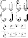
IL-12 is required for DC maturation but not for iRBC uptake. (A) Uptake of iRBC by splenic DCs at day 0 and day 4 p.i. The histograms represent CFSE expression by gated CD11c+ cells cultured alone (solid gray area) or with CFSE-labeled iRBC (lines). Splenic CD11c+ DCs were purified from WT (solid lines), IL-15−/− (dashed lines), and IL-12p40−/− (dotted lines) mice. (B) Kinetics of iRBC and nRBC uptake by splenic DCs from naïve and P. chabaudi AS-infected mice. The data were pooled from three experiments, each containing three or four mice per group. The asterisks indicate significant differences between iRBC and nRBC uptake at the specified time points: *, P < 0.05; **, P < 0.01; and ***, P < 0.001. (C) MFI of CD40, CD80, and CD86 expression by gated CD11c+ cells from naïve or day 4 infected mice. The data are representative of two experiments, each containing three or four mice per group. The asterisks indicate significant differences between WT and IL-12p40−/− DCs: *, P < 0.05, and **, P < 0.01. The error bars indicate SEM.
FIG. 2.
FIG. 2.
IL-12 deficiency impairs IFN-γ production but increases IL-2 production by DCs during blood-stage malaria. (A) Splenic CD11c+ DCs from naïve or day 4 infected WT, IL-15−/−, and IL-12p40−/− mice were stimulated in vitro without (Medium) or with iRBC. The supernatants were analyzed for IL-12p70, IFN-γ, IL-2, and IL-10 by ELISA. The asterisks indicate significant differences between WT and cytokine-deficient DCs: *, P < 0.05; **, P < 0.01; and ***, P < 0.001. (B) Intracellular IFN-γ expression by iRBC-stimulated splenic DCs from day 4 infected WT, IL-15−/−, and IL-12p40−/− mice. The cells were gated on the DX5-negative population, and the percentages of IFN-γ expression by CD11c+ DCs are shown. (C) Intracellular IL-2 expression by splenic CD11c+ DCs from naïve or day 7 infected WT and IL-12p40−/− mice. For panels B and C, appropriate isotype staining controls are shown. The data are representative of two experiments, each containing three or four mice per group. The error bars indicate SEM.
FIG. 3.
FIG. 3.
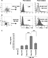
Malaria-activated DCs promote high IFN-γ production by resting NK cells. CD11c+ DCs from day 5 infected WT mice and DX5+ NK cells from naïve WT mice were cultured separately or together for 24 to 36 h at 37°C and analyzed for IFN-γ production. (A and B) Intracellular IFN-γ expression by gated populations of CD11c+ DX5 DC or DX5+ CD3 NK cells isolated from infected and naïve WT mice, respectively. (Right) IFN-γ expression by CD11c+ DX5 DCs and DX5+ CD3 NK cells before culture (Pre Culture) and after culture alone (lines) or together (shaded areas) (Post Culture). The data are representative of four experiments giving similar results. (C) Secreted levels of IFN-γ were determined by ELISA in single cultures of NK cells from naïve WT mice or DCs from infected WT mice (Day 5 DC) and in cocultures of WT NK cells with DCs from naïve WT mice (Day 0 DC) or day 5 p.i. (Day 5 DC). The DCs were separated from NK cells by using a transwell insert (Transwell). The data are representative of four experiments, each with three or four replicates per group. The asterisks indicate significant differences compared with DCs from day 5 infected WT mice (Day 5 DC): ***, P < 0.001. The error bars indicate SEM.
FIG. 4.
FIG. 4.
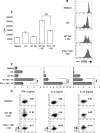
IL-12 and IL-2 are key cytokines in DC-mediated stimulation of NK cells. Splenic DCs from day 5 infected WT, IL-15−/−, or IL-12−/− mice were cocultured for 24 to 36 h at 37°C with NK cells from naïve WT mice. (A) Intracellular IFN-γ expression by WT, IL-15−/−, or IL-12−/− DCs and WT NK cells after coculture. Each panel displays composite histograms of IFN-γ signals from gated CD11c+ DX5 DC or DX5+ CD3 NK cell populations. The lines represent expression by DCs, and the shaded histograms represent NK cells. The percentages of DC or NK cells expressing IFN-γ are shown. The data are representative of four experiments showing similar results. (B) IFN-γ levels in the coculture supernatants were quantified by ELISA. The data are representative of four experiments, each containing three or four mice per group. The asterisks indicate a significant difference between WT and IL-12p40−/− DCs: ***, P < 0.001. (C) WT, IL-15−/−, or IL-12−/− DCs were cocultured with WT NK cells, and IL-2 activity was blocked using a neutralizing anti-IL-2 MAb. Control wells were treated with isotype control IgG MAb. IFN-γ levels were quantified by ELISA after 36 h of coculture. The data are representative of two experiments, each containing three or four replicates per group. The asterisks indicate significant differences between DCs treated with isotype control MAb and DCs treated with anti-IL-2 MAb: **, P < 0.01. The error bars indicate SEM.
FIG. 5.
FIG. 5.
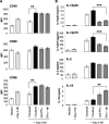
Malaria-activated NK cells stimulate DCs to mature and secrete cytokines. Splenic NK cells from day 5 infected WT mice were cocultured for 18 to 24 h at 37°C with naïve WT DCs exposed to iRBC before coculture. DCs were analyzed for costimulatory molecule expression (A) and cytokine production (B). To determine the role of IFN-γ in these interactions, DCs were cocultured with NK cells from IFN-γ−/− mice or with WT NK cells in the presence of anti-IFN-γ MAb or isotype control MAb (data not shown). DCs were also cocultured with NK cells in wells with a transwell insert to prevent cell-cell contact. (A) Cells were gated on the CD11c+ signal and analyzed for CD40, CD80, and CD86 expression (MFI) after culture with medium or coculture with NK cells from naïve mice (Day 0 NK) as controls or coculture with WT or IFN-γ−/− NK cells from day 5 infected mice (Day 5 NK). The asterisks indicate a significant difference between DCs separated from day 5 NK cells by a transwell insert (Transwell) and DCs cocultured with day 5 NK cells (Coculture): **, P < 0.01. (B) Cytokine levels in supernatants of DCs cultured alone or with day 0 or day 5 NK cells were determined by ELISA. The data are representative of three experiments, each containing three or four replicates per group. The asterisks indicate significant differences between WT NK cells (Coculture) and IFN-γ−/− NK cells: **, P < 0.01 and ***, P < 0.001. The error bars indicate SEM.
FIG. 6.
FIG. 6.
NK cell-activated DCs prime CD4+ T cells in an IFN-γ-dependent mechanism. Splenic NK cells from day 5 infected WT or IFN-γ−/− mice were cocultured with DCs from naïve WT mice for 24 h and then plated with freshly isolated CD4+ T cells from naïve mice. After 5 days, the cells were harvested, restimulated in wells coated with anti-CD3 MAb for 48 h, and analyzed for proliferation (A and B) and cytokine production (C and D). CD4+ T cells cultured alone (Medium) or with DCs or WT NK cells separately were included as controls. (A) DCs activated by WT NK cells induced significantly higher levels of T-cell proliferation than did DCs cultured with IFN-γ−/− NK cells. The asterisks indicate a significant difference between WT NK cells and IFN-γ−/− NK cells cocultured with DCs: **, P < 0.01. (B) The CFSE dilution in gated CD4+ T cells following coculture with medium, DCs alone, or DCs stimulated with WT or IFN-γ−/− NK cells from infected mice was determined by flow cytometry. (C) DCs activated by WT NK cells induced higher levels of IFN-γ production by CD4+ T cells. The asterisks indicate significant differences in cytokine production by CD4+ T cells primed by DCs cocultured with malaria-activated WT versus IFN-γ−/− NK cells: **, P < 0.01, and ***, P < 0.001. (D) Intracellular cytokine expression by gated CD4+ T cells as determined by flow cytometry. The percentages of cells expressing each cytokine are indicated. The data are representative of two or three experiments, each containing three or four replicates per group. The error bars indicate SEM.
FIG. 7.
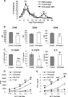
FIG. 7.
Depletion of NK cells leads to increased parasitemia and impaired DC maturation and Th1 priming. B6 mice were injected intravenously with sterile PBS, control IgG, or anti-asialo-GM1 Ab at days −7, −4, 0, and 2 p.i. (A) Courses of parasitemia in PBS-treated mice and in mice treated with control rabbit IgG or anti-asialo-GM1 Ab. Mice were infected intraperitoneally with 106 P. chabaudi AS iRBC. (B) Splenic CD11c+ DCs from day 5 infected mice, either control or NK cell depleted, were analyzed by flow cytometry for costimulatory molecule expression. The cells were gated on the CD11c+ signal and analyzed for CD40, CD80, and CD86 expression (MFI). (C) IL-12p40, IL-12p70, and IL-2 production by splenic DCs from infected mice in response to iRBC in vitro. In panels D and E, CD11+ DCs from naïve mice (Day 0 DC) or from day 5 infected mice treated with control IgG or anti-asialo-GM1 Ab were cocultured with CD4+ T cells from naïve mice for 36 to 48 h, and the proliferation (D) and IFN-γ production (E) were determined. As a control, CD4+ T cells were cultured with iRBC alone (T cell). In panels A to E, the data are representative of two independent experiments, each with four mice per group. The asterisks indicate significant differences between DCs from control mice versus DCs from mice treated with anti-asialo-GM1Ab: *, P < 0.05; **, P < 0.01; and ***, P < 0.001. The error bars indicate SEM.

References

    1. Ahvazi, B. C., P. Jacobs, and M. M. Stevenson. 1995. Role of macrophage-derived nitric oxide in suppression of lymphocyte proliferation during blood-stage malaria. J. Leukoc. Biol. 5823-31. - PubMed
    1. Alli, R. S., and A. Khar. 2004. Interleukin-12 secreted by mature dendritic cells mediates activation of NK cell function. FEBS Lett. 55971-76. - PubMed
    1. Artavanis-Tsakonas, K., K. Eleme, K. L. McQueen, N. W. Cheng, P. Parham, D. M. Davis, and E. M. Riley. 2003. Activation of a subset of human NK cells upon contact with Plasmodium falciparum-infected erythrocytes. J. Immunol. 1715396-5405. - PubMed
    1. Artavanis-Tsakonas, K., and E. M. Riley. 2002. Innate immune response to malaria: rapid induction of IFN-gamma from human NK cells by live Plasmodium falciparum-infected erythrocytes. J. Immunol. 1692956-2963. - PubMed
    1. Banchereau, J., F. Briere, C. Caux, J. Davoust, S. Lebecque, Y. J. Liu, B. Pulendran, and K. Palucka. 2000. Immunobiology of dendritic cells. Annu. Rev. Immunol. 18767-811. - PubMed

Publication types