. 2008 Dec 1;22(23):3320-34.
doi: 10.1101/gad.1726608. Epub 2008 Nov 17.

YAP regulates neural progenitor cell number via the TEA domain transcription factor

Affiliations

YAP regulates neural progenitor cell number via the TEA domain transcription factor

Xinwei Cao et al. Genes Dev. .

Abstract

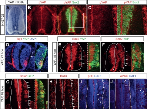
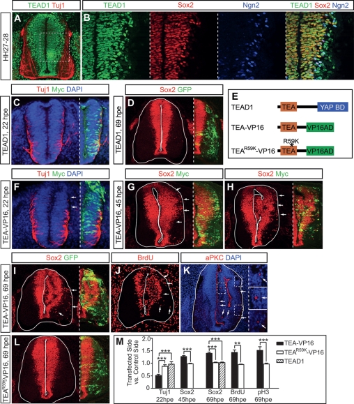
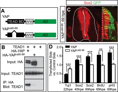
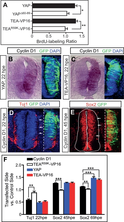
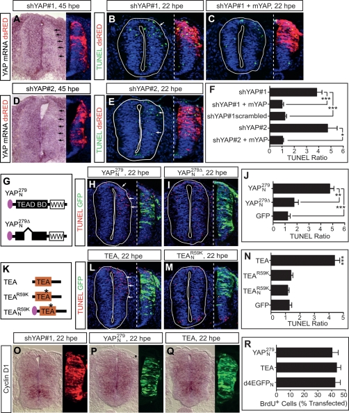
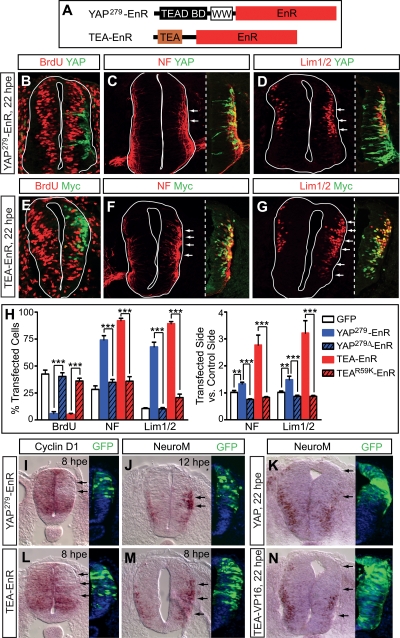
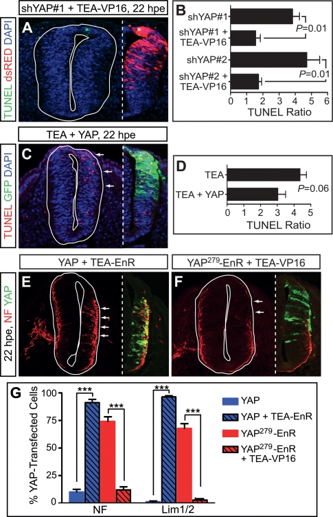
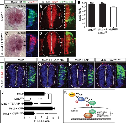
Tight control of cell proliferation is essential for proper growth during development and for tissue homeostasis in mature animals. The evolutionarily conserved Hippo pathway restrains proliferation through a kinase cascade that culminates in the inhibition of the transcriptional coactivator YAP. Unphosphorylated YAP activates genes involved in cell proliferation and survival by interacting with a DNA-binding factor. Here we show that during vertebrate neural tube development, the TEA domain transcription factor (TEAD) is the cognate DNA-binding partner of YAP. YAP and TEAD gain of function causes marked expansion of the neural progenitor population, partly owing to their ability to promote cell cycle progression by inducing cyclin D1 and to inhibit differentiation by suppressing NeuroM. Their loss of function results in increased apoptosis, whereas repressing their target genes leads to premature neuronal differentiation. Inhibiting the upstream kinases of the Hippo pathway also causes neural progenitor overproliferation. Thus, the Hippo pathway plays critical roles in regulating neural progenitor cell number by affecting proliferation, fate choice, and cell survival.


Figures

Figure 1.
Figure 1.
Overexpressing YAP in the neural tube results in reduced differentiation and a marked expansion of the progenitor population. (A) In situ hybridization shows YAP expression in the ventricular zone of HH27–28 chick neural tube. (B,C) Phosphorylated YAP (pYAP) was detected in neural progenitor cells marked by Sox2 immunostaining. The boxed area in B is shown at higher magnification in C. (D–G) Transfecting YAP caused a reduction in Tuj1+ neurons (D, arrows) and an increase in Sox2+ progenitor cells (E–G, arrows). In all cases, the right side of the neural tube was transfected. Images on the left of the dashed lines show the full neural tube cross sections, those on the right only the transfected side. (D–F) Overexpressed YAP was detected by a YAP antibody, which was not sensitive enough to detect endogenous YAP. (G) At 69 hpe, the transfected side was marked by cotransfected GFP. (H–J) Ectopic progenitor cells continued to proliferate at 69 hpe (shown in H). They divided at the mantle zone (I, arrows) and formed tumor-like rosettes with foci of aPKC+ adherens junctions (J, arrows; areas in dashed squares are shown at higher magnification in the images to the right of the dashed line).
Figure 2.
Figure 2.
Overexpressing a transcriptionally active form of TEAD1 increases neural progenitor cell number. (A,B) TEAD1 expression pattern examined by immunostaining. The region examined in B corresponds to that within the dashed square in A. (C,D) Overexpressing TEAD1 did not significantly affect differentiation (C) or progenitor cell number (D). (E) Domain structures of TEAD1 and derivatives. Mammalian and avian TEAD proteins are composed of an N-terminal TEA domain and a C-terminal YAP-binding domain (YAP BD) connected by a linker region. (VP16AD) The activation domain of VP16. (F–K) Reduced neuronal differentiation (F, arrows) and increased progenitor cells (G–J, arrows) upon TEA-VP16 expression. Transfected side showed severe dysplasia with disruptions of ventricular surface and the formation of tumor-like rosettes (K, arrows; areas in dashed squares are shown at higher magnification in the images right of the dashed line). (L) TEAR59K-VP16 did not have a strong effect on progenitor cell number. (C,F–H) TEAD constructs have an N-terminal Myc tag and were detected by a Myc antibody. (D,I,L) At 69 hpe, the transfected side was marked by cotransfected GFP. (M) Quantifications comparing the transfected side with the control side. In this and all following graphs, results are mean ± SEM. (*) P < 0.05; (**) P < 0.01; (***) P < 0.001.
Figure 3.
Figure 3.
YAPΔ60–89 cannot interact with TEAD and fails to increase neural progenitor cell number. (A) Domain structures of YAP and the deletion construct. Chick YAP is composed of an N-terminal TEAD-binding domain (TEAD BD), a central WW domain, and a C-terminal activation domain (AD). (B) A coimmunoprecipitation experiment shows that YAPΔ60–89 could not interact with TEAD1. 293T cells were transfected with TEAD1 and HA-YAP or HA-YAPΔ60–89. Cell lysates were immunoprecipitated (IP) with an HA antibody and blotted with a TEAD1 antibody. (C) Overexpressing YAPΔ60–89 did not significantly increase neural progenitor cells. The transfected side was marked by cotransfected GFP. (D) Quantifications comparing the effects of misexpressing YAP and YAPΔ60–89.
Figure 4.
Figure 4.
YAP and TEA-VP16 overexpression promotes cell cycle progression. (A) Overexpressing YAP and TEA-VP16 accelerated the cell cycle. (B,C) In situ hybridization (ISH) reveals up-regulation of cyclin D1 by YAP and TEA-VP16 transfection. YAP and TEA-VP16 cDNAs are followed by IRES-GFP to monitor their expression. The image next to the ISH photograph shows GFP expression in an adjacent section. (D,E) Overexpressing cyclin D1 reduced differentiation (D, arrows) and increased progenitor cell number (E, arrows). Cyclin D1 cDNA is followed by IRES-GFP to monitor its expression. (F) Quantifications comparing the effect of cyclin D1 overexpression with that of YAP and TEA-VP16. Neural tubes transfected with TEAR59K-VP16 serve as the baseline.
Figure 5.
Figure 5.
YAP and TEAD loss of function in the neural tube leads to increased cell death. (A) ISH shows reduced YAP mRNA level in shYAP#1-transfected neural tube (arrows). The shRNA vector also encodes dsRED to mark transfected cells. The image next to the ISH photograph shows dsRED expression in an adjacent section. Blue signals are DAPI. (B,C) TUNEL assay detected increased cell death in shYAP#1-transfected side (B, arrows). The cell death phenotype was rescued by cotransfecting mYAP (C). (D,E) shYAP#2 reduced YAP mRNA level (D, arrows) and caused increased cell death (E, arrows). (F) Quantifications of the cell death phenotype. For each embryo, the sum of TUNEL signals in the transfected side (four to 10 sections per embryo) was divided by that in the control side to obtain a TUNEL ratio. (G) Domain structures of YAP dominant-negative constructs. The pink oval represents NLS. (H–J) YAPN279, but not YAPN279Δ, caused increased cell death (H, arrows). The transfected side was marked by cotransfected GFP. (K) Domain structures of TEAD dominant-negative constructs. (L–N) TEA, but not TEAR59K or TEANR59K, triggered increased cell death (arrows in L). The transfected side was marked by cotransfected GFP. (O–Q) ISH shows inhibiting YAP and TEAD did not significantly affect cyclin D1 expression. Transfection efficiency was monitored by the expression of dsRED encoded in the shRNA vector (O) or cotransfected GFP (P,Q) in adjacent sections. (R) Inhibiting YAP and TEAD with YAPN279 and TEA, respectively, did not significantly affect BrdU incorporation during a 2-h labeling period. Transfected embryos were harvested at 22 hpe. Neural tubes transfected with d4EGFPN were used as the control.
Figure 6.
Figure 6.
Repressing YAP and TEAD downstream genes leads to cell cycle exit and neuronal differentiation. (A) Domain structures of YAP279-EnR and TEA-EnR. EnR: the repression domain of Engrailed. (B–H) Repressing YAP and TEAD target genes through YAP279-EnR and TEA-EnR, respectively, promoted cell cycle exit (B,E,H) and neuronal differentiation (C,D,F,G, arrows, H). YAP279-EnR was detected by a YAP antibody. TEA-EnR has a Myc tag and was detected by a Myc antibody. (I–N) ISH shows that YAP279-EnR and TEA-EnR repressed cyclin D1 (I,L, arrows) and induced NeuroM (J,M, arrows), whereas overexpressing YAP and TEA-VP16 reduced NeuroM mRNA levels (K,N, arrows). Transfection efficiency was monitored by GFP expression—either from the cotransfected GFP-expressing plasmids (I,J,L,M) or the downstream IRES-GFP (K,N)—in adjacent sections.
Figure 7.
Figure 7.
Epistasis analyses of YAP and TEAD. (A,B) TUNEL assays show that cotransfecting TEA-VP16 with YAP shRNAs rescued the cell death phenotype. Transfected cells were marked by dsRED expressed from the shRNA vector. TUNEL ratios are shYAP#1: 3.8 ± 0.4, n = 12; shYAP#1 + TEA-VP16: 1.6 ± 0.3, n = 4; shYAP#2: 4.7 ± 0.8, n = 9; shYAP#2 + TEA-VP16: 1.7 ± 0.2, n = 6. (C,D) Cotransfecting YAP with TEA could not rescue the cell death phenotype (arrows in C). YAP cDNA is followed by IRES-GFP to mark transfected cells. TUNEL ratios are TEA: 4.4 ± 0.4, n = 6; TEA + YAP: 3.1 ± 0.4, n = 11. (E–G) Cotransfecting TEA-EnR with YAP increased neuronal differentiation (E, arrows), whereas cotransfecting TEA-VP16 with YAP279-EnR reduced it (F, arrows). In both cases, the phenotype resembled that of the single transfection of the TEAD variant and opposite to that of the YAP variant. A YAP antibody was used to label YAP- and YAP279-EnR-transfected cells.
Figure 8.
Figure 8.
Mst1/2 and Lats1/2 regulate neural progenitor proliferation and survival. (A–E) Inhibiting Mst1/2 with Mst2KD and Lats1/2 with shLats1 and Lats2KD caused an up-regulation of cyclin D1 expression at 22 hpe (A,C, arrows) as well as a widening of the Sox2+ progenitor zone and the appearance of ectopic Sox2+ cells in the mantle zone at 69 hpe (B,D, arrows). Blue signals are DAPI. (E) “Sox2 ratio” was obtained by dividing Sox2 area in the transfected side by that in the control side. Neural tubes transfected with dsRED was used as the baseline for statistics analysis. (F–J) Overexpressing Mst2 induced cell death (arrows in F). Cotransfection of TEA-VP16 and YAPS126A, but not YAP, reduced Mst2-induced cell death. Transfected side was marked by cotransfected GFP. TUNEL ratios are Mst2: 3.5 ± 0.3, n = 5; Mst2KD: 0.98 ± 0.1, n = 4; Mst2 + TEA-VP16: 1.39 ± 0.08, n = 4; Mst2 + YAP: 4.9 ± 0.4, n = 5; Mst2 + YAPS126A: 1.7 ± 0.2, n = 5. (K) A simplified model illustrating that the Hippo pathway regulates neural progenitor cell number through TEAD. Mst kinases phosphorylate and activate Lats kinases, which phosphorylate YAP and inhibit its nuclear translocation. Unphosphorylated YAP enters the nucleus and interacts with the DNA-binding factor TEAD. Together, they activate genes that promote proliferation, survival, and the progenitor fate.

Similar articles

Cited by

References

    1. Anbanandam A., Albarado D.C., Nguyen C.T., Halder G., Gao X., Veeraraghavan S. Insights into transcription enhancer factor 1 (TEF-1) activity from the solution structure of the TEA domain. Proc. Natl. Acad. Sci. 2006;103:17225–17230. - PMC - PubMed
    1. Azakie A., Larkin S.B., Farrance I.K., Grenningloh G., Ordahl C.P. DTEF-1, a novel member of the transcription enhancer factor-1 (TEF-1) multigene family. J. Biol. Chem. 1996;271:8260–8265. - PubMed
    1. Bylund M., Andersson E., Novitch B.G., Muhr J. Vertebrate neurogenesis is counteracted by Sox1-3 activity. Nat. Neurosci. 2003;6:1162–1168. - PubMed
    1. Camargo F.D., Gokhale S., Johnnidis J.B., Fu D., Bell G.W., Jaenisch R., Brummelkamp T.R. YAP1 increases organ size and expands undifferentiated progenitor cells. Curr. Biol. 2007;17:1–7. - PubMed
    1. Campbell S., Inamdar M., Rodrigues V., Raghavan V., Palazzolo M., Chovnick A. The scalloped gene encodes a novel, evolutionarily conserved transcription factor required for sensory organ differentiation in Drosophila. Genes & Dev. 1992;6:367–379. - PubMed

Publication types

MeSH terms