. 2008 Nov 17;183(4):597-606.
doi: 10.1083/jcb.200804041.

HP1-beta is required for development of the cerebral neocortex and neuromuscular junctions

Affiliations

HP1-beta is required for development of the cerebral neocortex and neuromuscular junctions

Rebecca Aucott et al. J Cell Biol. .

Abstract

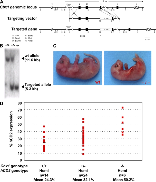
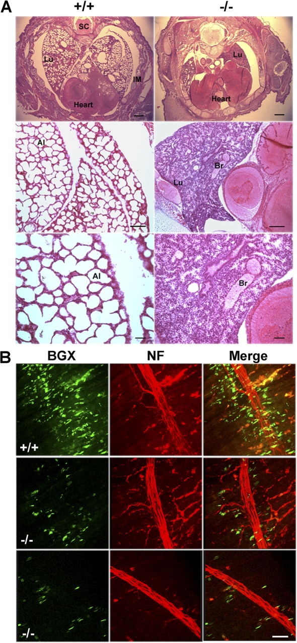
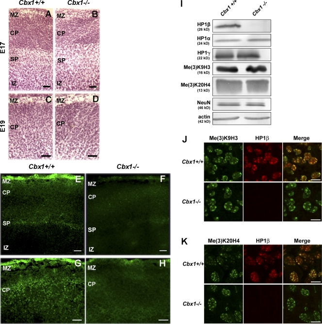
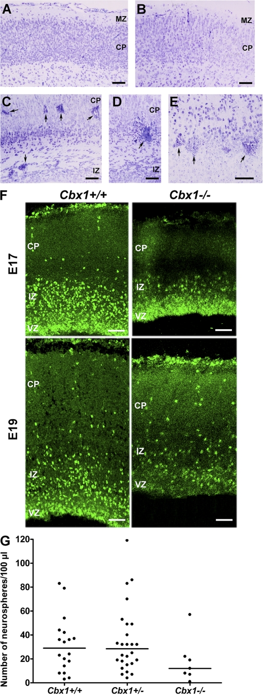
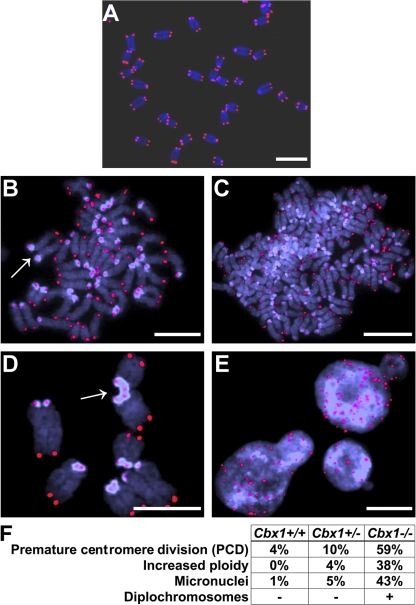
HP1 proteins are thought to be modulators of chromatin organization in all mammals, yet their exact physiological function remains unknown. In a first attempt to elucidate the function of these proteins in vivo, we disrupted the murine Cbx1 gene, which encodes the HP1-beta isotype, and show that the Cbx1(-/-) -null mutation leads to perinatal lethality. The newborn mice succumbed to acute respiratory failure, whose likely cause is the defective development of neuromuscular junctions within the endplate of the diaphragm. We also observe aberrant cerebral cortex development in Cbx1(-/-) mutant brains, which have reduced proliferation of neuronal precursors, widespread cell death, and edema. In vitro cultures of neurospheres from Cbx1(-/-) mutant brains reveal a dramatic genomic instability. Our results demonstrate that HP1 proteins are not functionally redundant and that they are likely to regulate lineage-specific changes in heterochromatin organization.


Figures

Figure 1.
Figure 1.
Cbx1 gene function is essential, and its product, HP1-β, is a modifier of PEV. (A) The relevant regions in the wild-type Cbx1 locus (top; see Materials and methods), the TK-Neor targeting vector (middle), and the targeted gene (bottom). Coding regions are depicted by closed boxes. Noncoding regions are denoted by striped boxes. The DNA probe used to screen for targeting events is shown as a shaded rectangle. (B) Southern blot authentication of germline transmission of the Cbx1 mutation. BamHI digest of genomic DNA produces an 11.6-kb fragment for the wild-type (wt) allele and a diagnostic 5.3-kb fragment for the targeted allele. (C) Images of wild-type (left) and Cbx1−/− (right) neonates. (D) Scatter plots showing the results of flow cytometry analysis of the proportion of transgenic embryonic DP (CD4+CD8+) thymocytes that express hCD2 taken from the three Cbx1 genotypes. Each point represents the result from a single embryo. The mean expression of hCD2 in embryonic thymocytes from Cbx1+/− animals is higher than that for Cbx1+/+ animals, but not significantly. The mean expression of hCD2 in thymocytes taken from Cbx1−/− animals is significantly different from expression in thymocytes taken from the parental genotypes (P < 0.001). Black lines represent the means.
Figure 2.
Figure 2.
Cbx1−/− neonates do not inflate their lungs, and E19 Cbx1−/− embryos exhibit reduced clusters of postsynaptic AChRs within diaphragm muscle. (A) Hematoxylin and eosin–stained transverse sections through the midbody of wild-type (left) and Cbx1−/− (right) neonates showing lack of inflation of the lungs in the Cbx1−/− neonates. Al, alveolus; Br, bronchiole; IM, intercostal muscle; Lu, lung; SC, spinal cord. Bars: (top) 450 μm; (middle) 150 μm; (bottom) 50 μm. (B) Staining of E19 wild-type diaphragms (top) with antibodies to NF (red) shows that the diaphragm is clearly innervated. Bungarotoxin-positive (BGX; green) AChRs are clustered around the nerve and its branches. The clusters of bungarotoxin-positive AChRs are much reduced in E19 Cbx1−/− diaphragms (middle and bottom), and, in some cases, the branching of the innervating nerve is also reduced (bottom; see red NF staining). Bar, 25 μm.
Figure 3.
Figure 3.
Immunohistochemical and Western blot analysis of Cbx1−/− brains. (A–D) Hematoxylin and eosin–stained sagittal sections of E17 (A and B) and E19 (C and D) neocortices correspond to the antibody-stained cortices in E–H. Genotypes of the embryos are marked above the photographs. (E–H) α-NeuN–stained E17 and E19 neocortices. The staining of the wild-type E17 cortex with the α-NeuN antibody (E) detects a layer of SP cells that separates the CP from the intermediate zone (IZ). These cells are very weakly stained in the Cbx1−/− neocortex (F). Similarly, CP cells are strongly stained with the α-NeuN antibody in the E19 neocortex (G), but such staining of CP cells is reduced to background levels in the E19 Cbx1−/− neocortex (H). MZ, marginal zone. (I) HP1-β protein is not detected in extracts from Cbx1−/− brains using an antibody to the C terminus of HP1-β; an N-terminal antibody also fails to detect HP1-β in the same way (Fig. S1, available at http://www.jcb.org/cgi/content/full/jcb.200804041/DC1). The levels of HP1-α, HP1-γ, Me(3)K9H3, Me(3)K20H4, and NeuN are not significantly changed in Cbx1−/− compared with wild-type brain extracts. The bottom panel is the actin-loading control. Identical results were obtained using whole embryo extracts (not depicted). (J) Me(3)K9H3 heterochromatic distribution is not affected by the Cbx1−/− mutation. Cbx1−/− and Cbx1+/+ CP neurons show identical Me(3)K9H3 staining patterns. (K) Me(3)K20H4 heterochromatic distribution is not affected by the Cbx1−/− mutation. Cbx1−/− and Cbx1+/+ CP neurons show identical Me(3)K20H4 staining patterns. Bars: (A–H) 100 μm; (J and K) 10 μm.
Figure 4.
Figure 4.
Cbx1−/− mutants exhibit defective cerebral corticogenesis and reduced proliferation of neuronal precursors. (A and B) Nissl staining of the E19 wild-type neocortex (A) shows the typical ordered arrangement of CP cells. In the E19 Cbx1+/− neocortex (B), the ordered arrangement of CP cells is disturbed, and the boundaries at the top and bottom edges of the CP layer are blurred. (C–E) In the E19 Cbx1−/− neocortex, the lamination of the neocortex is severely disturbed with patches of edema (C and D, arrows) and clusters of dying cells (E, arrows) scattered through the cortical layers. (F) Proliferating neural progenitors are depleted in the VZ of Cbx1−/− brains. On E17, the layer of pKi-67–positive proliferating neural progenitors is thicker in the wild-type VZ compared with the Cbx1−/− VZ. On E19, the pool of proliferating progenitors of the Cbx1−/− VZ is essentially exhausted compared with the wild-type E19 VZ. (G) There is a trend toward fewer neurospheres from cultures of Cbx1−/− brains compared with wild-type and Cbx1+/− brains (P < 0.04); each dot represents the result from a single embryo. There is no significant difference in the numbers of neurospheres from the wild-type and Cbx1+/− brains. Black lines represent the medians. IZ, intermediate zone; MZ, marginal zone. Bars: (A–E) 60 μm; (F) 80 μm.
Figure 5.
Figure 5.
Cbx1−/− neurospheres exhibit an increased genomic instability. (A) A mitotic chromosome spread showing the normal situation in which sister chromatids are paired. (B–E) Chromosomes from the Cbx1−/− neurosphere cells exhibit a variety of aberrations, including unpaired sister chromatids that have undergone PCD (B, arrow), increased ploidy (C), diplochromosomes (D, arrow), and micronuclei (E). Telomeres (red signals) in all panels were labeled by a specific PNA probe. (F) The table shows that there is a highly significant increase (P < 0.001) in PCD, polyploidy, diplochromosomes, and micronuclei between Cbx1−/− and the other two genotypes (Cbx1+/− and Cbx1+/+). There is only a borderline significant difference between Cbx1+/− and Cbx1+/+. Bars, 5 μm.

References

    1. Ball, L.J., N.V. Murzina, R.W. Broadhurst, A.R. Raine, S.J. Archer, F.J. Stott, A.G. Murzin, P.B. Singh, P.J. Domaille, and E.D. Laue. 1997. Structure of the chromatin binding (chromo) domain from mouse modifier protein 1. EMBO J. 16:2473–2481. - PMC - PubMed
    1. Cheutin, T., A.J. McNairn, T. Jenuwein, D.M. Gilbert, P.B. Singh, and T. Misteli. 2003. Maintenance of stable heterochromatin domains by dynamic HP1 binding. Science. 299:721–725. - PubMed
    1. Cheutin, T., S.A. Gorski, K.M. May, P.B. Singh, and T. Misteli. 2004. In vivo dynamics of Swi6 in yeast: evidence for a stochastic model of heterochromatin. Mol. Cell. Biol. 24:3157–3167. - PMC - PubMed
    1. Collombet, J.M., C. Masqueliez, E. Four, M.F. Burckhart, D. Bernabé, D. Baubichon, and G. Lallement. 2006. Early reduction of NeuN antigenicity induced by soman poisoning in mice can be used to predict delayed neuronal degeneration in the hippocampus. Neurosci. Lett. 398:337–342. - PubMed
    1. Cordelires, F., and J. Jackson. 2007. 3D Object Counter. National Institutes of Health, Baltimore, MD. http://rsb.info.nih.gov/ij/plugins/track/objects.html (accessed August, 2008).

Publication types

MeSH terms