Review
. 2008 Oct;8(5):242-53.
doi: 10.1124/mi.8.5.9.

Inside information: the unique features of visceral sensation

Affiliations
Review

Inside information: the unique features of visceral sensation

David R Robinson et al. Mol Interv. 2008 Oct.

Abstract

Most of what is written and believed about pain and nociceptors originates from studies of the "somatic" (non-visceral) sensory system. As a result, the unique features of visceral pain are often overlooked. In the clinic, the management of visceral pain is typically poor, and drugs that are used with some efficacy to treat somatic pain often present unwanted effects on the viscera. For these reasons, a better understanding of visceral sensory neurons-particularly visceral nociceptors-is required. This review provides evidence of functional, morphological, and biochemical differences between visceral and non-visceral afferents, with a focus on potential nociceptive roles, and also considers some of the potential mechanisms of visceral mechanosensation.


Figures
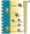
Figure 1
Figure 1. Functional neuroanatomy of the visceral sensory system
The gut is depicted here as an example of the sensory innervation of the visceral system. Vagal afferent neurons, which do not innervate the urinary bladder or the distal gut, have cell bodies in the nodose ganglia (NG) bilaterally and travel alongside parasympathetic efferent pathways to organs in the thoracic and abdominal cavities. Once in the gut wall, vagal afferent fibers innervate neurons in the myenteric or submucosal plexus (M/SP), circular and longitudinal muscle layers, and the mucosa. Pelvic afferent neurons also travel alongside parasympathetic efferent pathways, but their cell bodies are in dorsal root ganglia (DRG). Other spinal nerves (e.g., greater splanchnic) travel alongside sympathetic efferent pathways, have cell bodies in DRG, and pass through prevertebral ganglia (e.g., the celiac ganglion, CG). Intestinofugal afferents (purple) synapse onto efferent sympathetic neurons in prevertebral ganglia, such as the inferior mesenteric ganglion (IMG) and have their cell bodies in M/SP. Afferent fibers of the intrinsic (or enteric) nervous system, termed intrinsic primary afferent neurons (IPANs), synapse onto intestinofugal fibers, either directly, or via interneurons (i). Rectospinal fibers (blue) have cell bodies in the myenteric plexus or muscle layers, with axons terminating in the spinal cord (CNS). Note that not all these nerves and fibers will terminate in the same areas of the gut, and inputs to the spinal cord may traverse a number of different levels; this figure has been simplified for clarity.
Figure 2
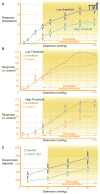
Figure 2. The viscera are innervated by low- and high-threshold mechanoreceptors
Hollow viscera are exquisitely sensitive to distension, a phenomenon that can be observed experimentally by recording the electrical activity induced in pelvic nerve afferent fibers (mechanoreceptors) during distension of the visceral organ under study. A) Low-threshold mechanoreceptors (blue) detect both non-noxious and noxious distension pressures, whereas high-threshold mechanoreceptors (green) only respond to noxious distention pressures. Data are recorded from distension of the rat bladder. B) Mechanoreceptors (of either the high- or low-threshold type) in the pelvic nerve can be sensitized by the addition of irritants (e.g., xylenes) into the bladder. C) Mice lacking the TRPV1 receptor show impaired visceral nociception. The visceromotor response (electrical activity recorded in the abdominal musculature) produced during distension of the colon to a noxious pressure (e.g., 60 mmHg) is lower in mice that do not have functional TRPV1 receptors compared to wild-type controls. Even at the highest tested colonic distension pressure, TRPV1 knockout mice only show a level of response equivalent to that of the wild-type mice at 30 mmHg, the pressure at which this stimulus is likely to be noxious (dashed red line). Panels A and B are adapted with permission from (64), and panel C is adapted with permission from (48).
Figure 3
Figure 3. The colon is innervated by five different types of mechanosensory afferent
Five different types of afferent fiber have been reported in the colon: serosal, mesenteric, muscular, mucosal, and muscular/mucosal. Each has characteristic response criteria based on a protocol of probing, stretching, and stroking the colonic wall, and each has a different putative functional role. Most serosal afferents are mechanosensitive and, given that their thresholds for activation are higher than would be expected physiologically, are thought to signal short, sharp events (e.g., muscle contraction). Found only in the splanchnic afferent innervation, mesenteric afferents are predominantly found closely associated with blood vessels and likely signal twisting of the colon wall and some changes in mesenteric blood pressure, with a potential role in inflammation. Muscular afferents, named for their termination in the circular and longitudinal muscle layers of the gut, respond directly to circumferential stretch with a low threshold of activation (though can code into the noxious range). Muscular afferents exhibit slightly different properties depending upon the nerve (splanchnic or pelvic) in which they are found, but generally are considered to contribute to sustained filling, bloating, or distending sensations. Mucosal afferents are stretch-insensitive (at least circular stretch-insensitive) and respond to fine probing and stoking of the mucosal membrane. This suggests a role in providing feedback from physiological stimuli such as the normal passing of fecal material through the gastrointestinal tract. Finally, muscular/mucosal afferents, so named for their ability to detect both circular stretch and fine mucosal stroking, are a class of mechanoreceptor that, in mouse colon at least, are only found in the pelvic innervation. Presumably, these fibers provide a combination of the information that is transmitted from the muscular and mucosal fibers described above. For examples of the responses seen for each of these mechanosensitive fiber types, please see reference (53).

References

    1. Grundy D. Signalling the state of the digestive tract. Auton Neurosci. 2006;125:76–80. - PubMed
    1. Blackshaw LA, Brookes SJ, Grundy D, Schemann M. Sensory transmission in the gastrointestinal tract. Neurogastroenterol Motil. 2007;19(Suppl 1):1–19. - PubMed
    1. Cervero F. Sensory innervation of the viscera: Peripheral basis of visceral pain. Physiol Rev. 1994;74:95–138. - PubMed
    1. Sengupta JN, Gebhart GF. Gastrointestinal afferent fibers and sensation. In: Johnson LR, Alpers DH, Christensen J, Jacobson ED, Walsh JH, editors. Physiology of the Gastrointestinal Tract. Raven Press; New York: 1994. pp. 483–519.
    1. Sherrington CS. Qualitative difference of spinal reflex corresponding with qualitative difference of cutaneous stimulus. J Physiol (Lond) 1903;30:39–46. - PMC - PubMed

MeSH terms