

Chronic blockade of phosphatidylinositol 3-kinase in the nucleus tractus solitarii is prohypertensive in the spontaneously hypertensive rat
- PMID: 19015400
- PMCID: PMC2735392
- DOI: 10.1161/HYPERTENSIONAHA.108.122341
Chronic blockade of phosphatidylinositol 3-kinase in the nucleus tractus solitarii is prohypertensive in the spontaneously hypertensive rat
Abstract
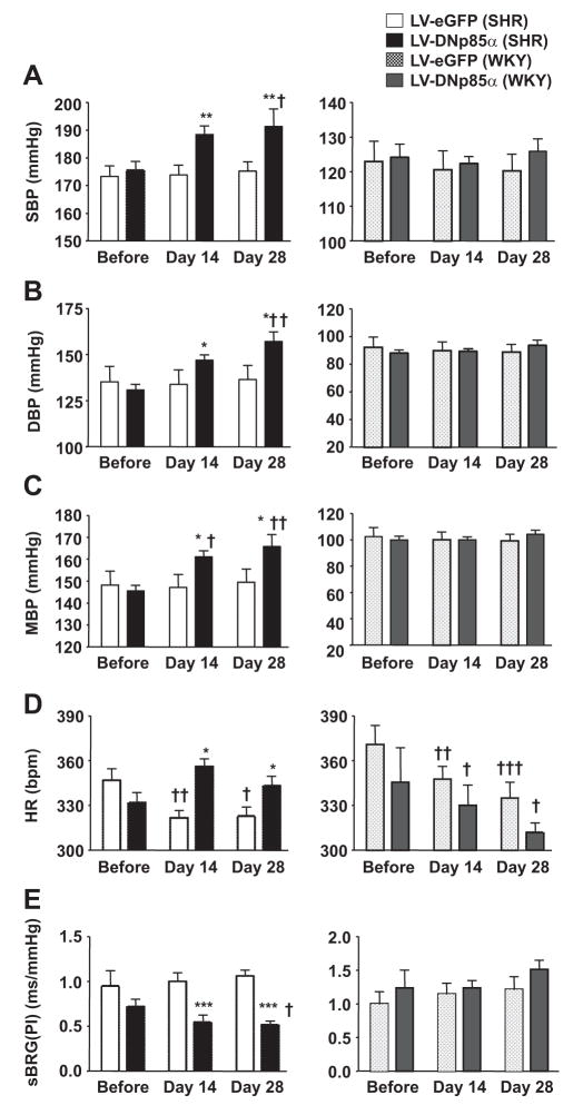
Phosphatidylinositol 3-kinase (PI3K) within brain stem neurons has been implicated in hypertension in the spontaneously hypertensive rat (SHR). Previously, we demonstrated elevated expression of PI3K subunits in rostral ventrolateral medulla and paraventricular nucleus of SHRs compared with Wistar-Kyoto rats. Here, we considered expression levels of PI3K in the nucleus tractus solitarii, a pivotal region in reflex regulation of arterial pressure, and determined its functional role for arterial pressure homeostasis in SHRs and Wistar-Kyoto rats. We found elevated mRNA levels of p110beta and p110delta catalytic PI3K subunits in the nucleus tractus solitarii of adult (12 to 14 weeks old) SHRs relative to the age-matched Wistar-Kyoto rats (fold differences relative to beta-actin: 1.7+/-0.2 versus 1.01+/-0.08 for p110beta, n=6, P<0.05; 1.62+/-0.15 versus 1.02+/-0.1 for p110delta, n=6, P<0.05). After chronic blockade of PI3K signaling in the nucleus tractus solitarii by lentiviral-mediated expression of a mutant form of p85alpha, systolic pressure increased from 175+/-3 mm Hg to 191+/-6 mm Hg (P<0.01) in SHRs but not in Wistar-Kyoto rats. In addition, heart rate increased (from 331+/-6 to 342+/-6 bpm; P<0.05) and spontaneous baroreflex gain decreased (from 0.7+/-0.07 to 0.5+/-0.04 ms/mm Hg; P<0.001) in the SHRs. Thus, PI3K signaling in the nucleus tractus solitarii of SHR restrains arterial pressure in this animal model of neurogenic hypertension.
Figures


References
-
- Carretero OA, Oparil S. Essential hypertension. Part I: definition and etiology. Circulation. 2000;101:329–335. - PubMed
-
- Touyz RM. Molecular and cellular mechanisms regulating vascular function and structure–implications in the pathogenesis of hypertension. Can J Cardiol. 2000;16:1137–1146. - PubMed
-
- Oudit GY, Crackower MA, Backx PH, Penninger JM. The role of ACE2 in cardiovascular physiology. Trends Cardiovasc Med. 2003;13:93–101. - PubMed
-
- Takeda K, Bunag RD. Chronic propranolol treatment inhibits sympathetic nerve activity and keeps blood pressure from rising in spontaneously hypertensive rats. Hypertension. 1980;2:228–235. - PubMed
-
- Arribas SM, Alonso MJ, Marin J, Fernandes F, Llergo JL, Sanchez-Ferrer CF, Salaices M. Noradrenergic transmission in the tail artery of hypertensive rats transgenic for the mouse renin gene Ren-2. J Auton Pharmacol. 1996;16:69–77. - PubMed
Publication types
MeSH terms
Substances
Grants and funding
LinkOut - more resources
Full Text Sources
Medical
Molecular Biology Databases
