. 2008 Nov 25;105(47):18261-6.
doi: 10.1073/pnas.0810357105. Epub 2008 Nov 17.

Revisiting the mechanism of macrolide-antibiotic resistance mediated by ribosomal protein L22

Affiliations

Revisiting the mechanism of macrolide-antibiotic resistance mediated by ribosomal protein L22

Sean D Moore et al. Proc Natl Acad Sci U S A. .

Abstract

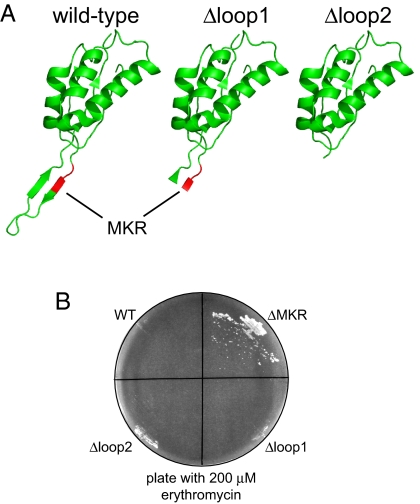
Bacterial antibiotic resistance can occur by many mechanisms. An intriguing class of mutants is resistant to macrolide antibiotics even though these drugs still bind to their targets. For example, a 3-residue deletion (DeltaMKR) in ribosomal protein L22 distorts a loop that forms a constriction in the ribosome exit tunnel, apparently allowing nascent-chain egress and translation in the presence of bound macrolides. Here, however, we demonstrate that DeltaMKR and wild-type ribosomes show comparable macrolide sensitivity in vitro. In Escherichia coli, we find that this mutation reduces antibiotic occupancy of the target site on ribosomes in a manner largely dependent on the AcrAB-TolC efflux system. We propose a model for antibiotic resistance in which DeltaMKR ribosomes alter the translation of specific proteins, possibly via changes in programmed stalling, and modify the cell envelope in a manner that lowers steady-state macrolide levels.


Conflict of interest statement

The authors declare no conflict of interest.

Figures

Fig. 1.
Fig. 1.
Ribosomal protein L22 and erythromycin inhibition of translation in vitro. (A) Crystal structure of L22 protein (green; M82K83R84 is shown in red), L4 protein (blue), and 23S and 5S RNA (orange) from the 50S subunit of the E. coli ribosome (PDB entry 2AWB). The exit tunnel is visible through the center of the subunit. (B) SDS-PAGE of proteins from purified ribosomes with wild-type L22, an L22-titin fusion, or a ΔMKR L22-titin fusion. Wild-type L22 (12 kDa) is not resolved from other ribosomal protein in the Left lane. The L22 fusion proteins are visible as separate bands (Center and Right lanes). (C) Autoradiograms of translation reactions containing 100 nM wild-type or ΔMKR ribosomes and increasing amounts of erythromycin. (D) Integrated band intensities from the autoradiogram shown in the Top and Middle of C are plotted as a percentage of the intensity of the band from the reaction without erythromycin. These data were fit to a quadratic binding equation to obtain the inhibition constant (Ki).
Fig. 2.
Fig. 2.
Erythromycin sensitivity increases when drug efflux is blocked. (A) Erythromycin resistance of strains containing wild-type L22-titin (triangles) or ΔMKR L22-titin (circles). The turbidity (600 nm) of 16 h cultures grown at 37 °C in the presence of erythromycin (dashed lines) or erythromycin and 30 μg/ml PAβN (solid lines) is plotted as a percentage of the value of a culture grown without drug. (B) Growth at 37 °C of cultures containing the wild-type L22-titin fusion in the absence of erythromycin (open circles), after addition of 200 μM erythromycin (filled circles), or after addition of 200 μM erythromycin and 30 μg/ml PAβN (filled triangles). (C) Growth of cultures containing the ΔMKR mutation in L22 with the same symbols as in B. (D) Strains containing wild-type (circles) or ΔMKR L22 (filled circles) and an induced ermC gene were grown in medium with 30 μg/ml PAβN to early log phase and erythromycin was added to 200 μM at the time indicated by the arrows. (E) ErmC methylation of A2058 in 23S RNA assayed by primer extension. An oligonucleotide complementary to 23S rRNA bases 2066–2102 was used to prime a reverse-transcription reaction containing dideoxy-ATP and RNA purified from strains containing wild-type or ΔMKR L22-titin fusions grown with or without induction of ermC. Reaction products were electrophoresed on a urea-acrylamide gel. In the presence of template RNA, reverse transcription proceeded until blocked by ErmC-mediated methylation of A2058 or until incorporation of dideoxy-ATP at the position corresponding to U2041.
Fig. 3.
Fig. 3.
Reduced erythromycin binding to ribosomes with ΔMKR L22 in vivo. Cultures were treated with dimethyl sulfate (DMS), reactions were quenched, and total RNA was purified and used in primer-extension assays to detect base modifications near the erythromycin-binding site in 23S rRNA. (A) Gel of primer-extension products from the following RNA templates: (Lane 1) Non-DMS-treated ErmC-modified template (product terminates at the position corresponding to A2058). (Lanes 2 and 3) Templates from cultures grown without erythromycin. (Lanes 4 and 5) Templates from cultures grown with 200 μM erythromycin. (Lanes 6 and 7) Templates from cultures grown with 200 μM erythromycin and 30 μg/ml PAβN. (B) Plots of the ratio of band intensities corresponding to A2058 (filled bars) or A2059 (hatched bars) in A divided by the intensity of A2062.
Fig. 4.
Fig. 4.
Extended L22 loop deletions. (A) Ribbon drawings of E. coli L22 with the M82-K83-R84 residues marked in red (PDB 2AWB). The Δloop1 and Δloop2 deletions remove residues 85–95 and 82–100, respectively. (B) Growth of strains on a plate containing 200 μM erythromycin.
Fig. 5.
Fig. 5.
Contribution of AcrAB-TolC to erythromycin resistance. The TolC or AcrA/AcrB components of the AcrAB-TolC efflux pump were deleted from strains with wild-type or ΔMKR ribosomes. (A) Erythromycin resistance of cells lacking tolC with wild-type (triangles) or ΔMKR (circles) ribosomes. (B) Erythromycin resistance of cells lacking acrA/acrB with wild-type (triangles) or ΔMKR (circles) ribosomes. Inhibition plots of tolC+ acrAB+ cells are shown for comparison (dashed lines).

References

    1. Vester B, Douthwaite S. Macrolide resistance conferred by base substitutions in 23S rRNA. Antimicrob Agents Chemother. 2001;45:1–12. - PMC - PubMed
    1. Roberts MC. Update on macrolide-lincosamide-streptogramin, ketolide, and oxazolidinone resistance genes. FEMS Microbiol Lett. 2008;282:147–159. - PubMed
    1. Schlünzen F, et al. Structural basis for the interaction of antibiotics with the peptidyl transferase centre in eubacteria. Nature. 2001;413:814–821. - PubMed
    1. Tu D, Blaha G, Moore PB, Steitz TA. Structures of MLSBK antibiotics bound to mutated large ribosomal subunits provide a structural explanation for resistance. Cell. 2005;121:257–270. - PubMed
    1. Contreras A, Vázquez D. Cooperative and antagonistic interactions of peptidyl-tRNA and antibiotics with bacterial ribosomes. Eur J Biochem. 1977;74:539–547. - PubMed

Publication types

MeSH terms

LinkOut - more resources