

doi: 10.1073/pnas.0808936105.
Epub 2008 Nov 17.
The structure of corepressor Dax-1 bound to its target nuclear receptor LRH-1
Affiliations
- PMID: 19015525
- PMCID: PMC2587556
- DOI: 10.1073/pnas.0808936105
Item in Clipboard
The structure of corepressor Dax-1 bound to its target nuclear receptor LRH-1
Proc Natl Acad Sci U S A.
.
Abstract
The Dax-1 protein is an enigmatic nuclear receptor that lacks an expected DNA binding domain, yet functions as a potent corepressor of nuclear receptors. Here we report the structure of Dax-1 bound to one of its targets, liver receptor homolog 1 (LRH-1). Unexpectedly, Dax-1 binds to LRH-1 using a new module, a repressor helix built from a family conserved sequence motif, PCFXXLP. Mutations in this repressor helix that are linked with human endocrine disorders dissociate the complex and attenuate Dax-1 function. The structure of the Dax-1:LRH-1 complex provides the molecular mechanism for the function of Dax-1 as a potent transcriptional repressor.
Conflict of interest statement
The authors declare no conflict of interest.
Figures
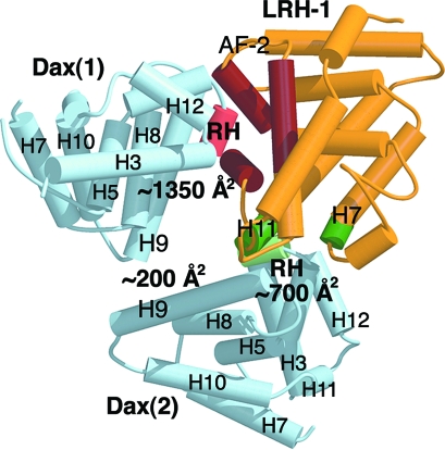
The three-dimensional structure of the Dax-1:LRH-1 complex. LRH-1 is shown in yellow; the two Dax-1 molecules are shown in blue. The structural elements involved in binding of the first Dax-1 to LRH-1 are shown in rose (RH site) and dark red (AF-2 region). The structural elements at the second Dax-1-LRH-1 interface are shown in light and dark green and are indicated.
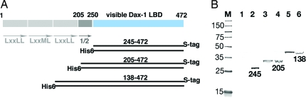
Structural determinants of Dax-1 dimerization. (A) Schematic representation of Dax-1. The N-terminal domain of Dax-1 (gray) has 3 and 1/2 structural repeats (indicated by arrows); the 1/2 repeat is the N-terminal to the visible Dax-1 LBD (blue). Pairs of coexpressed Dax-1 fragments 245–472, 205–472, and 138–472 with either His6 or S tags are shown as black bars. (B) Western blot analysis showing formation of Dax-1 homodimers. Coexpressed fragments were purified using Ni-NTA matrix and analyzed by Western blot using the S-tagged antibody. Lanes 2, 4, and 6 correspond to the soluble bacterial extracts; lanes 1, 3, and 5 correspond to the purified Dax-1 proteins.
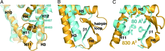
Structural comparison of LRH-1 (yellow) and Dax-1 (blue) LBDs. (A) The AF-2 sites of LRH-1 and Dax-1. Helix H12 of LRH-1 is in an “active” conformation, whereas helix H12 of Dax-1 is in an “inactive” conformation and is docked in the coactivator groove; the residues participating in the docking interactions are indicated. Helix H11 of Dax-1 is rotated by ≈45° compared with its counterpart in LRH-1. (B) Helices H3 and H5-H6 of LRH-1 and Dax-1. Helix H3 of Dax-1 is shifted toward the core of the LBD. Helix H5 of Dax-1 is shorter and is followed by a short β-strand (indicated). (C) The ligand-binding pockets of LRH-1 and Dax-1. Residues filling the putative ligand-binding cavity of Dax-1 are shown as stick models.
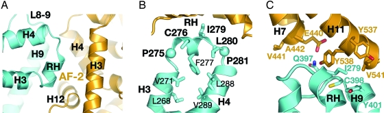
Binding interactions between Dax-1 (blue) and LRH-1 (yellow). (A) A magnified view of the primary Dax-1-LRH-1 interface indicating the structural elements involved in binding. (B) Architecture of the RH site. The repressor helix is bordered by two conserved Pro residues, P275 and P281 (shown as spheres). The helical conformation of the RH site is supported by multiple intramolecular interactions. Residues participating in binding with LRH-1 are shown in bold. (C) A magnified view of the secondary Dax-1-LRH-1 interface indicating the residues participating in binding interactions.
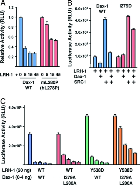
Regulatory interactions between Dax-1 and LRH-1. (A) Repression of LRH-1 by Dax-1 in HepG2 cells. Luciferase activity was measured after cotransfection of vectors encoding the Aro-Luc reporter, mLRH-1, and either wild-type or mutant mDax-1. Mutation L280P in the RH site of mDax-1 (L278P in hDax-1) resulted in diminished repression by Dax-1. (B) Repression of LRH-1 by Dax-1 in the presence of coactivator SRC-1. LRH-1 and Dax-1 variants were cotransfected into HepG2 cells with or without SRC-1, as indicated by “+.” Dax-1 is a potent repressor of LRH-1 that overpowers activation by SRC-1; however, a single mutation I279D in the RH site abrogates repression by Dax-1. (C) Diminished repression of Dax-1 by parallel mutations at the Dax-1 RH site and at the secondary binding site of LRH-1. Either wild-type or I279A/L280A Dax-1 RH mutant was cotransfected into HepG2 cells with either wild-type or mutant Y538D LRH-1.
References
-
- Lalli E, Sassone-Corsi P. DAX-1, an unusual orphan nuclear receptor at the crossroads of steroidogenic function and sexual differentiation. Mol Endocrinol. 2003;17:1445–1453. - PubMed
-
- Bardoni B, et al. A dosage-sensitive locus at chromosome Xp21 is involved in male to female sex reversal. Nat Genet. 1994;7:497–501. - PubMed
Publication types
MeSH terms
Substances
Associated data
- Actions
Grants and funding
LinkOut - more resources
Full Text Sources
Molecular Biology Databases
