. 2008 Nov 25;105(47):18460-5.
doi: 10.1073/pnas.0809850105. Epub 2008 Nov 17.

IL-6 controls Th17 immunity in vivo by inhibiting the conversion of conventional T cells into Foxp3+ regulatory T cells

Affiliations

IL-6 controls Th17 immunity in vivo by inhibiting the conversion of conventional T cells into Foxp3+ regulatory T cells

Thomas Korn et al. Proc Natl Acad Sci U S A. .

Abstract

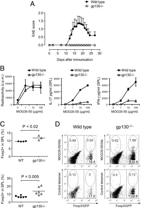
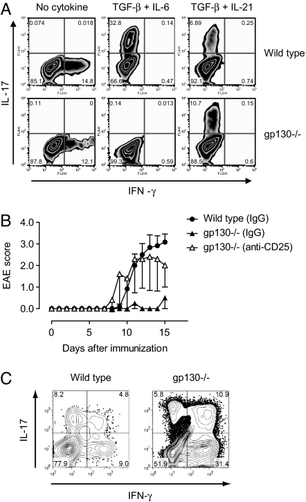
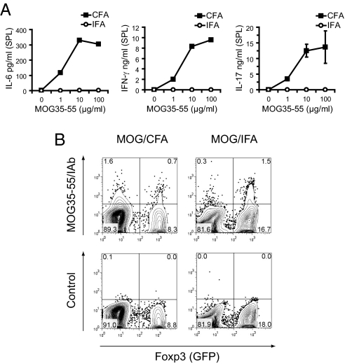
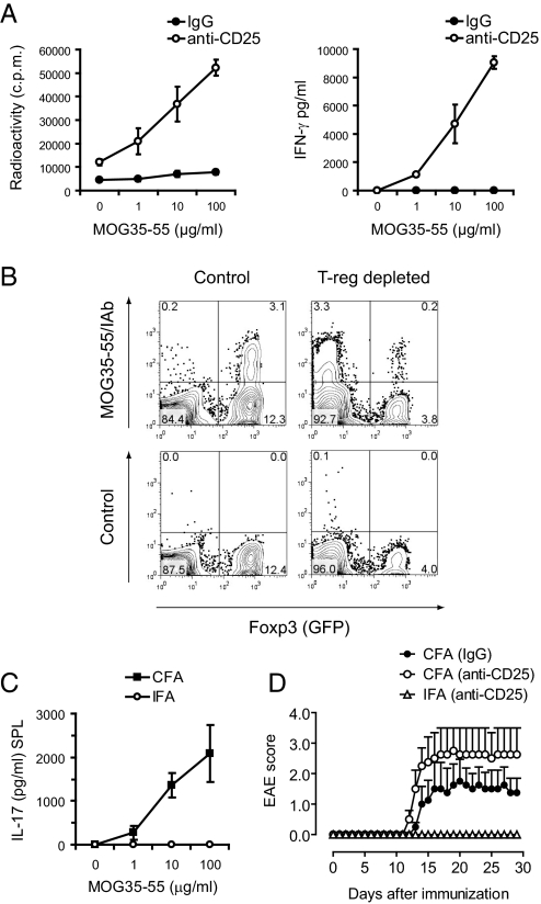
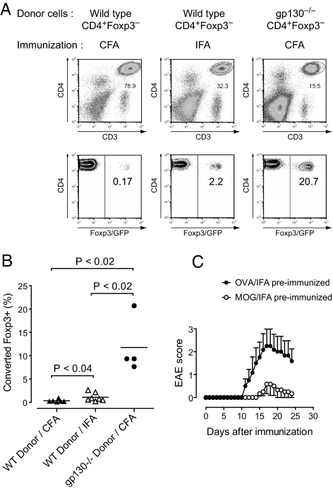
The conditions leading to the induction of adaptive Foxp3(+) regulatory T cells (T-regs) from peripheral T cells in vivo are incompletely understood. Here, we show that unresponsiveness of T cells to IL-6 by T cell-selective deletion of gp130 or immunization of wild-type mice with antigen in incomplete Freund's adjuvant (IFA), which fails to induce IL-6, promotes the conversion of peripheral CD4(+) T cells into adaptive Foxp3(+) T-regs. Thus, both T cell-conditional gp130 knockout (KO) mice immunized with MOG35-55 in complete Freund's adjuvant (CFA) and wild-type mice immunized with MOG35-55 in IFA develop overwhelming antigen-specific T-reg responses and are protected from experimental autoimmune encephalomyelitis (EAE). Depletion of T-regs restores T helper (Th)17 responses and clinical EAE in MOG/CFA-immunized T cell-conditional gp130 KO mice, but not in MOG/IFA-immunized wild-type mice. We conclude that in the absence of T-regs, IL-6 signaling is dispensable for the induction of Th17 cells, and alternative pathways exist to induce Th17 cells and EAE in the absence of IL-6 signaling. However, IL-6 signaling is dominant in inhibiting the conversion of conventional T cells into Foxp3(+) T-regs in vivo, and in the absence of IL-6 signaling, no other cytokine can substitute in inhibiting T-reg conversion. These data identify IL-6 as an important target to modulate autoimmune responses and chronic inflammation.


Conflict of interest statement

The authors declare no conflict of interest.

Figures

Fig. 1.
Fig. 1.
Unresponsiveness of T cells to IL-6 confers resistance to EAE caused by lack of Th17 cells and an increased T-reg response. (A) Wild-type or T cell-conditional gp130−/− mice on the Foxp3gfp.KI background were immunized with MOG35-55/CFA plus pertussis toxin and followed for signs of EAE (mean clinical score ± SEM, n = 10). (B) On day 10 after immunization, splenocytes were isolated and restimulated with MOG35-55 in vitro. The proliferative response was measured by [3H]thymidine incorporation, and the cytokine production in 48-h culture supernatants was determined by ELISA. Mean of triplicate cultures is shown. (C) Frequency of Foxp3+ T-regs in the splenic CD4+ T cell compartments of MOG35-55/CFA-immunized wild-type and gp130−/− mice as determined by the expression of Foxp3/GFP ex vivo (Upper) and after in vitro restimulation with MOG35-55 (Lower). (D) MOG35-55/IAb tetramer staining in MOG35-55-stimulated splenocytes from in vivo-sensitized wild-type and gp130−/− mice. The splenocytes were isolated on day 11 after immunization with MOG35-55/CFA followed by restimulation in vitro for 4 days. The gate was set on blasting CD4+ T cells. Representative cytograms are shown.
Fig. 2.
Fig. 2.
The combination of TGF-β plus IL-21 induces Th17 cells in gp130−/− mice. (A) Naïve T cells were purified from wild-type or gp130−/− mice and differentiated in vitro with either TGF-β plus IL-6 or TGF-β plus IL-21. The frequency of IL-17- and IFN-γ-positive T cells was determined by intracellular cytokine staining. (B) gp130−/− mice were either treated with control IgG (n = 3) or depleted of T-regs by treatment with a monoclonal antibody to CD25 (PC61, 2 × 0.5 mg) (8) 5 and 3 days before immunization with MOG/CFA (n = 5). As further control group, T-reg-competent wild-type mice were included (n = 6). The mean EAE score of each group is shown. Data represent 1 of 3 independent experiments. (C) At the peak of disease, mononuclear cells were isolated from the CNS of wild-type animals and T-reg-depleted gp130−/− mice followed by stimulation with PMA/ionomycin and intracellular cytokine staining for IL-17 and IFN-γ. The numbers in the quadrants of the cytograms indicate percentages of cytokine-positive cells in the CNS-derived CD4+ T cell compartment. One representative experiment is shown. Because gp130−/− mice that were not depleted of T-regs did not develop EAE, the T cellular infiltrate into the CNS of these mice was insufficient to perform intracellular cytokine staining.
Fig. 3.
Fig. 3.
Immunization with MOG35-55 in IFA results in the generation/expansion of MOG35-55 specific Foxp3+ T-regs and does not support the induction of Th17 cells. Wild-type Foxp3gfp.KI mice were immunized with MOG35-55 emulsified in CFA vs. IFA. After 10 days, splenocytes were isolated and stimulated with MOG35-55. Supernatants were analyzed for the indicated cytokines by cytometric bead array (A), and the fractions of MOG35-55-specific CD4+ T cells in the Foxp3 and Foxp3+ compartments were determined by tetramer staining (B).
Fig. 4.
Fig. 4.
Immunization with MOG/IFA fails to induce Th17 cells in vivo. (A) Foxp3gfp.KI mice were treated with control IgG or depleted of CD4+CD25+ T-regs by i.p. administration of a monoclonal antibody to CD25 followed by immunization with MOG/IFA. Splenic recall cultures were tested for antigen-specific proliferation and cytokine production by ELISA. Mean ± SD of triplicate cultures is shown. (B) The fraction of MOG35-55 specific T cells in the Foxp3 and Foxp3+ compartments of MOG/IFA-immunized control and T-reg-depleted Foxp3gfp.KI mice was measured by tetramer staining. (C) Wild-type Foxp3gfp.KI mice were depleted of T-regs and immunized with MOG/CFA or MOG/IFA. After 10 days, the antigen-specific IL-17 response was tested in splenocytes by ELISA. Mean ± SD of triplicate cultures is shown. (D) In a parallel experiment, T-reg-depleted and MOG/CFA (n = 4, control) vs. MOG/IFA (n = 7) immunized mice were monitored for EAE. Mean clinical score ± SEM is shown.
Fig. 5.
Fig. 5.
IL-6 inhibits the conversion of conventional Foxp3 T cells into Foxp3+ T-regs and blocks the generation of antigen-specific T-regs in vivo. (A) CD4+Foxp3 T cells from naïve wild-type or gp130−/− mice on the Foxp3gfp.KI background were purified by flow cytometry and transferred into Rag1-deficient recipients followed by immunization with MOG/CFA or MOG/IFA as indicated. On day 20 after immunization, splenocytes were isolated, and the CD4+ T cell compartment was assessed for expression of Foxp3. (B) Fraction of converted Foxp3+ T cells in the transferred wild-type or gp130-deficient T cell populations after immunization with MOG35-55/CFA or MOG35-55/IFA as indicated. Statistical analysis was performed by using Student's t test. (C) Immunization with MOG35-55/IFA induces antigen-specific tolerance. Wild-type mice were either preimmunized with OVA323–339/IFA (control) or MOG35-55/IFA. After 7 days, both groups were rechallenged with MOG35-55/CFA plus pertussis toxin. Mean clinical score ± SEM (n = 6 in each group).

References

    1. Kim JM, Rasmussen JP, Rudensky AY. Regulatory T cells prevent catastrophic autoimmunity throughout the lifespan of mice. Nat Immunol. 2007;8:191–197. - PubMed
    1. Fontenot JD, et al. Regulatory T cell lineage specification by the forkhead transcription factor foxp3. Immunity. 2005;22:329–341. - PubMed
    1. Knoechel B, Lohr J, Kahn E, Bluestone JA, Abbas AK. Sequential development of interleukin 2-dependent effector and regulatory T cells in response to endogenous systemic antigen. J Exp Med. 2005;202:1375–1386. - PMC - PubMed
    1. Bluestone JA, Abbas AK. Natural versus adaptive regulatory T cells. Nat Rev Immunol. 2003;3:253–257. - PubMed
    1. Kamanaka M, et al. Expression of interleukin-10 in intestinal lymphocytes detected by an interleukin-10 reporter knockin tiger mouse. Immunity. 2006;25:941–952. - PubMed

Publication types

MeSH terms

LinkOut - more resources