. 2008 Nov 25;105(47):18349-54.
doi: 10.1073/pnas.0806270105. Epub 2008 Nov 17.

Molecular structural basis for polymorphism in Alzheimer's beta-amyloid fibrils

Affiliations

Molecular structural basis for polymorphism in Alzheimer's beta-amyloid fibrils

Anant K Paravastu et al. Proc Natl Acad Sci U S A. .

Abstract

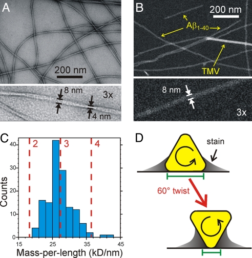
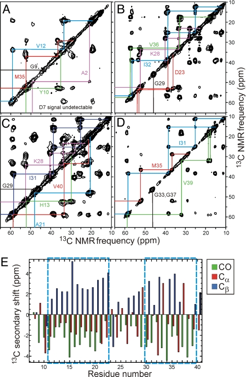
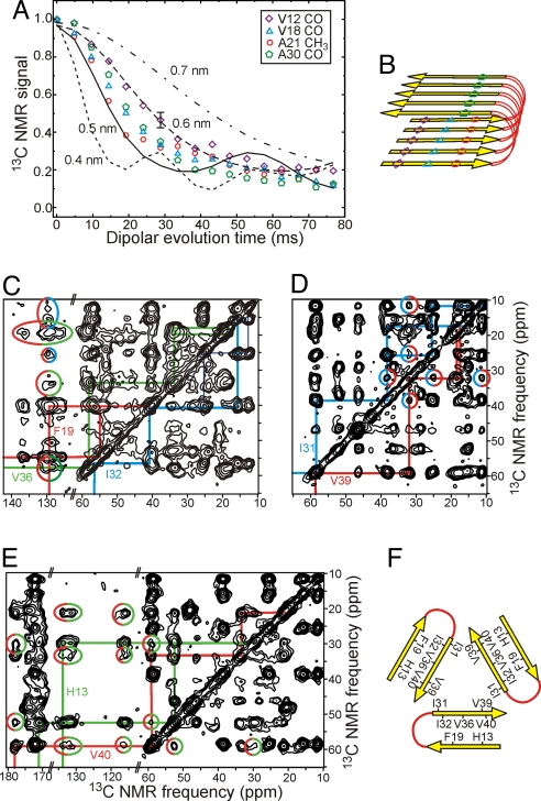
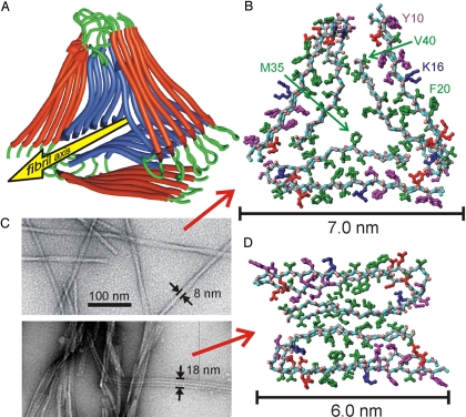
We describe a full structural model for amyloid fibrils formed by the 40-residue beta-amyloid peptide associated with Alzheimer's disease (Abeta(1-40)), based on numerous constraints from solid state NMR and electron microscopy. This model applies specifically to fibrils with a periodically twisted morphology, with twist period equal to 120 +/- 20 nm (defined as the distance between apparent minima in fibril width in negatively stained transmission electron microscope images). The structure has threefold symmetry about the fibril growth axis, implied by mass-per-length data and the observation of a single set of (13)C NMR signals. Comparison with a previously reported model for Abeta(1-40) fibrils with a qualitatively different, striated ribbon morphology reveals the molecular basis for polymorphism. At the molecular level, the 2 Abeta(1-40) fibril morphologies differ in overall symmetry (twofold vs. threefold), the conformation of non-beta-strand segments, and certain quaternary contacts. Both morphologies contain in-register parallel beta-sheets, constructed from nearly the same beta-strand segments. Because twisted and striated ribbon morphologies are also observed for amyloid fibrils formed by other polypeptides, such as the amylin peptide associated with type 2 diabetes, these structural variations may have general implications.


Conflict of interest statement

The authors declare no conflict of interest.

Figures

Fig. 1.
Fig. 1.
Electron microscopy. (A) Negatively stained TEM image of Aβ1–40 fibrils, showing a periodically twisted morphology with apparent width modulation. (B) STEM image of the same fibrils, with tobacco mosaic virus (TMV) included for mass density calibration. (C) Mass-per-length histogram, extracted from multiple STEM images, indicating a structure comprised of 3 cross-β units. (D) Schematic explanation of the enhanced width modulation in negatively stained TEM images of fibrils with approximately triangular cross-sections. Green bars represent the apparent widths at 2 positions of twist, as measured from the approximate inner edges of the stain.
Fig. 2.
Fig. 2.
Solid state NMR. (A–D) 2D 13C NMR spectra of Aβ1–40 fibril samples A–D, respectively, with chemical shift assignment paths for the indicated residues. Strong, sharp cross-peaks are observed for residues 10–40, indicating a rigid, ordered structure, whereas cross-peaks for A2, D7, and G9 are weak or absent because of dynamic disorder of the N-terminal segment. The absence of splittings of the NMR lines indicates that all Aβ1–40 molecules have equivalent conformations and structural environments, implying threefold symmetry. (E) Secondary 13C chemical shifts (i.e., deviations from random coil values) determined from 2D 13C NMR spectra. Dashed boxes indicate segments in which the secondary shifts indicate β-strands. Note that Cα secondary shifts for glycines (residues 25, 29, 33, 37, and 38) are not correlated with secondary structure.
Fig. 3.
Fig. 3.
Tertiary and quaternary constraints. (A) Measurements of intermolecular 13C-13C magnetic dipole–dipole couplings in samples E–G using the PITHIRDS-CT solid state NMR technique. Comparison with numerical simulations (lines) for linear chains of 13C nuclei with specified spacings indicates intermolecular distances of ≈0.5 nm for V12 CO, A21 CH3, and V18 CO sites, implying an in-register parallel β-sheet structure. (B) Schematic representation of the secondary and tertiary structure implied by the PITHIRDS-CT data and data in Fig. 2. (C–E) 2D 13C NMR spectra of samples H, D, and C, obtained with 0.5, 1.5, and 1.0-s RAD exchange periods, respectively. Cross-peaks enclosed in color-coded ellipses indicate F19/I32, F19/V36, I31/V39, and H13/V40 quaternary contacts. (F) Schematic representation of the quaternary structure implied by these long-range interresidue cross-peaks.
Fig. 4.
Fig. 4.
Experimentally based structural models. (A) Ribbon representation of the lowest-energy model for fibrils with the twisted morphology in Fig. 1. Modeling calculations assume threefold symmetry, consistent with STEM data and solid state NMR spectra, and are constrained by specific secondary, tertiary, and quaternary structural data from solid state NMR. (B) Atomic representation, viewed down the fibril axis. Hydrophobic, polar, negatively-charged, and positively charged amino acid sidechains are green, magenta, red, and blue, respectively. Backbone nitrogen and carbonyl oxygen atoms are cyan and pink. Unstructured N-terminal residues 1–8 are omitted. (C) Comparison of twisted (Upper) and striated ribbon (Lower) fibril morphologies in negatively stained TEM images. (D) Atomic representation of a model for striated ribbon fibrils developed previously by Petkova et al. (4, 15).

References

    1. Fowler DM, Koulov AV, Balch WE, Kelly JW. Functional amyloid: From bacteria to humans. Trends BiochemSci. 2007;32:217–224. - PubMed
    1. Chiti F, Dobson CM. Protein misfolding, functional amyloid, and human disease. Annu Rev Biochem. 2006;75:333–366. - PubMed
    1. Tycko R. Molecular structure of amyloid fibrils: Insights from solid state NMR. Q Rev Biophys. 2006;39:1–55. - PubMed
    1. Petkova AT, et al. Self-propagating, molecular-level polymorphism in Alzheimer's β-amyloid fibrils. Science. 2005;307:262–265. - PubMed
    1. Wetzel R, Shivaprasad S, Williams AD. Plasticity of amyloid fibrils. Biochemistry. 2007;46:1–10. - PMC - PubMed

Publication types

MeSH terms

Substances