. 2008 Nov 25;105(47):18296-301.
doi: 10.1073/pnas.0803279105. Epub 2008 Nov 17.

Hierarchical mechanisms build the DNA-binding specificity of FUSE binding protein

Affiliations

Hierarchical mechanisms build the DNA-binding specificity of FUSE binding protein

Lawrence R Benjamin et al. Proc Natl Acad Sci U S A. .

Abstract

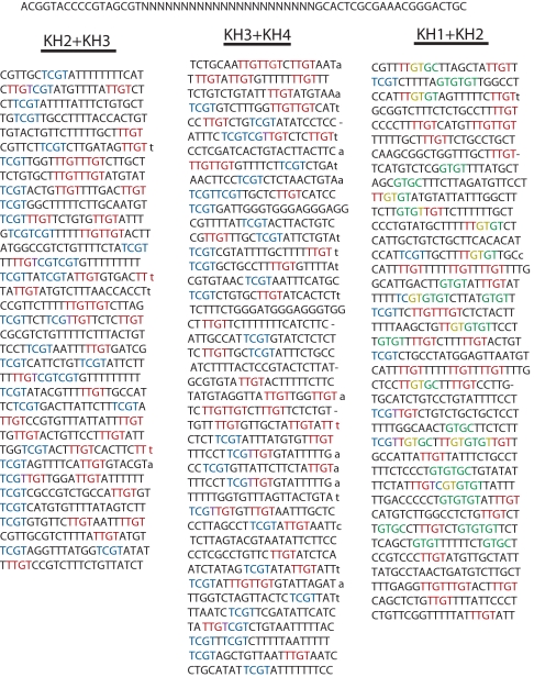
The far upstream element (FUSE) binding protein (FBP), a single-stranded nucleic acid binding protein, is recruited to the c-myc promoter after melting of FUSE by transcriptionally generated dynamic supercoils. Via interactions with TFIIH and FBP-interacting repressor (FIR), FBP modulates c-myc transcription. Here, we investigate the contributions of FBP's 4 K Homology (KH) domains to sequence selectivity. EMSA and missing contact point analysis revealed that FBP contacts 4 separate patches spanning a large segment of FUSE. A SELEX procedure using paired KH-domains defined the preferred subsequences for each KH domain. Unexpectedly, there was also a strong selection for the noncontacted residues between these subsequences, showing that the contact points must be optimally presented in a backbone that minimizes secondary structure. Strategic mutation of contact points defined in this study disabled FUSE activity in vivo. Because the biological specificity of FBP is tuned at several layers: (i) accessibility of the site; (ii) supercoil-driven melting; (iii) presentation of unhindered bases for recognition; and (iv) modular interaction of KH-domains with cognate bases, the FBP-FIR system and sequence-specific, single-strand DNA binding proteins in general are likely to prove versatile tools for adjusting gene expression.


Conflict of interest statement

The authors declare no conflict of interest.

Figures

Fig. 1.
Fig. 1.
EMSA scanning of FBP binding with FUSE oligonucleotides. (A) The eight 27-mer single-stranded probes used in this gel-mobility shift assay are aligned with the FUSE region upstream of the c-myc P2 promoter. The KH3 element (5′-ATTTTT-3′) and the KH4 element (5′-TATTCC-3′) reported from the NMR study (5) are shown as boldface italic. (B) A total of 0.5 pmol of each FUSE probe was incubated with 5 pmol of each of affinity-purified GST-FBP(KH2+KH3) or GST-FBP(KH3+KH4), or with 1.25 pmol of full-length FBP. Binding reactions lacking protein are shown at Left. (C) A diagram showing how FBP's KH domains are situated in the full-length protein.
Fig. 2.
Fig. 2.
SELEX-based enrichment of binding sites in ssDNA. (Upper) Diagram of method. (Lower) Sequences selected by paired KH domains. (KH3+KH4) Sequences of the input degenerate oligonucleotide and “winners” after 7 rounds of binding to GST-FBP(KH3+KH4). (KH2+KH3) Winners after 6 rounds of binding to GST-FBP(KH2+KH3). (KH1+KH2) Winners after 4 rounds of binding to GST-FBP(KH1+KH2). Mutations in the nonrandomized flanking sequence are shown in lowercase; deletions are indicated with dashes. Some output sequences were longer than the 22 bases randomized. Selected tetrads TTGT are red, TTGT are blue, GTGT and GTGC are green ([KH1+KH2] only); overlapping tetrads are purple or yellow.
Fig. 3.
Fig. 3.
Determination of the Optimal Spacing Between KH-domains by EMSA. (A) Sequence of spacer oligonucleotides. (B and C) A standard oligonucleotide was end-labeled and gel-shifted by an excess of (B) GST-FBP(KH3+KH4) (compare lanes 1 and 2) or (C) GST-FBP(KH2+KH3). Unlabeled competitor oligonucleotides (17 μM) in excess relative to the protein (≈40 nM) were added to most binding reactions (lanes 3–16), before protein. “Spacer4,” (lane 8) competed most effectively among the spacer oligonucleotides (lanes 6–14). All spacer oligonucleotides competed for protein binding more effectively than control oligonucleotides containing a single (lanes 4, 5, 15, and 16) or no (lane 3) consensus elements. Shifted bands were quantified by Imagequant software, version 1.2, and charted in Fig. S3.
Fig. 4.
Fig. 4.
Missing-base interference footprinting. (A) Preparative gel shift of FBP-FUSE (Left) (3 μmol of FBP; ≈0.1 μmol of probe), and FIR-FUSE or FIR-FBP-FUSE (Right) (0.5 μmol of FIR ± 29 nmol of FBP; ≈0.1 μmol of probe). (Left) A longer exposure than shown at Right. Probe was recovered from all bands and processed through the remainder of the assay (SI Methods). (B) Interference footprint of FBP on FUSE. Compare samples that bound FBP (lane 3) with free (lane 4) and input (lane 2) DNA. Regions with strong interference (thick bars) and weak interference (thin bars) are shown on both the gel and the sequence (5′ to 3′ starting at the bottom). No other interfering regions were seen on a 20% gel (data not shown). (C) Interference footprints of FIR alone (annotated in red, lane 3) and FIR+FBP (green, lane 6) on FUSE 87-mer; FBP unsupershifted by FIR is also shown (blue, lane 5). The EMSAs for lanes 2–3 and lanes 5–7 used the same input probe (lane 4). Reinforcing (“hypersensitive”) sites are marked with bullets. The same sample was run on 7% (Left) and 20% (Right) denaturing PA gels. (D) Comparison of footprints. Strongly footprinted regions from B are shown in gray.
Fig. 5.
Fig. 5.
Double point mutation in FUSE reduces FBP binding in vitro and FUSE function in vivo. (A) Schematic of pMT2 (+FUSE) with pMT2(mutFUSE) mutation sites indicated (partial FUSE sequence is shown). In pMT2(-FUSE), bacterial sequence replaces FUSE to maintain the length. (B) In vitro competition for FBP binding by (wt)FUSE 87-mer and (mut)FUSE 87-mer oligonucleotides. A small amount of end-labeled wtFUSE87-mer was gel-shifted by an excess of FBP (compare lanes 1 and 11). Included in the binding reactions were increasing amounts: 1× (> [FBP]), 3×, 10×, or 30× unlabeled wtFUSE87-mer (lanes 2–5); or 1×, 3×, 10×, 30×, or 100× unlabeled mut-FUSE87-mer (lanes 6–10). Comparing the quantified (ImageQuant) shifts in lanes 2 and 8, wtFUSE binds FBP ≈15-fold more strongly than mut-FUSE87-mer. (C) Histograms of flow cytometry testing FUSE function. EGFP expression was measured 10 h after induction with 60 μM Zn2+ (x axis). The histogram for uninduced pMT2(FUSE) is overlaid in green.

References

    1. Smogorzewska A, de Lange T. Regulation of telomerase by telomeric proteins. Annu Rev Biochem. 2004;73:177–208. - PubMed
    1. Kouzine F, Sanford S, Elisha-Feil Z, Levens D. The functional response of upstream DNA to dynamic supercoiling in vivo. Nat Struct Mol Biol. 2008;15:146–154. - PubMed
    1. Lavelle C. DNA torsional stress propagates through chromatin fiber and participates in transcriptional regulation. Nat Struct Mol Biol. 2008;15:123–125. - PubMed
    1. Iftode C, Daniely Y, Borowiec JA. Replication protein A (RPA): The eukaryotic SSB. Crit Rev Biochem Mol Biol. 1999;34:141–180. - PubMed
    1. Braddock DT, Louis JM, Baber JL, Levens D, Clore GM. Structure and dynamics of KH domains from FBP bound to single-stranded DNA. Nature. 2002;415:1051–1056. - PubMed

Publication types