. 2009 Jan;181(1):313-22.
doi: 10.1534/genetics.108.091538. Epub 2008 Nov 17.

Formation and longevity of chimeric and duplicate genes in Drosophila melanogaster

Affiliations

Formation and longevity of chimeric and duplicate genes in Drosophila melanogaster

Rebekah L Rogers et al. Genetics. 2009 Jan.

Abstract

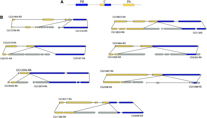
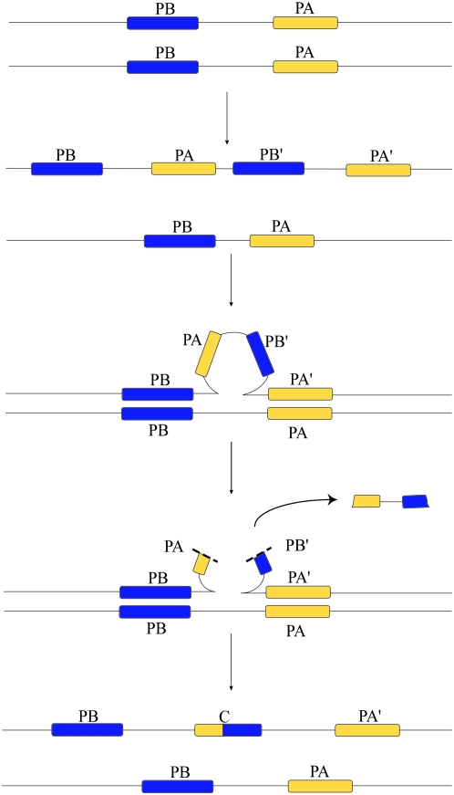
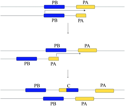
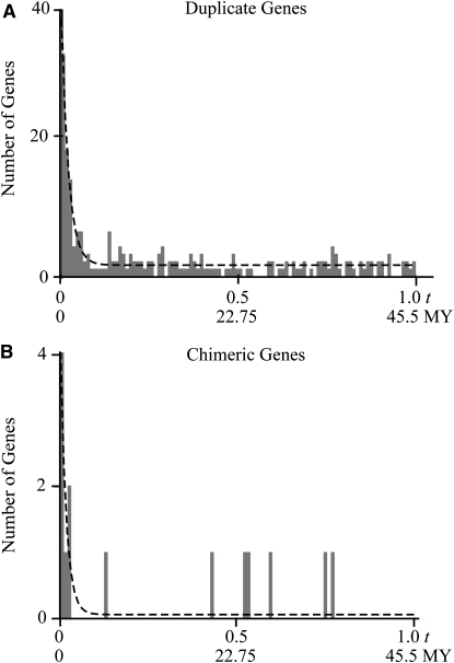
Historically, duplicate genes have been regarded as a major source of novel genetic material. However, recent work suggests that chimeric genes formed through the fusion of pieces of different genes may also contribute to the evolution of novel functions. To compare the contribution of chimeric and duplicate genes to genome evolution, we measured their prevalence and persistence within Drosophila melanogaster. We find that approximately 80.4 duplicates form per million years, but most are rapidly eliminated from the genome, leaving only 4.1% to be preserved by natural selection. Chimeras form at a comparatively modest rate of approximately 11.4 per million years but follow a similar pattern of decay, with ultimately only 1.4% of chimeras preserved. We propose two mechanisms of chimeric gene formation, which rely entirely on local, DNA-based mutations to explain the structure and placement of the youngest chimeric genes observed. One involves imprecise excision of an unpaired duplication during large-loop mismatch repair, while the other invokes a process akin to replication slippage to form a chimeric gene in a single event. Our results paint a dynamic picture of both chimeras and duplicate genes within the genome and suggest that chimeric genes contribute substantially to genomic novelty.


Figures

F<sc>igure</sc> 1.—
Figure 1.—
Placement and structure of chimeric genes. (A) The seven youngest chimeric genes were all found in close proximity with their parental genes. The parental gene that contributed the 5′ end is found downstream, and the parental gene that contributed to the 3′ end is upstream. (B) Genomic sequences were aligned to find the breakpoints of chimera formation. Breakpoints fall in exons for six of the seven youngest chimeric genes. Portions of the chimera that align to parental gene A are highlighted in yellow, and portions aligning to parental gene B are highlighted in blue. Portions of parental genes that do not align to the chimera are shown in gray. Structures are adapted from FlyBase (http://flybase.org). Gene structure was largely conserved with one intron loss in CG12592 and one intron gain in CG18217.
F<sc>igure</sc> 2.—
Figure 2.—
Large-loop mismatch repair mechanism of chimera formation. A short segmental duplication occurs on a given chromosome, copying genes PB and PA. During meiosis or mitosis, the duplicated region pairs with an unduplicated chromatid. The outer genes PB and PA′ pair with the unduplicated genes on the opposite chromatid, producing a loop of unpaired DNA, initiating large-loop mismatch repair. Imprecise excision of the loop creates a chimeric gene with the observed structure.
F<sc>igure</sc> 3.—
Figure 3.—
Replication slippage-based mechanism of chimera formation. During the synthesis of the parental gene PA, replication stalls. While searching for the proper template to resume replication, genes misalign as shown so that PA pairs with PB. New DNA is synthesized on the misaligned strand, producing a chimeric gene with the structure and placement observed in the seven youngest chimeras.
F<sc>igure</sc> 4.—
Figure 4.—
The age distribution for (A) duplicate and (B) chimeric genes fitted with our model of birth (formation), death (decay), and preservation. Histogram bins show the observed distribution of t values, while dashed lines show the maximum-likelihood (ML) model fit. The rate of formation λ scales the overall number of duplicates/chimeras observed, but does not affect the shape of the distribution. The rate of decay μ determines how quickly the distribution drops to a constant threshold, while both μ and the rate of preservation ν together determine the height of the constant tail. ML estimates of rate parameters are shown in Table 2.
F<sc>igure</sc> 5.—
Figure 5.—
Phylogenetic distribution of chimeric genes found in Drosophila melanogaster. Phylogenetic locations were determined through parsimony. Gene names are placed on the branch in which each chimera was formed. Phylogeny and t were consistent for almost all genes considered. Eight chimeric genes (††: CG18853, CG32318, CG31864, CG12592, CG31904, CG31687, CG18217, and CG31668) are specific to D. melanogaster. Putative losses (see results) are not shown.

References

    1. Adams, M. D., S. E. Celniker, R. A. Holt, C. A. Evans, J. D. Gocayne et al., 2000. The genome sequence of Drosophila melanogaster. Science 287 2185–2195. - PubMed
    1. Altschul, S. F., W. Gish, W. Miller, E. W. Myers and D. J. Lipman, 1990. Basic local alignment search tool. J. Mol. Biol. 215 403–410. - PubMed
    1. Alvarez, A. D., W. Shi, B. A. Wilson and J. B. Skeath, 2003. Pannier and pointedP2 act sequentially to regulate Drosophila heart development. Development 130 3015–3026. - PubMed
    1. Bogarad, L. D., and M. W. Deem, 1999. A hierarchical approach to protein molecular evolution. Proc. Natl. Acad. Sci. USA 96 2591–2595. - PMC - PubMed
    1. Brown, S., and J. Castelli-Gair Hombria, 2000. Drosophila grain encodes a GATA transcription factor required for cell rearrangement during morphogenesis. Development 127 4867–4876. - PubMed

Publication types

LinkOut - more resources