Comparative Study
. 2009 Feb;181(2):473-82.
doi: 10.1534/genetics.107.080515. Epub 2008 Nov 17.

The Caenorhabditis elegans ing-3 gene regulates ionizing radiation-induced germ-cell apoptosis in a p53-associated pathway

Affiliations
Comparative Study

The Caenorhabditis elegans ing-3 gene regulates ionizing radiation-induced germ-cell apoptosis in a p53-associated pathway

Jingjing Luo et al. Genetics. 2009 Feb.

Abstract

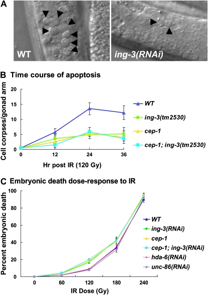
The inhibitor of growth (ING) family of type II tumor suppressors are encoded by five genes in mammals and by three genes in Caenorhabditis elegans. All ING proteins contain a highly conserved plant homeodomain (PHD) zinc finger. ING proteins are activated by stresses, including ionizing radiation, leading to the activation of p53. ING proteins in mammals and yeast have recently been shown to read the histone code in a methylation-sensitive manner to regulate gene expression. Here we identify and characterize ing-3, the C. elegans gene with the highest sequence identity to the human ING3 gene. ING-3 colocalizes with chromatin in embryos, the germline, and somatic cells. The ing-3 gene is part of an operon but is also transcribed from its own promoter. Both ing-3(RNAi) and ing-3 mutant strains demonstrate that the gene likely functions in concert with the C. elegans p53 homolog, cep-1, to induce germ-cell apoptosis in response to ionizing radiation. Somatically, the ing-3 mutant has a weak kinker uncoordinated (kinker Unc) phenotype, indicating a possible neuronal function.


Figures

F<sc>igure</sc> 1.—
Figure 1.—
ing-3 protein and gene structure. (A) Diagram of the predicted ING-3 protein, indicating the PHD, leucine zipper-like domain (LZL), the NLS, and the LID. The corresponding regions and amino acid similarities with human ING-3 are shown. (B) The ing-3 gene is the second gene in a predicted operon that includes mitochondrial dehydrogenase (Y51H1A.3) and histone deacetylase hda-6 and mcd-1. (C) The six exons of the ing-3 gene are indicated by yellow boxes while green boxes represent the 5′ and 3′ untranslated regions. The relative exon positions on cosmid Y51H1A are indicated. The position of the alleles ttTi5439 and tm2530 are shown in red, and the blue box indicates the region used to raise the antisera. The region used to generate dsRNA for RNAi is marked with red arrows.
F<sc>igure</sc> 2.—
Figure 2.—
Reporters driven by the operon or internal promoter give different expression patterns. (A) The 5′ genomic sequence (600 bp) of the first gene of this operon (Y51H1A.3) was used as the “operon promoter,” while the 880 bp between ing-3 and Y51H1A.3 was used as the “internal promoter.” (B) GFP and (C) LacZ indicate that the operon promoter was widely expressed in L1 larva, especially in the pharynx. (D) The operon promoter showed GFP expression in the adult vulva, spermatheca (asterisks), neurons, and epidermal cells, among others. (E) Differential interference contrast image corresponding to D. The internal promoter expressed GFP (F) and LacZ (G) in the L1 intestine, the limits of which are indicated by asterisks. The signal between the asterisks in F represents gut auto-fluorescence. Note higher expression in the anterior intestinal cells.
F<sc>igure</sc> 3.—
Figure 3.—
Immunolocalization of ING-3. (A) Mouse polyclonal anti-ING-3 recognized endogenous ING-3 as a single 45-kDa band on Western blots of wild-type gravid hermaphrodites. This band was absent in the two ing-3 mutants but was present in cep-1 lysates. Actin was used as the loading control. (B) ING-3 colocalized with chromatin in the newly fertilized embryo (top left) but is absent from ing-3(tm2530) (top right). ING-3 was also found on chromatin at mitosis in developing embryos (arrows, bottom row). (C) Immunostaining of dissected wild-type adult gonads (left-most column) shows that ING-3 overlapped DAPI. Lower levels were present in the mitotic region (I) compared to the transition zone (II). High ING-3 persisted into the early pachytene region (III), but then decreased as cells proceeded through meiosis. Low levels, mainly on chromosomes, were seen at diplotene (IV). Higher magnification of the early pachytene section of the gonad (columns 2–4) showed extensive overlap between ING-3 and DAPI. The nuclear signal disappeared when antisera was preincubated with the immunizing peptide (peptide block) or in ing-3(tm2530). Thus the low level of ING-3 staining between cells likely represents background. The right-most column shows the staining pattern for ING-3 in the anterior nuclei of a dissected gut.
F<sc>igure</sc> 4.—
Figure 4.—
Germ-cell apoptosis and embryonic death following IR. (A) L4 stage larvae were exposed to 120 Gy of IR and the number of apoptotic germ cells (arrowheads) was scored 24 hr later. Animals treated with ing-3(RNAi) showed a significantly reduced number of apoptotic germ cells. (B) Time course of accumulation of cell corpses following 120 Gy of IR. Similar to wild type and cep-1, apoptosis plateaus by 24 hr in ing-3 and ing-3; cep-1, indicating that ing-3 is blocking rather than delaying cell death. (C) Dose-response curve of wild type and mutants vs. embryonic inviability of the 8- to 22-hr brood following the indicated levels of IR. Error bars represent 1 standard deviation in B and C.

References

    1. Bienz, M., 2006. The PHD finger, a nuclear protein-interaction domain. Trends Biochem. Sci. 31 35–40. - PubMed
    1. Blumenthal, T., and K. Steward, 1997. RNA processing and gene structure, pp. 117–146 in C. elegans II, edited by D. L. Riddle, T. Blumenthal, B. J. Meyer and J. R. Priess. Cold Spring Harbor Laboratory Press, Cold Spring Harbor, NY. - PubMed
    1. Brenner, S., 1974. The genetics of Caenorhabditis elegans. Genetics 77 71–94. - PMC - PubMed
    1. Cheung, K. J., Jr, J. A. Bush, W. Jia, and G. Li, 2000. Expression of the novel tumour suppressor p33(ING1) is independent of p53. Br. J. Cancer 83 1468–1472. - PMC - PubMed
    1. Clark, S. G., X. Lu and H. R. Horvitz, 1994. The Caenorhabditis elegans locus lin-15, a negative regulator of a tyrosine kinase signaling pathway, encodes two different proteins. Genetics 137 987–997. - PMC - PubMed

Publication types

MeSH terms