Review
. 2009 Sep-Oct;28(5):785-815.
doi: 10.1002/mas.20203.

Probing protein structure by amino acid-specific covalent labeling and mass spectrometry

Affiliations
Review

Probing protein structure by amino acid-specific covalent labeling and mass spectrometry

Vanessa Leah Mendoza et al. Mass Spectrom Rev. 2009 Sep-Oct.

Abstract

For many years, amino acid-specific covalent labeling has been a valuable tool to study protein structure and protein interactions, especially for systems that are difficult to study by other means. These covalent labeling methods typically map protein structure and interactions by measuring the differential reactivity of amino acid side chains. The reactivity of amino acids in proteins generally depends on the accessibility of the side chain to the reagent, the inherent reactivity of the label and the reactivity of the amino acid side chain. Peptide mass mapping with ESI- or MALDI-MS and peptide sequencing with tandem MS are typically employed to identify modification sites to provide site-specific structural information. In this review, we describe the reagents that are most commonly used in these residue-specific modification reactions, details about the proper use of these covalent labeling reagents, and information about the specific biochemical problems that have been addressed with covalent labeling strategies.


Figures

Figure 1
Figure 1
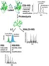
Analytical scheme showing covalent labeling with MS detection. The label modifies solvent accessible amino acids. ESI- or MALDI-MS can be used to calculate the extent of modification. The modified protein can then be subjected to proteolysis followed by either LC-MS/MS or MALDI-MS to determine the peptide fragments and/or the specific amino acids that have been modified.
Figure 2
Figure 2
(A). Peptide mass mapping. Labeled peptides are identified from a mass spectrum that shows unmodified and modified fragments. (B) In tandem mass spectra unmodified and modified product ions can be used to identify the specific amino acid that has been modified.
Figure 2
Figure 2
(A). Peptide mass mapping. Labeled peptides are identified from a mass spectrum that shows unmodified and modified fragments. (B) In tandem mass spectra unmodified and modified product ions can be used to identify the specific amino acid that has been modified.
Figure 3
Figure 3
Analytical scheme showing a two-step differential chemical modification. Free and bound proteins are first treated with a low molar excess of the label followed by reaction with a high molar excess of an isotopically-labeled form of the modification reagent. Relative reactivities are calculated from the ratio of the intensities of the modified and isotopically-labeled peptides. Modification of two residues is illustrated. The residue in Peptide ● has similar reactivity in both forms of the protein. The residue in Peptide ▲ is less accessible in the native protein and has a reduced reactivity in this form.
Figure 4
Figure 4
Plot of unmodified peptide (or protein) as a function of time (or reagent concentration) showing that a more buried residue His-B (black) react more slowly and therefore has a shallower slope in the kinetics plot compared to a more exposed residue like His-A (gray).
Figure 5
Figure 5
A 2nd order kinetics plot for the peptide fragment 49–60 from β-2-microglobulin being modified by sulfo-N-hydroxysuccinamide acetate. Linear region (black) indicates unchanged protein structure. Deviation from linearity (gray) suggests a protein structural change due to the modification.
Figure 6
Figure 6
Interaction site of CD4 with gp120 as derived from the crystal structure, PDB code 1GC1. Phe43 and Arg59 of CD4 interact with gp120 while Arg58 does not.
Figure 7
Figure 7
Ung:Ugi interface (PDB code 1UGH) showing the interactions of Glu28 and Glu31 of Ugi with Ser247, His268 and Arg276 of Ung.
Figure 8
Figure 8
Tubulin cysteines in positively-charged and negatively-charged environments (PDB code 1JFF). (A). Cys241 and Cys246 are within 5 Å of Arg320. (B). Cys129 is within 6 Å of Glu3.
Figure 9
Figure 9
Crystal structure of human catechol O-methyltransferase in the presence of S-adenosyl-L-methionine (AdoMet) (PDB code 3BWY). Cys68 and Cys 94 are in the AdoMet binding site.
Figure 10
Figure 10
Crystal structure of the insulin dimer (PDB code 4INS). The ε-nitrogen (NE) and δ-nitrogen (ND) of His-10 are both accessible. His-5 participates in the insulin core structure and shows an accessible surface for either the ε-nitrogen (NE) or the δ-nitrogen (ND), depending on the monomer.
Figure 11
Figure 11
Crystal structures of the p70 subunit of human replication protein A. (A) Structure of DBD-AB (PDB code 1JMC). Lys263 and Lys343 are found in the binding clefts of DBD-A and DBD-B, respectively, and are protected from modification by direct contact with the single-stranded (ss) piece of DNA. Lys183 and Lys259 are also protected from modification when ssDNA is present, even though they are not bound to the ssDNA. (B) Structure of DBD-C (PDB code 1L1O). Lys489, Lys577, and Lys588 are located in the binding cleft of DBD-C and are protected from modification when ssDNA is present.
Figure 12
Figure 12
Crystal structure of the E9:Im9 complex (PDB code 1EMV). Lys89 and Lys97 are directly involved in the interaction. Lys76 and Lys81 are located in an α-helical region near the interaction surface. Lys55 and Lys63 do not interact with Im9, and the region of E9 containing these residues possibly undergoes a conformational change upon Im9 binding.
Figure 13
Figure 13
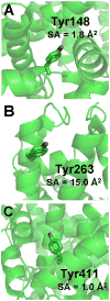
Selected tyrosine residues from human serum albumin (PDB code 1AO6). (A) Tyr148 showed high reactivity despite its low solvent accessibility. (B) Tyr263 has high solvent accessibility but showed low reactivity. (C) Tyr411 exhibited different reactivity towards iodine and tetranitromethane. The authors estimated the the solvent accessibility (SA) of CE1 and CE2 atoms of the tyrosine phenol ring (ortho positions to hydroxy group) with the program Joy. This program uses Lee and Richards method to calculate SA. A probe radius of 1.4 Å, representing the van der Waals sphere of water, was used.
Scheme 1
Scheme 1
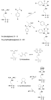
Modification reactions of arginine by vicinal dicarbonyl compounds.
Scheme 2
Scheme 2
Reaction showing how borate can stabilize adducts that are formed by arginine with the various dicarbonyl compounds.
Scheme 3
Scheme 3
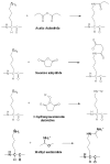
Modification reactions of carboxyl groups.
Scheme 4
Scheme 4
Modification reactions of cysteine residues.
Scheme 5
Scheme 5
Modification reactions of histidine using DEPC.
Scheme 6
Scheme 6
Modification reactions of lysine residues.
Scheme 7
Scheme 7
Modification reactions of tryptophan.
Scheme 8
Scheme 8
Modification reactions of tyrosine residues.

References

    1. Akashi S, Shirouzu M, Terada T, Ito Y, Yokohama S, Takio K. Characterization of the structural difference between active and inactive forms of the Ras protein by chemical modification followed by mass spectrometric peptide mapping. Anal Biochem. 1997;248:15–25. - PubMed
    1. Akinsiku OT, Yu ET, Fabris D. Mass spectrometric investigation of protein alkylation by the RNA footprinting probe kethoxal. J Mass Spectrom. 2005;40:1372–1381. - PubMed
    1. Apuy JL, Chen XH, Russell DH, Baldwin TO, Giedroc DP. Ratiometric pulsed Alkylation/Mass spectrometry of the cysteine pairs in individual zinc fingers of MRE-Binding transcription factor-1 (MTF-1) as a probe of zinc chelate stability. Biochemistry. 2001;40:15164–15175. - PubMed
    1. Apuy JL, Park ZY, Swartz PD, Dangott LJ, Russell DH, Baldwin TO. Pulsed-alkylation mass spectrometry for the study of protein folding and dynamics: Development and application to the study of a folding/unfolding intermediate of bacterial luciferaset. Biochemistry. 2001;40:15153–15163. - PubMed
    1. Azim-Zadeh O, Hillebrecht A, Linne U, Marahiel MA, Klebe G, Lingelbach K, Nyalwidhe J. Use of biotin derivatives to probe conformational changes in proteins. J Biol Chem. 2007;282:21609–21617. - PubMed

Publication types

LinkOut - more resources