Case Reports
. 2009 Jul;19(3):459-66.
doi: 10.1111/j.1750-3639.2008.00231.x. Epub 2008 Oct 31.

Molecular changes in white matter adjacent to an active demyelinating lesion in early multiple sclerosis

Affiliations
Case Reports

Molecular changes in white matter adjacent to an active demyelinating lesion in early multiple sclerosis

Thomas Zeis et al. Brain Pathol. 2009 Jul.

Abstract

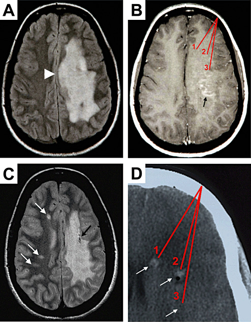
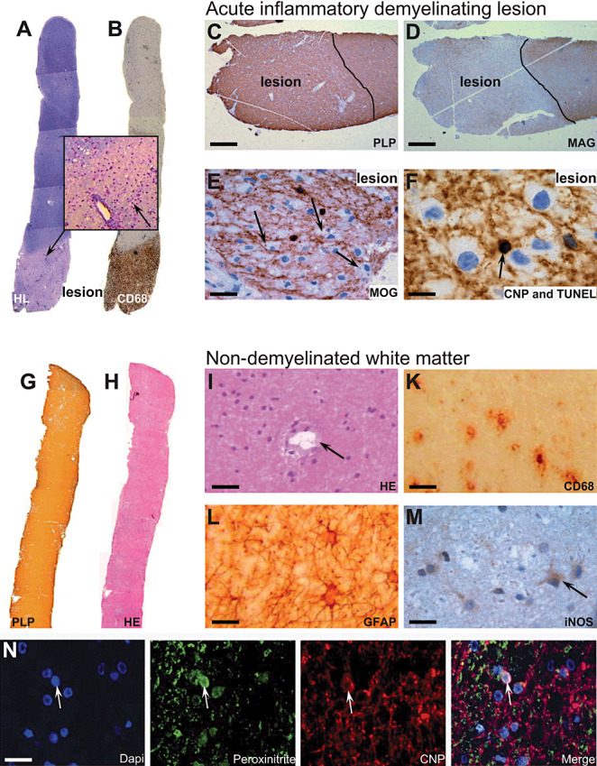
A stereotactic biopsy of a 17-year-old woman revealed an active inflammatory demyelinating lesion compatible with pattern III multiple sclerosis (MS) according to Lucchinetti et al. The biopsy included a white matter region distant from the active inflammatory demyelinating lesion with abnormal MRI signal, lacking histopathological signs of demyelination and/or oligodendrocyte apoptosis. Expression analysis of this area revealed a strong up-regulation of neuronal nitric oxide synthase (nNOS). Furthermore, detection of nitrotyrosine provided evidence for reactive nitrogen species (RNS)-mediated damage to oligodendrocytes. Concomitantly, genes involved in neuroprotection against oxidative stress such as heme oxygenase 1 were up-regulated. Even though a single case report, this study shows earliest molecular changes in white matter surrounding an actively demyelinating lesion during the first manifestation of MS, pointing toward a more widespread pathological process. Therapeutic targeting of the identified mechanisms of tissue injury might be crucial to prevent further lesion formation or secondary tissue damage.


Figures

Figure 1
Figure 1
MRI analysis of the index patient. A T2‐weighted MRI during the first manifestation displays a large confluent hyperintense lesion in the subcortical white matter of left hemisphere (A) with mass effect (arrowhead). Gadolinium‐enhanced T1‐weighted MRI revealed a punctate, garland‐like enhancement in the subcortical white matter (B, arrow). Red lines mark schematically the three target regions from which the stereotactic biopsy specimens were taken (B). A routine follow‐up MRI investigation after 5 years revealed three new white matter lesions in the contralateral hemisphere (C, white arrows). Black arrow points to tissue destruction by one of the stereotactic brain biopsies (C). A CT picture (D) illustrates the three target regions (1, 2, 3) of the biopsy specimens taken from the frontal subcortical white matter (arrows). Note, CT signal enhancement by bleedings caused by the excision of tissue is visible (D, arrows 1 and 2). Note that the origin of all biopsy specimens was in regions of abnormal T1, T2, and CT signals, but only some biopsy specimens contained inflammatory demyelinating lesion, whereas others contained histomorphologically normal appearing white matter only.
Figure 2
Figure 2
Immunohistochemical analysis of lesion and non‐demyelinated white matter tissue. Immunopathological examination of paraffin‐embedded biopsies containing lesion tissue (AF) revealed a relatively sharp bordered lesion by Holmes/Luxol staining (A) with a dense infiltrate of CD68‐positive macrophages (B). Inset of (A) shows dense infiltrating cells around a blood vessel (arrow). Staining for PLP in the acute lesion still appeared normal (C), whereas a specific absence of MAG was detected (D). Furthermore, MOG‐positive myelin debris was detected within macrophages (E, arrows). TUNEL staining revealed apoptotic oligodendrocytes, co‐stained by CNPase, within the lesion (F, arrow). Examination of the fresh‐frozen biopsies used for RNA analysis did not show any signs of infiltrates or lesion formation (GM). Staining by anti‐PLP showed normal myelin (G), haematoxylin staining revealed normal cellularity (H) and blood vessels without infiltrating cells (I, arrow). However, a number of CD68‐positive cells, histopathological microglia, were detected (K). Furthermore, activated astrocytes could be detected throughout the white matter (L). Staining for iNOS in non‐demyelinated white matter distant to lesion revealed low expression levels in cells resembling microglia or astrocytes (M, arrow). Immunofluorescent colocalization for nitrotyrosine showed an accumulation in CNPase‐positive oligodendrocytes (N, arrow). Bars: C, D: 100 µm; E: 30 µm; F: 25 µm; I, K: 50 µm; L, M, N: 25 µm.

References

    1. Barnett MH, Prineas JW (2004) Relapsing and remitting multiple sclerosis: pathology of the newly forming lesion. Ann Neurol 55:458–468. - PubMed
    1. Bruck W, Bitsch A, Kolenda H, Bruck Y, Stiefel M, Lassmann H (1997) Inflammatory central nervous system demyelination: correlation of magnetic resonance imaging findings with lesion pathology. Ann Neurol 42:783–793. - PubMed
    1. Cannella B, Raine CS (2004) Multiple sclerosis: cytokine receptors on oligodendrocytes predict innate regulation. Ann Neurol 55:46–57. - PubMed
    1. Catania MV, Aronica E, Yankaya B, Troost D (2001) Increased expression of neuronal nitric oxide synthase spliced variants in reactive astrocytes of amyotrophic lateral sclerosis human spinal cord. J Neurosci 21:RC148. - PMC - PubMed
    1. Chitnis T, Najafian N, Benou C, Salama AD, Grusby MJ, Sayegh MH, Khoury SJ (2001) Effect of targeted disruption of STAT4 and STAT6 on the induction of experimental autoimmune encephalomyelitis. J Clin Invest 108:739–747. - PMC - PubMed

Publication types

MeSH terms

Substances