. 2008 Oct;99(10):1884-91.
doi: 10.1111/j.1349-7006.2008.00901.x.

Identification of mitochondrial F(1)F(0)-ATP synthase interacting with galectin-3 in colon cancer cells

Affiliations

Identification of mitochondrial F(1)F(0)-ATP synthase interacting with galectin-3 in colon cancer cells

Duck-Woo Kim et al. Cancer Sci. 2008 Oct.

Abstract

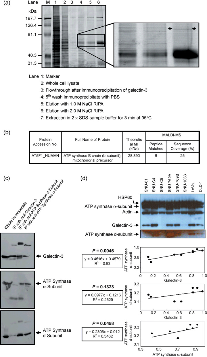
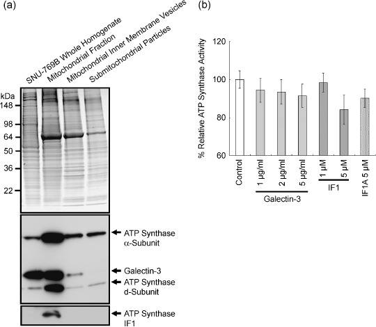
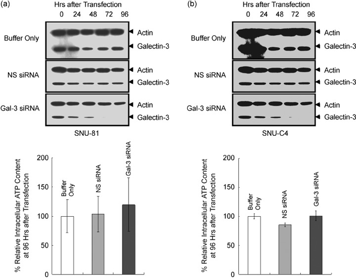
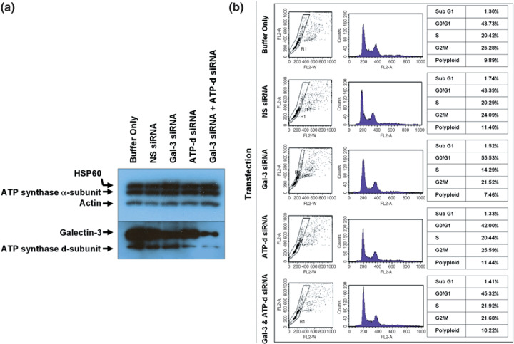
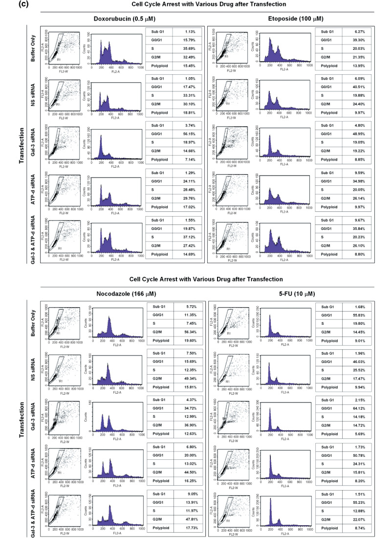
To evaluate the effect of galectin-3 in cell cycle regulation of colon cancer cells, we looked for binding molecules interacting with galectin-3 and examined the changes in cell cycle by suppressing galectin-3 and the binding molecule. To identify target molecules interacting with galectin-3, we analyzed immunoprecipitate of the anti-galectin-3 antibody obtained from human colon cancer cell line, using matrix-assisted laser desorption ionization-mass spectrometry. We validated subcellular localization of galectin-3 and ATP synthase identified, and ATP synthase activity was determined in the presence of galectin-3. Cell cycle regulation was monitored after galectin-3 siRNA transfection. ATP synthase b-subunit was identified in immunoprecipitate of the anti-galectin-3 antibody. Galectin-3 and ATP synthase were co-isolated in the inner membrane vesicles of mitochondria. Galectin-3 has an inhibitory activity against ATP synthase, and intracellular ATP content showed increasing tendency after galectin-3 suppression. Suppression of galectin-3 resulted in G0/G1 progression of human colon cancer cells arrested at S, S/G2 and G2/M phase in the presence of doxorubicin, and etoposide or nocodazole, respectively. Compared to cells in which ATP synthase d-subunit was suppressed alone, sub-G1 fraction caused by etoposide or nocodazole was decreased in cells with galectin-3 suppression alone. In conclusion, galectin-3 co-localized with ATP synthase in the inner membrane of mitochondria and has an inhibitory effect on ATP synthase in human colon cancer cells. In the presence of cell cycle synchronizing drugs, doxorubicin, etoposide, or nocodazole, suppression of galectin-3 induced cell cycle progression to G0/G1 phase.


Figures

Figure 1
Figure 1
Identification of mitochondrial F1F0‐ATP synthase (ATP synthase) b‐subunit in the antigalectin‐3 antibody immunoprecipitate. (a) Sodium dodecylsulfate–polyacrylamide gel electrophoresis (SDS–PAGE) of the galectin‐3 antibody immunoprecipitate. The galectin‐3 immunoprecipitate was fractionated using a salt gradient, as shown in the figure, and subjected to SDS‐PAGE. (b) Identification of the ATP synthase b‐subunit in the immunoprecipitate by matrix‐assisted laser desorption ionization–time of flight analysis. After staining the SDS‐PAGE gel, the protein band specified in (a) was excised and in‐gel digested to determine the peptide mass. The protein was successfully identified as the ATP synthase b‐subunit, a mitochondrial precursor. (c) Interactions of the ATP synthase d‐subunit with galectin‐3. Immunoprecipitates of the antigalectin‐3 antibody were probed with commercially available anti‐ATP synthase α‐ or d‐subunit antibodies, and vice‐versa. Immunoreactive signals of the ATP synthase d‐subunit were clearly detected in immunoprecipitates of antigalectin‐3 and anti‐ATP synthase α‐subunit antibodies. (d) Positive correlation of galectin‐3 expression with ATP synthase α‐ and d‐subunits in eight individual human colon cancer cell lines. Expression of galectin‐3 and ATP synthase subunits was normalized with actin and heat shock protein (HSP) 60, respectively. OD, arbitrary unit.
Figure 2
Figure 2
Copurification of galectin‐3 with ATP synthase, and the inhibitory effect of galectin‐3 on ATP synthase activity. (a) Copurification of galectin‐3 with ATP synthase. Mitochondria were isolated initially from whole homogenates of SNU‐769B, and inner‐membrane vesicles of mitochondria and submitochondrial particles were prepared. The upper panel depicts the sodium dodecylsulfate–polyacrylamide gel electrophoresis gel image after staining, and the lower panel represents western blot results. The levels of ATP synthase α‐ and d‐subunits, IF1, and galectin‐3 were determined in each isolation step. Galectin‐3 was additionally detected in the inner‐membrane vesicles (lower panel). (b) Inhibitory effect of galectin‐3 on ATP synthase activity. The control activity of SNU‐769A was defined as 100%, and the percentage relative ATP synthase activities have been determined in the presence of either ATP synthase inhibitor or galectin‐3. Enzyme activity was decreased by up to 8% in the presence of 2 µg/mL galectin‐3.
Figure 3
Figure 3
Effects of galectin‐3 suppression on intracellular ATP content of SNU‐81. (a) Suppression of galectin‐3 after small interfering RNA (siRNA) transfection. The human colon cancer cell line SNU‐81 was transfected with siRNA specific for galectin‐3. Galectin‐3 siRNA transfection resulted in suppression of both of galectin‐3 and ATP synthase d‐subunit. (b) Intracellular ATP content after galectin‐3 suppression. The intracellular ATP level in cells transfected with galectin‐3 siRNA increased slightly compared to the controls, but data were not statistically significant.
Figure 4
Figure 4
Effect of ATP synthase and galectin‐3 suppression on SNU‐81 cell‐cycle arrest. (a) Suppression of galectin‐3 and ATP synthase d‐subunit after small interfering RNA transfection. (b) There was no significant effect of galectin‐3 or ATP synthase d‐subunit expression on cell‐cycle progression. (c) Cell‐cycle arrest caused by doxorubicin, etoposide, and nocodazole treatment, and the effect of galectin‐3 and ATP synthase d‐subunit suppression.
Figure 4
Figure 4
Effect of ATP synthase and galectin‐3 suppression on SNU‐81 cell‐cycle arrest. (a) Suppression of galectin‐3 and ATP synthase d‐subunit after small interfering RNA transfection. (b) There was no significant effect of galectin‐3 or ATP synthase d‐subunit expression on cell‐cycle progression. (c) Cell‐cycle arrest caused by doxorubicin, etoposide, and nocodazole treatment, and the effect of galectin‐3 and ATP synthase d‐subunit suppression.

Similar articles

Cited by

References

    1. Barondes SH, Castronovo V, Cooper DN et al . Galectins: a family of animal β‐galactoside‐binding lectins. Cell 1994; 76: 597–8. - PubMed
    1. Dumic J, Dabelic S, Flogel M. Galectin‐3: an open‐ended story. Biochimica et Biophysica Acta 2006; 1760: 616–35. - PubMed
    1. Ruebel KH, Jin L, Qian X et al . Effects of DNA methylation on galectin‐3 expression in pituitary tumors. Cancer Res 2005; 65: 1136–40. - PubMed
    1. Paz A, Haklai R, Elad‐Sfadia G et al . Galectin‐1 binds oncogenic H‐Ras to mediate Ras membrane anchorage and cell transformation. Oncogene 2001; 20: 7486–93. - PubMed
    1. Takenaka Y, Fukumori T, Yoshii T et al . Nuclear export of phosphorylated galectin‐3 regulates its antiapoptotic activity in response to chemotherapeutic drugs. Mol Cell Biol 2004; 24: 4395–406. - PMC - PubMed

Publication types

MeSH terms

Substances

LinkOut - more resources