. 2008 Oct;99(10):2019-27.
doi: 10.1111/j.1349-7006.2008.00910.x.

Apoptosis induced by methylene-blue-mediated photodynamic therapy in melanomas and the involvement of mitochondrial dysfunction revealed by proteomics

Affiliations

Apoptosis induced by methylene-blue-mediated photodynamic therapy in melanomas and the involvement of mitochondrial dysfunction revealed by proteomics

Yongjun Chen et al. Cancer Sci. 2008 Oct.

Abstract

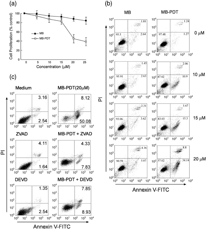
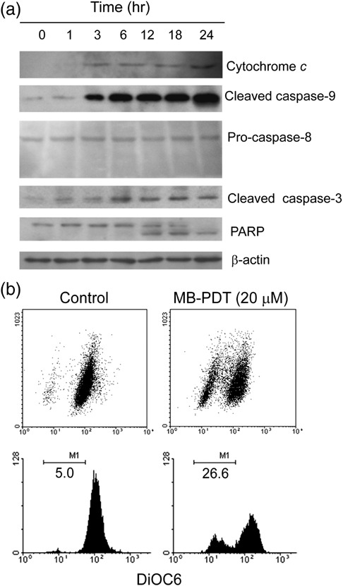
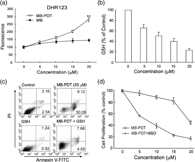
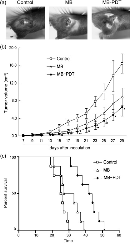
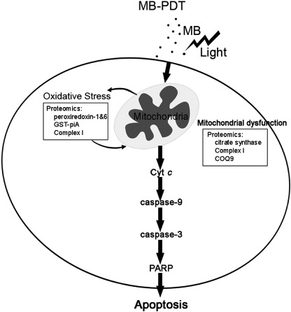
Methylene blue (MB) is a widely studied agent currently under investigation for its properties relating to photodynamic therapy (PDT). Recent studies have demonstrated that MB exhibits profound phototoxicity affecting a variety of tumor cell lines. However, the mechanistic explanation for methylene-blue-mediated photodynamic therapy (MB-PDT) in the context of melanoma therapy is still obscure. In the present study, B16F1 melanoma cells were treated by MB-PDT under different conditions, and thereafter subjected to cell viability detection assays. MB-PDT could induce intense apoptotic cell death through a series of steps beginning with the photochemical generation of reactive oxygen species that activate the caspase-9/caspase-3 apoptosis pathway. Blocking activation of caspase-3 and induction of oxidative stress by caspase inhibitor and by glutathione, respectively, markedly reduced apoptotic cell death in vitro. Importantly, proteomics study defining altered protein expression in treated cells suggests the involvement of several mitochondrial proteins playing important roles in electron transfer chain, implying mitochondrial dysfunction during the treatment. Furthermore, a transplantable mouse melanoma model was utilized to estimate the effectiveness of MB-PDT in vivo. The treated mice displayed decreased tumor size and prolonged survival days, which was associated with enhanced apoptotic cell death. These results, offering solid evidence of the induction of mitochondria-related apoptosis in tumor cells, reveal new aspects of MB-PDT having potential to be a palliative treatment of melanoma.


Figures

Figure 1
Figure 1
Photodynamic therapy (PDT) with methylene blue (MB) induces apoptosis in B16F1 cells. (a) Cell viability was determined by 3‐(4,5‐dimethylthiazol‐2‐yl)‐2,5‐diphenyl tetrazolium bromide (MTT) assay 18 h after treatment of MB‐PDT (ranging from 0 to 25 µM). Results are expressed as the percentage of viable cells compared with light alone‐treated control (mean ± SD, n = 3). (b) Apoptosis in B16F1 cells was assessed at 6 h post‐treatment by annexin V‐FITC/PI binding and measured by flow cytometry analysis. Numbers indicate the percentage of cells in each quadrant. (c) B16F1 cells were pretreated with 50 µM benzyloxycarbonyl‐Val‐Ala‐Asp (Z‐VAD‐FMK) or the caspase inhibitor Z‐DEVD‐FMK for 1 h followed by treatment with MB‐PDT, then apoptosis was determined by flow cytometry at 6 h post‐treatment.
Figure 2
Figure 2
Methylene‐blue‐mediated photodynamic therapy (MB‐PDT)‐mediated apoptosis is effected through the intrinsic apoptotic pathway. (a) The time‐dependent apoptosis was measured in B16F1 cells. For this analysis, cells were treated with MB‐PDT (20 µM), and then harvested at 1, 3, 6, 12, 18 and 24 h post‐PDT, respectively. The blots were probed with specific antibodies as shown. (b) Treated cells were stained with fluorochrome dye DiOC6 to determine the mitochondrial membrane potential (Δψm ) as indicated. Similar results were obtained in three independent experiments.
Figure 3
Figure 3
Methylene‐blue‐mediated photodynamic therapy (MB‐PDT)‐mediated apoptosis is oxidative stress dependent. (a) Reactive oxygen species (ROS) production was measured in treated cells labeled with DHR as indicated. Dose‐dependent increase in intracellular ROS production (mean ± SD) was shown. (b) Total cellular glutathione (GSH) levels after a 4‐h treatment of B16F1 cells with MB‐PDT. (c) Apoptosis was assessed at 6 h in B16F1 cells treated with 20 mM GSH for 1 h followed by treatment with MB‐PDT. (d) B16F1 cells were incubated with 10 µM DL‐buthionine (S,R)‐sulfoximine (BSO), followed by treatment with MB‐PDT. After the indicated time, cell death was measured by 3‐(4,5‐dimethylthiazol‐2‐yl)‐2,5‐diphenyl tetrazolium bromide (MTT) assay. Data are expressed as percent of the control.
Figure 4
Figure 4
Image of two‐dimensional (2‐D) gel stained with Coomassie Blue R‐250. B16F1 cells were treated with methylene‐blue‐mediated photodynamic therapy (MB‐PDT) (20 µM). 6 h after irradiation, the treated cells were lyzed in 4% CHAPS buffer and assayed for protein concentration. 1 mg of total protein was prepared for two‐dimensional gel separation. (a) Representative example of gel image from MB‐PDT treated group showing the entire pI range 3–10 (non‐linear) and molecular masses of 10–100 kDa. (b) Differential protein expression analysis with zoomed images of selected gel areas. The number in each zoomed image presents the reduction fold‐change compared with MB‐PDT. The selected subset of proteins, as listed in Table 1, is shown.
Figure 5
Figure 5
Methylene‐blue‐mediated photodynamic therapy (MB‐PDT) delayed tumor growth in mouse melanoma model. Mice with tumor were treated with the following treatments weekly for three weeks: light alone, MB alone, and MB‐PDT. Tumor volume was determined by direct measurement of tumor dimensions, using calipers. Results are presented as the mean tumor volume ± SEM (n = 8). (a) Morphology contrast of tumors from the three treated groups. (b) Tumor growth curve was expressed as the mean tumor diameters and calculated tumor volumes. (c) Survival plot of melanoma‐bearing mice after various treatments. The time of death, or when mice became moribund, was recorded and plotted as percentage of survival.
Figure 6
Figure 6
Methylene‐blue‐mediated photodynamic therapy (MB‐PDT) induces apoptotic cell death in vivo. Samples were gained from treated mice as described above. (a) In situ detection of apoptotic cells by fluorescence microscopy using the terminal 2′‐deoxyuridine‐5′‐triphosphate (dUTP) nick end labeling (TUNEL) assay. Bright‐field image (up), TUNEL reaction fluorescence (middle) and merge of both (down) sections from melanomas treated by MB‐PDT as indicated. (b) Immunohistochemical staining of melanoma sections using anti‐cleaved caspase‐3 antibody. Shown is 1 representative experiment of 3. Original magnifications: up ×100; down ×200.
Figure 7
Figure 7
Methylene‐blue‐mediated photodynamic therapy (MB‐PDT) mediated induction of apoptosis by mitochondria‐dependent pathway. MB seems to selectively bind to mitochondria, where it produces reactive oxygen species (ROS) after excitation in combination with light. Then ROS act on mitochondria to induce an imbalance between ROS production and antioxidant capacity, which can be proved by proteomics study of alterations of proteins, such as peroxiredoxin‐1 and 6, GST‐piA, complex I‐49 kDa and COQ9. Consequently, MB‐PDT mediated oxidative stress causes impairment of mitochondrial integrity and function, resulting in release of cytochrome c and loss of mitochondrial membrane potential (Δψm ), which ultimately leads to apoptotic cell death and tumor destruction.

References

    1. Dougherty TJ, Gomer CJ, Henderson BW et al . Photodynamic therapy. J Natl Cancer Inst 1998; 90: 889–905. - PMC - PubMed
    1. Almeida RD, Manadas BJ, Carvalho AP et al . Intracellular signaling mechanisms in photodynamic therapy. Biochim Biophys Acta 2004; 1704: 59–86. - PubMed
    1. Hopper C. Photodynamic therapy: a clinical reality in the treatment of cancer. Lancet Oncol 2000; 1: 212–19. - PubMed
    1. Dolmans DE, Fukumura D, Jain RK. Photodynamic therapy for cancer. Nat Rev Cancer 2003; 3: 380–7. - PubMed
    1. Chudnovsky Y, Khavari PA, Adams AE. Melanoma genetics and the development of rational therapeutics. J Clin Invest 2005; 115: 813–24. - PMC - PubMed

Publication types

MeSH terms