. 2008 Oct;99(10):2055-61.
doi: 10.1111/j.1349-7006.2008.00905.x.

Antisense hypoxia-inducible factor 1alpha gene therapy enhances the therapeutic efficacy of doxorubicin to combat hepatocellular carcinoma

Affiliations

Antisense hypoxia-inducible factor 1alpha gene therapy enhances the therapeutic efficacy of doxorubicin to combat hepatocellular carcinoma

Fengjun Liu et al. Cancer Sci. 2008 Oct.

Abstract

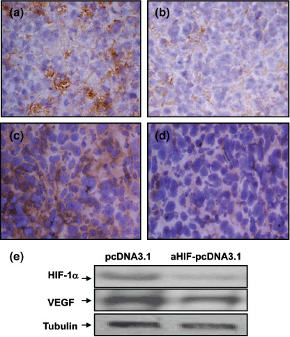
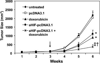
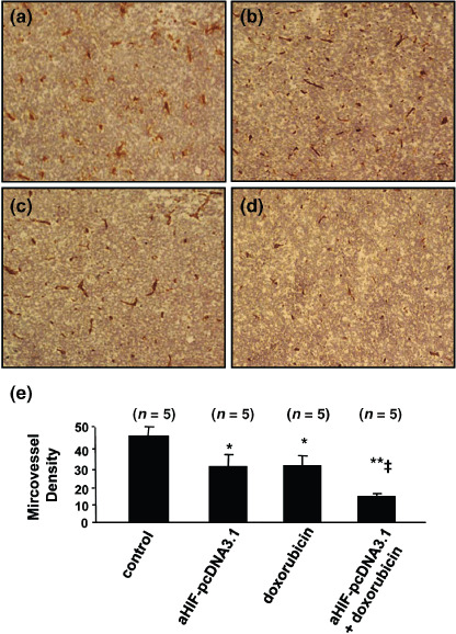
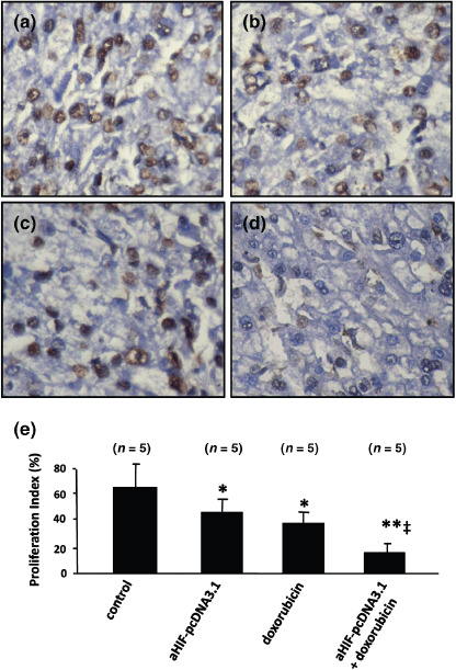
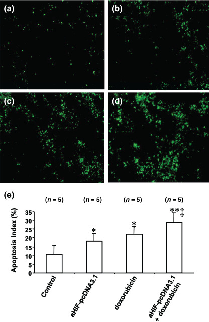
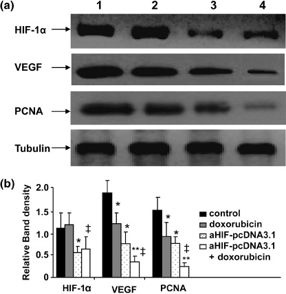
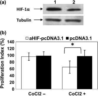
Hepatocellular carcinoma (HCC), one of the most common cancers worldwide, is resistant to anticancer drugs. Hypoxia is a major cause of tumor resistance to chemotherapy, and hypoxia-inducible factor (HIF)-1 is a key transcription factor in hypoxic responses. We have previously demonstrated that gene transfer of an antisense HIF-1alpha expression vector downregulates expression of HIF-1alpha and vascular endothelial growth factor (VEGF), and synergizes with immunotherapy to eradicate lymphomas. The aim of the present study was to determine whether gene transfer of antisense HIF-1alpha could enhance the therapeutic efficacy of doxorubicin to combat HCC. Both antisense HIF-1alpha therapy and doxorubicin suppressed the growth of subcutaneous human HepG2 tumors established in BALB/c nude mice, tumor angiogenesis, and cell proliferation, and induced tumor cell apoptosis. The combination therapy with antisense HIF-1alpha and doxorubicin was more effective in suppressing tumor growth, angiogenesis, and cell proliferation, and inducing cell apoptosis than the respective monotherapies. Gene transfer of antisense HIF-1alpha downregulated the expression of both HIF-1alpha and VEGF, whereas doxorubicin only downregulated VEGF expression. Antisense HIF-1alpha and doxorubicin synergized to downregulate VEGF expression. Both antisense HIF-1alpha and doxorubicin inhibited expression of proliferating cell nuclear antigen, and combined to exert even stronger inhibition of proliferating cell nuclear antigen expression. Antisense HIF-1alpha therapy warrants investigation as a therapeutic strategy to enhance the efficacy of doxorubicin for treating HCC.


Figures

Figure 1
Figure 1
Antisense hypoxia inducible factor (HIF)‐1α gene transfer downregulates expression of HIF‐1α and vascular endothelial growth factor (VEGF). Tumors of approximately 100 mm3 in volume were injected with either (a,c) pcDNA3.1, or (b,d) aHIF‐pcDNA3.1 plasmids. Representative tumor sections prepared 4 days after gene transfer, stained brown with antibodies against (a,b) HIF‐1α and (c,d) VEGF (×400 magnification). (e) Homogenates of tumors prepared 4 days after intratumoral injection of the pcDNA3.1 or aHIF‐pcDNA3.1 plasmids were western blotted with antibodies against HIF‐1α (upper panel), VEGF (middle panel), and tubulin (lower panel), as indicated.
Figure 2
Figure 2
Antisense hypoxia inducible factor (HIF)‐1α gene therapy synergizes with doxorubicin to suppress tumor growth. HepG2 hepatomas were established subcutaneously in the flanks of mice. When the tumors reached approximately 100 mm3 in volume (arrow), they were injected with the pcDNA3.1, and aHIF‐pcDNA3.1 plasmids, either alone or in combination with intraperitoneal injection of mice with doxorubicin, as indicated. Untreated tumors served as controls. The sizes (mm3) of tumors were monitored and recorded. *Significant and highly significant difference in tumor volumes compared with control. Significant difference compared with aHIF‐pcDNA3.1 or doxorubicin treatments.
Figure 3
Figure 3
Antisense hypoxia inducible factor (HIF)‐1α synergizes with doxorubicin to inhibit tumor angiogenesis. Representative tumor sections prepared 2 weeks after treatment from mice receiving (a) pcDNA3.1 (control), (b) aHIF‐pcDNA3.1, (c) doxorubicin, or (d) aHIF‐pcDNA3.1 + doxorubicin treatment. Tumor sections were stained with an anti‐CD31 antibody to view microvessels. (e) Tumor microvessels in sections were counted in blindly chosen random fields to record microvessel density. n, number of tumors assessed. *Significant difference in microvessel density for tumors treated with doxorubicin, or aHIF‐pcDNA3.1 versus control; **highly significant difference for tumors treated with doxorubicin + aHIF‐pcDNA3.1 versus control; significant difference for the combination therapy versus aHIF‐pcDNA3.1 or doxorubicin monotherapy.
Figure 4
Figure 4
Antisense hypoxia inducible factor (HIF)‐1α synergizes with doxorubicin to inhibit tumor cell proliferation. Representative tumor sections prepared 2 weeks after treatment from mice receiving (a) pcDNA3.1 (control), (b) aHIF‐pcDNA3.1, (c) doxorubicin, or (d) aHIF‐pcDNA3.1 + doxorubicin treatment. Tumor sections were stained with an anti‐Ki‐67 antibody to detect proliferating cells. (e) Cells expressing Ki‐67 were counted to calculate the proliferation index. n, number of tumors assessed. *Significant difference in the proliferation index for tumors treated with doxorubicin, or aHIF‐pcDNA3.1 versus control; **highly significant difference for tumors treated with doxorubicin + aHIF‐pcDNA3.1 versus control; significant difference for the combination therapy versus aHIF‐pcDNA3.1 or doxorubicin monotherapy.
Figure 5
Figure 5
Antisense hypoxia inducible factor (HIF)‐1α synergizes with doxorubicin to induce cell apoptosis. Representative tumor sections prepared 2 weeks after treatment from mice receiving (a) pcDNA3.1 (control), (b) aHIF‐pcDNA3.1, (c) doxorubicin, or (d) aHIF‐pcDNA3.1 + doxorubicin treatment. Tumor sections were stained with the terminal deoxynucleotidyl transferase biotin‐dUTP nick end labeling agent to view apoptotic cells. (e) Cells stained by the TUNEL agent were counted to calculate the apoptosis index. n, number of tumors assessed. *Significant difference in the apoptosis index for tumors treated with doxorubicin, or aHIF‐pcDNA3.1 versus control; **highly significant difference for tumors treated with doxorubicin + aHIF‐pcDNA3.1 versus control; and significant difference for the combination therapy versus aHIF‐pcDNA3.1 or doxorubicin monotherapy.
Figure 6
Figure 6
Antisense hypoxia inducible factor (HIF)‐1α and doxorubicin downregulate gene expression. (a) Homogenates of tumors from mice treated with pcDNA3.1 (lane 1), doxorubicin (lane 2), aHIF‐pcDNA3.1 (lane 3), or doxorubicin + aHIF‐pcDNA3.1 (lane 4) were western blotted with antibodies against HIF‐1α, vascular endothelial growth factor (VEGF), proliferating cell nuclear antigen (PCNA), and tubulin. The density of each band was measured and compared to that of the internal control tubulin. Significant difference in the band intensities of each protein for *pcDNA3.1 versus doxorubicin or aHIF‐pcDNA3.1; doxorubicin versus doxorubicin + aHIF‐pcDNA3.1; and **aHIF‐pcDNA3.1 versus doxorubicin + aHIF‐pcDNA3.1.
Figure 7
Figure 7
Antisense hypoxia inducible factor (HIF)‐1α gene transfection downregulates HIF‐1α expression and inhibits cell proliferation in vitro. (a) Lysates of HepG2 cells transfected with aHIF‐pcDNA3.1 (lane 2) and pcDNA3.1 (lane 1) were western blotted with antibodies against HIF‐1α and tubulin. (b) HepG2 cells transfected with aHIF‐pcDNA3.1 or pcDNA3.1 were cultured in the absence or presence of CoCl2 to mimic hypoxia. Untreated cells served as controls. Cell proliferation was assessed by the 3‐(4,5‐dimethylthiazol‐2‐yl)‐2,5‐diphenyl tetrazolium bromide method to calculate the proliferation index (% inhibition of cell proliferation). *Significant difference in the proliferation index.

References

    1. Befeler AS, Di Bisceglie AM. Hepatocellular carcinoma: diagnosis and treatment. Gastroenterology 2002; 122: 1609–19. - PubMed
    1. Parkin DM, Bray F, Ferlay J, Pisani P. Global cancer statistics, 2002. CA Cancer J Clin 2005; 55: 74–108. - PubMed
    1. Zhu AX. Systemic therapy of advanced hepatocellular carcinoma: how hopeful should we be? Oncologist 2006; 11: 790–800. - PubMed
    1. Sciarrino E, Simonetti RG, Le Moli S et al . Adriamycin treatment for hepatocellular carcinoma. Experience with 109 patients. Cancer 1985; 56: 2751–5. - PubMed
    1. Nerenstone SR, Ihde DC, Friedman MA. Clinical trials in primary hepatocellular carcinoma: current status and future directions. Cancer Treat Rev 1988; 15: 1–31. - PubMed

Publication types

MeSH terms

Substances