. 2009 Jan;11(1):170-82.
doi: 10.1111/j.1462-5822.2008.01247.x. Epub 2008 Oct 30.

Polarized response of endothelial cells to invasion by Aspergillus fumigatus

Affiliations

Polarized response of endothelial cells to invasion by Aspergillus fumigatus

Yasuki Kamai et al. Cell Microbiol. 2009 Jan.

Abstract

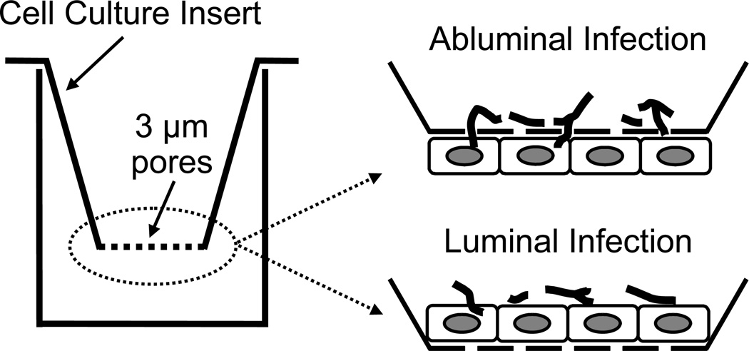
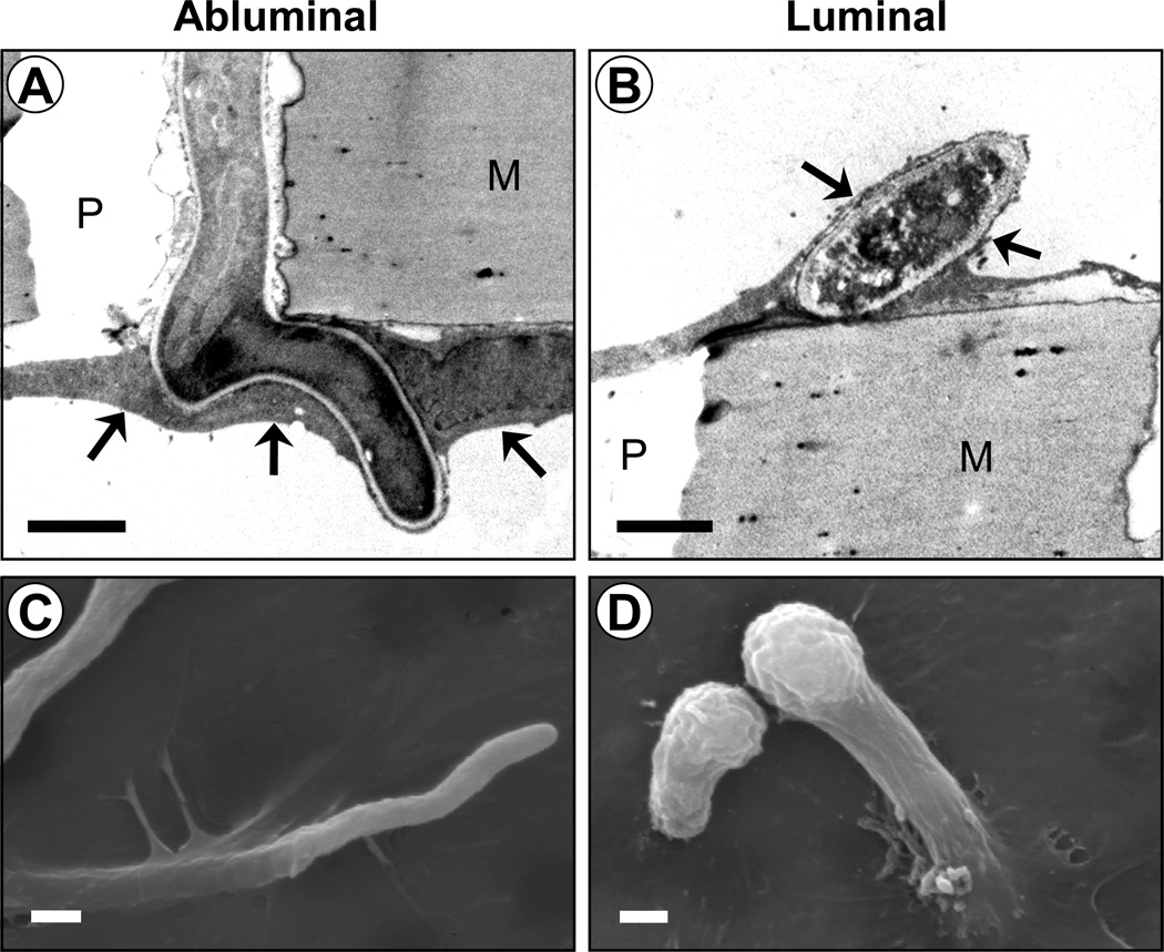
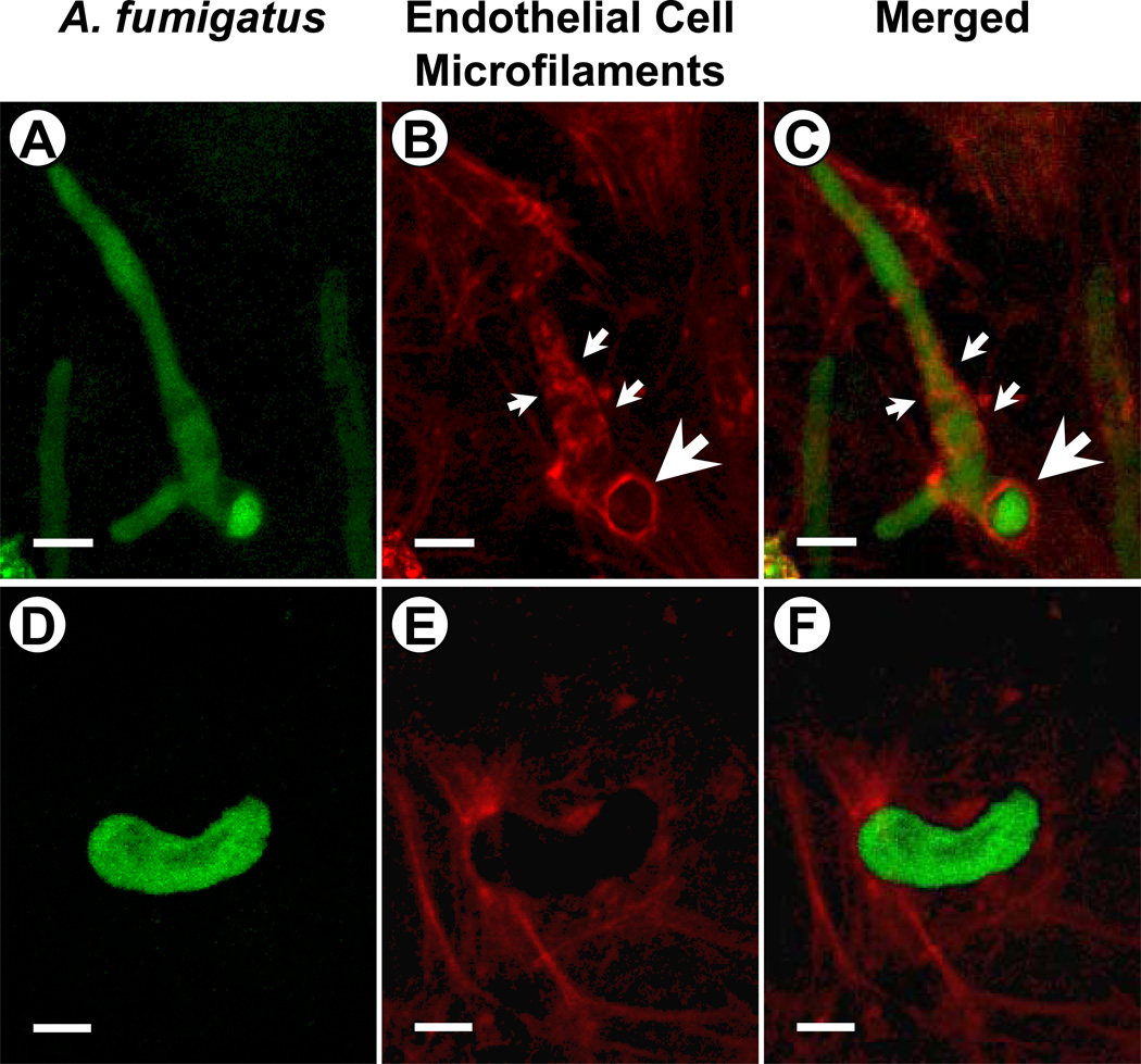
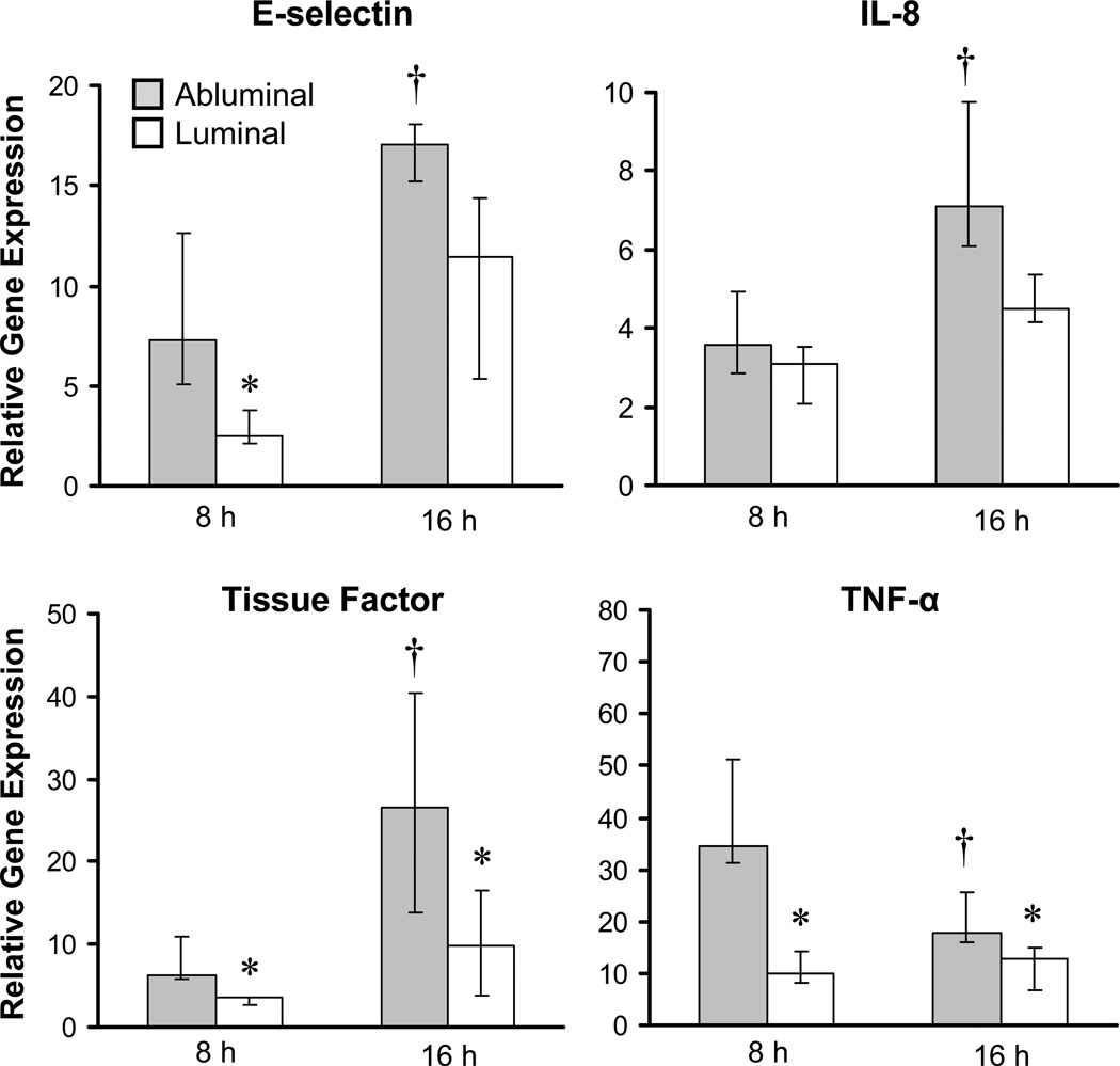
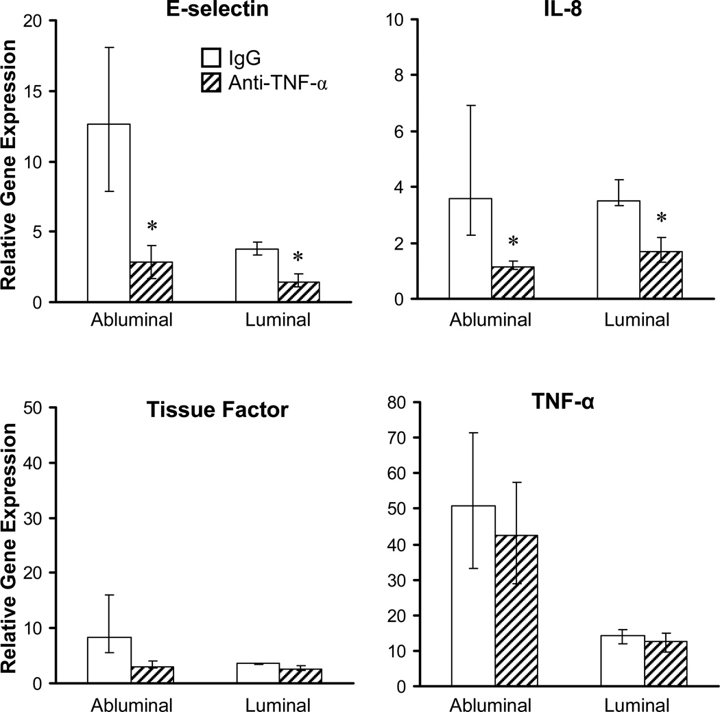
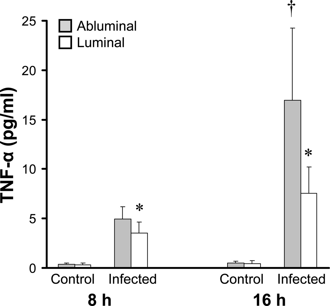
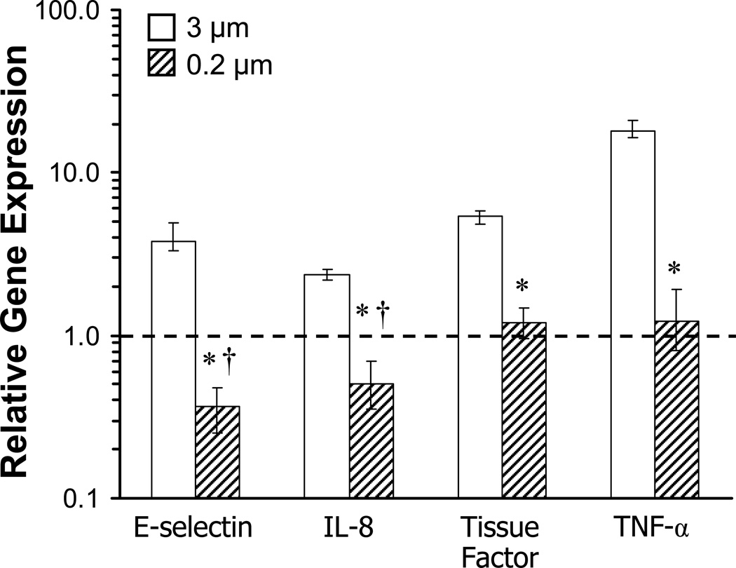
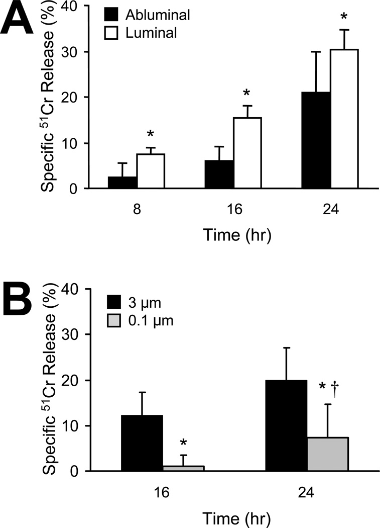
Hyphal invasion of blood vessels is a prominent feature of invasive aspergillosis. During invasive pulmonary aspergillosis, Aspergillus fumigatus hyphae invade the abluminal endothelial cell surface, whereas they invade the luminal endothelial cell surface during haematogenous dissemination. We investigated the endothelial cell response to abluminal and luminal infection with A. fumigatus hyphae in vitro. We found that these hyphae invaded the abluminal endothelial cell surface without inducing the formation of endothelial cell pseudopods. Also, the internalized hyphae were surrounded by a loose network of microfilaments. In contrast, A. fumigatus hyphae invaded the luminal endothelial cell surface by inducing by the formation of endothelial cell pseudopods. These endocytosed hyphae were surrounded by a tight network of microfilaments. Abluminal infection induced greater E-selectin, IL-8, tissue factor and TNF-alpha gene expression, but less endothelial cell damage than did luminal infection. Endothelial cell stimulation by infection of either surface was mediated by endothelial cell-derived TNF-alpha, and was not influenced by gliotoxin secreted by A. fumigatus. These differences in the endothelial cell response to abluminal versus luminal infection may contribute to differences in the pathogenesis of invasive versus haematogenously disseminated aspergillosis.


Figures

Fig. 1
Fig. 1
In vitro model of A. fumigatus infection of the abluminal and luminal endothelial cell surface. Abluminal endothelial cell invasion occurred 8 h after the A. fumigatus hyphae were added to the cell culture inserts, whereas luminal invasion occurred within 1 h after the hyphae were added to the cell culture inserts.
Fig. 2
Fig. 2
A. fumigatus hyphae invade the abluminal and luminal surface of endothelial cells by different mechanisms. Endothelial cells were infected with A. fumigatus hyphae via their abluminal surface for 8 h or their luminal surface for 1 h and then imaged by electron microscopy. Bars represent 2 µm. (A and B) Transmission electron micrographs of hyphae invading the abluminal surface (A) and luminal surface (B) of endothelial cells. Arrows in (A) indicate an endothelial cell that is being invaded by a hypha. Arrows in (B) indicate endothelial cell pseudopods surrounding an oblique section of a germling. M, membrane of the cell culture insert; P, pore of membrane of the cell culture insert. (C and D) Scanning electron micrographs of the luminal surface of the endothelial cells after abluminal (C) and luminal (D) infection.
Fig. 3
Fig. 3
Different patterns of microfilaments surround A. fumigatus hyphae during abluminal versus luminal invasion. Endothelial cells were incubated for 8 h (abluminal infection) and 1 h (luminal infection) with A. fumigatus hyphae expressing green fluorescent protein, stained for filamentous actin with Alexa Fluor 568-phalloidin, and then imaged by confocal microscopy. (A–C) Abluminal infection. Small arrows indicate the network of microfilaments surrounding an internalized hypha. Large arrows indicate a ring of microfilaments outlining a pore in the cell culture insert through which the organism has passed. (D–E) Luminal infection with a germling. Bars represent 5 µm.
Fig. 4
Fig. 4
Abluminal infection induces greater proinflammatory and procoagulant gene expression compared to luminal infection. Endothelial cells were infected abluminally or luminally with A. fumigatus hyphae for the indicated times, and then total endothelial cell RNA was extracted. E-selectin, IL-8, tissue factor, and TNF-α mRNA levels were measured using real-time RT-PCR. The expression of these genes was normalized to GAPDH, and results are expressed as the fold increase in expression relative to uninfected endothelial cells. Results are mean ± SD of at least 3 independent experiments. *P < 0.05 versus abluminal infection at the same time point; p < 0.015 vs 8 h of luminal infection.
Fig. 5
Fig. 5
Neutralization of TNF-α inhibits endothelial cell stimulation by A. fumigatus. Endothelial cells were infected abluminally and luminally with A. fumigatus hyphae for 8 h in the presence of either a neutralizing TNF-α monoclonal antibody or an isotype matched control antibody. Next, total endothelial cell RNA was collected, and mRNA levels of the indicated genes were measured using real-time RT-PCR. The expression of these genes was normalized to GAPDH, and results are expressed as the fold increase in expression relative to uninfected endothelial cells in the presence of the control antibody. Results are mean ± SD of 4 independent experiments. *P < 0.05 versus inhibition by the control antibody; P < 0.05 compared to abluminal infection.
Fig. 6
Fig. 6
Abluminal infection induces greater secretion of TNF-α compared to luminal infection. Endothelial cells were infected abluminally and luminally with A. fumigatus hyphae for the indicated times, after which the medium above and below the endothelial cells was collected. The concentration of TNF-α in the conditioned medium was determined by enzyme immunoassay. Results are the mean ± SD of at least 3 independent experiments, each performed in duplicate. *P < 0.05 compared to abluminal infection at the same time point, p < 0.001 compared to 8 h of luminal infection.
Fig. 7
Fig. 7
Effects of soluble factors released by A. fumigatus on endothelial cell stimulation. Endothelial cells were grown on the underside of cell culture inserts with either 3 µm or 0.2 µm pore size. When A. fumigatus hyphae were added to the upper side of the cell culture inserts, they could pass through the 3 µm pores, but not the 0.2 µm pores. After 16 h, total endothelial cell RNA was collected, and mRNA levels of the indicated genes were measured using real-time RT-PCR. The expression of these genes was normalized to GAPDH, and results are expressed as the fold change in expression relative to uninfected endothelial cells grown on cell culture inserts with the same pore size. Results are mean ± SD of 4 independent experiments. Dashed line indicates the level of gene expression in uninfected endothelial cells. *P < 0.01 compared to infected endothelial cells on 3 µm cell culture inserts. P < 0.02 compared to uninfected endothelial cells on 0.2 µm cell culture inserts.
Fig. 8
Fig. 8
Endothelial cell damage caused by A. fumigatus hyphae during abluminal and luminal infection. Endothelial cells were grown on the underside or upper side of cell culture inserts and loaded with 51Cr. They were incubated with A. fumigatus hyphae for indicated times, and the extent of endothelial cell damage was determined by the specific release of 51Cr into the medium. Results are means ± SD of at least 3 independent experiments, each performed in triplicate. (A) Time course of endothelial cell damage during abluminal and luminal infection. *P < 0.05 versus luminal infection. (B) Contribution of soluble factors released from A. fumigatus to endothelial cell damage during abluminal infection. Endothelial cells were grown on underside of cell culture inserts with 3 µm pores to permit abluminal invasion or with 0.1 µm pores to prevent abluminal invasion. A. fumigatus hyphae were added to the upper side of the inserts and incubated for the indicated times. *P < 0.05 versus cell culture inserts with 3 µm pores. P < 0.05 versus uninfected endothelial cells.

Similar articles

Cited by

References

    1. Allen LA, Aderem A. Molecular definition of distinct cytoskeletal structures involved in complement- and Fc receptor-mediated phagocytosis in macrophages. J Exp Med. 1996;184:627–637. - PMC - PubMed
    1. Barth PJ, Rossberg C, Koch S, Ramaswamy A. Pulmonary aspergillosis in an unselected autopsy series. Pathol Res Pract. 2000;196:73–80. - PubMed
    1. Birch M, Robson G, Law D, Denning DW. Evidence of multiple extracellular phospholipase activities of Aspergillus fumigatus. Infect Immun. 1996;64:751–755. - PMC - PubMed
    1. Bodey GP, Vartivarian S. Aspergillosis. Eur J Clin Microbiol Infect Dis. 1989;8:413–437. - PubMed
    1. Bok JW, Balajee SA, Marr KA, Andes D, Nielsen KF, Frisvad JC, Keller NP. LaeA, a regulator of morphogenetic fungal virulence factors. Eukaryot Cell. 2005;4:1574–1582. - PMC - PubMed

Publication types

MeSH terms

LinkOut - more resources