

Review
doi: 10.1111/j.1442-9071.2008.01823.x.
Epub 2008 Nov 5.
Clinical and research applications of anterior segment optical coherence tomography - a review
Affiliations
- PMID: 19016809
- PMCID: PMC2706099
- DOI: 10.1111/j.1442-9071.2008.01823.x
Item in Clipboard
Review
Clinical and research applications of anterior segment optical coherence tomography - a review
Clin Exp Ophthalmol.
2009 Jan.
Abstract
Optical coherence tomography (OCT) is being employed more and more often to image pathologies and surgical anatomy within the anterior segment, specifically in anterior chamber biometry, corneal pachymetric mapping, angle evaluation and high-resolution cross-sectional imaging. The cross-sectional imaging capability of OCT is similar to ultrasound, but its higher resolution allows OCT to measure and visualize very fine anatomic structures. No contact is required. In this review, we describe the utility and limitations of anterior segment OCT.
Figures
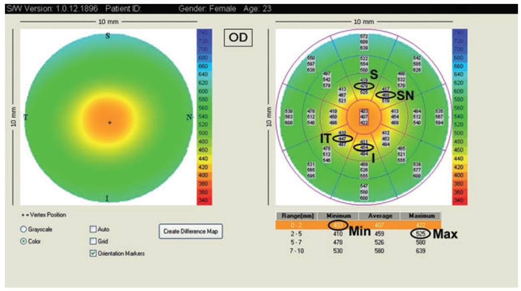
Visante optical coherence tomography pachymetry map and the parameters used to detect keratoconus. I, inferior; IT, inferotemporally; Max, maximum; Min, minimum; S, superior; SN, superonasal.
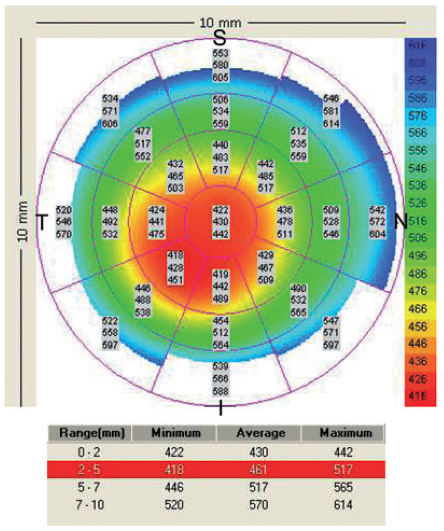
Visante pachymetry map of a keratoconic eye with a central thinning. I − S = 442 − 483 = −41 µm; IT − SN = 428 − 485 = −57 µm; Min = 422 µm; Min − Max = 422 − 517 = −99 µm. The pachymetry map was abnormal in two indices (IT–SN and Min); a third index was borderline (Min–Max), in that it fell below keratoconus diagnostic cut-off values. I, inferior; IT, inferotemporal; Max, maximum; Min, minimum; S, superior; SN, superonasal.
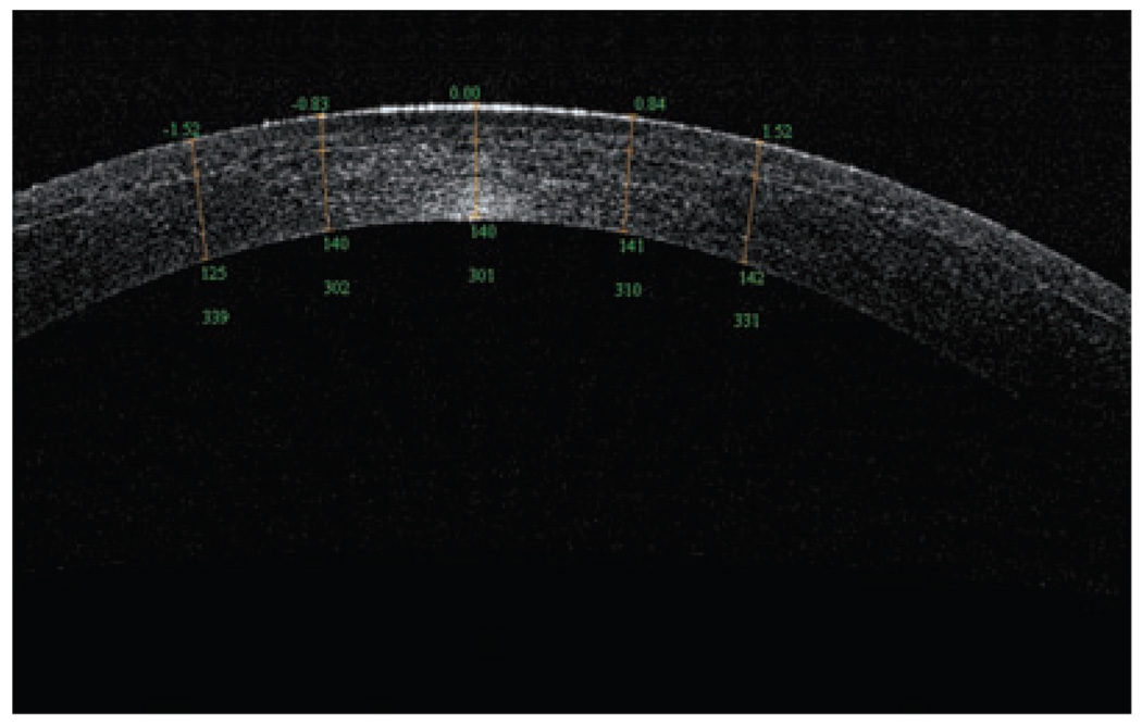
Optical coherence tomography scan (RTVue) of an Intra-laser flap, 1 week postoperative, with a 301-µm residual stromal bed.
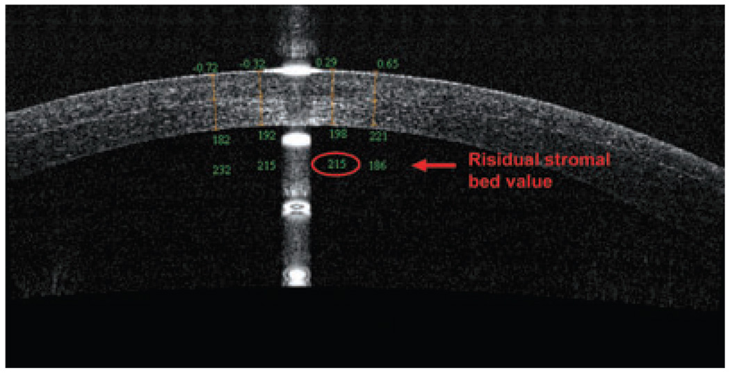
Optical coherence tomography scan (RTVue) of a right cornea with a thick flap and a thin residual stromal bed (<250 µm).
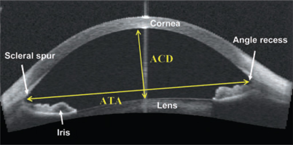
Visante optical coherence tomography of an anterior chamber measurement in a phakic patient. ACD, anterior chamber depth; ATA, angle-to-angle width.
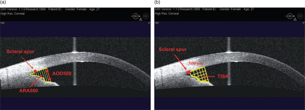
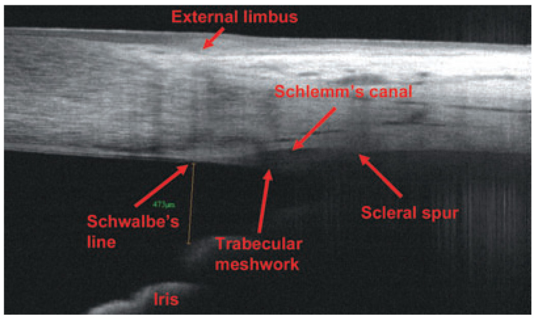
(a) Visante optical coherence tomography image of an anterior chamber angle: angle open distance (AOD) and angle recess area (ARA); (b) trabeculo-iris space area (TISA). Does not include the area posterior to the scleral spur.
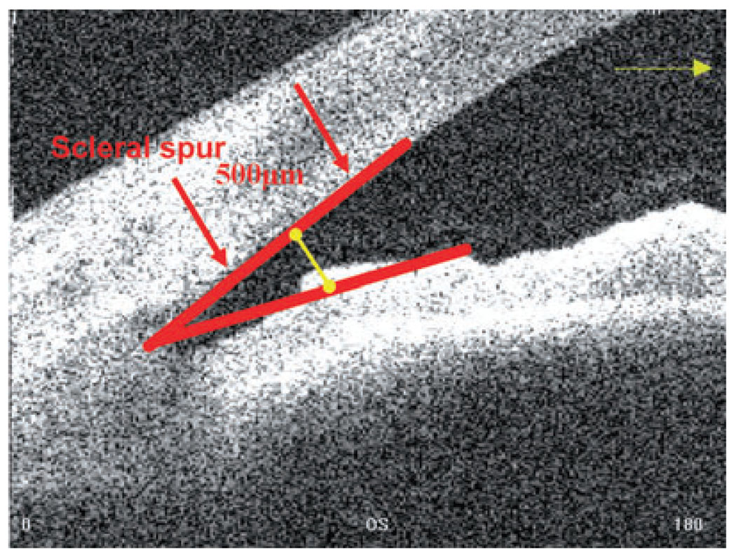
Anterior chamber angle, in degrees (Visante optical coherence tomography).
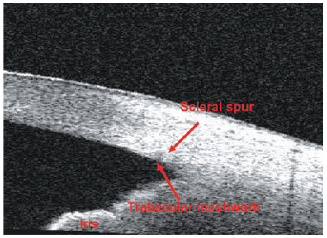
Time-domain optical coherence tomography (Visante) angle scan.
Fourier-domain optical coherence tomography (RTVue) angle scan.
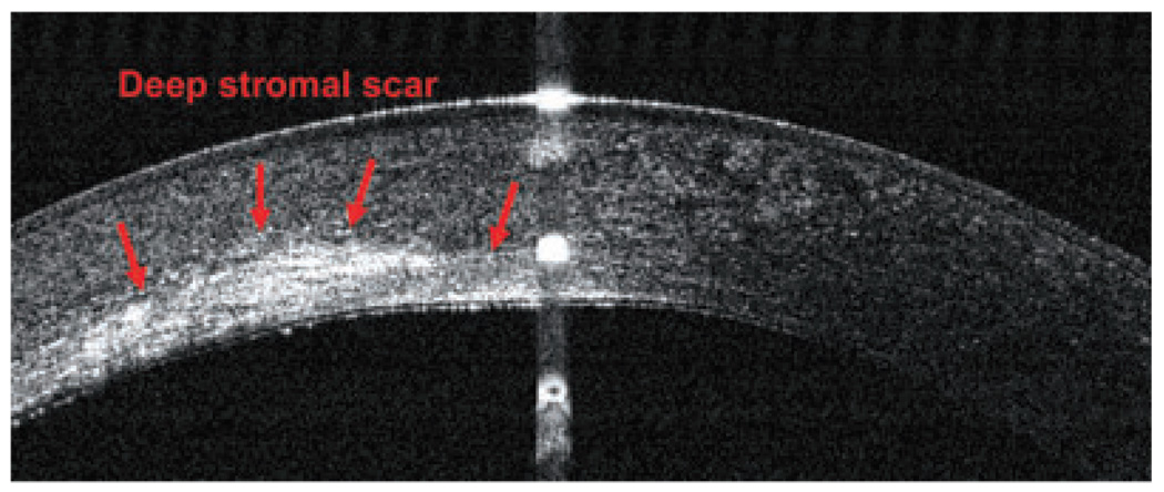
Optical coherence tomography image (RTVue) of a deep stromal scar in a left eye.
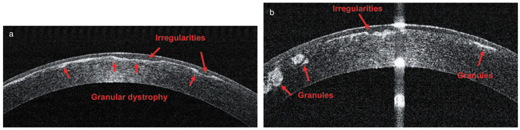
Granular dystrophy in both eyes of one patient: (a) right eye; (b) left eye (RTVue).
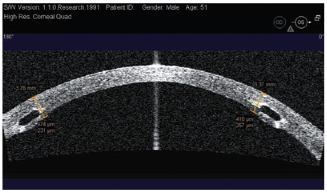
Horizontal Visante optical coherence tomography cross-section and measurement of the intrastromal ring depth. Both rings are placed at an adequate depth.
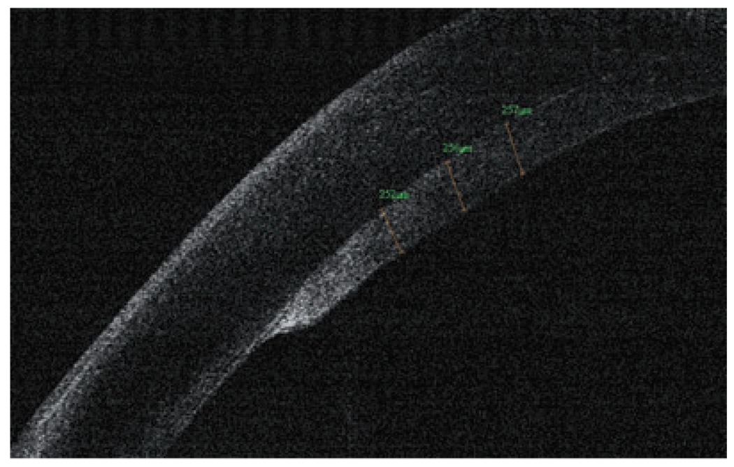
Optical coherence tomography graft measurement, 1 month after a successful descemet-stripping endothelial keratoplasty surgery (RTVue).
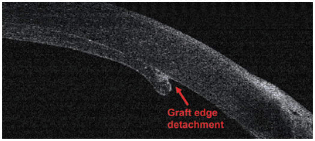
A small graft edge detachment after a descemet-stripping endothelial keratoplasty surgery (RTVue).
References
-
- Izatt JA, Hee MR, Swanson EA, et al. Micrometer-scale resolution imaging of the anterior eye in vivo with optical coherence tomography. Arch Ophthalmol. 1994;112:1584–1589. - PubMed
-
- Huang D, et al. Corneal Power Measurement with Optical Coherence Tomography; Paper presented at the Association for Research in Vision and Ophthalmology Meeting; Fort Lauderdale, FL. 2008.
-
- Ambrosio R, Jr, Alonso RS, Luz A, Coca Velarde LG. Corneal-thickness spatial profile and corneal-volume distribution: tomographic indices to detect keratoconus. J Cataract Refract Surg. 2006;32:1851–1859. - PubMed
-
- Khurana RN, Li Y, Tang M, Lai MM, Huang D. High-speed optical coherence tomography of corneal opacities. Ophthalmology. 2007;114:1278–1285. - PubMed
Publication types
MeSH terms
Grants and funding
LinkOut - more resources
Full Text Sources
Other Literature Sources
