Peripheral blood CD27+ IgG+ B cells rapidly proliferate and differentiate into immunoglobulin-secreting cells after exposure to low CD154 interaction
- PMID: 19016905
- PMCID: PMC2753896
- DOI: 10.1111/j.1365-2567.2008.02976.x
Peripheral blood CD27+ IgG+ B cells rapidly proliferate and differentiate into immunoglobulin-secreting cells after exposure to low CD154 interaction
Abstract
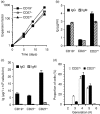
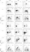
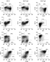
In vitro CD40 stimulation of human B cells isolated from lymphoid organs is dominated by memory B cells undergoing faster proliferation and higher differentiation than naive B cells. In contrast, we previously reported that blood memory B cells mainly differentiate into immunoglobulin-secreting cells in response to CD40 stimulation. However, variations in CD40-CD154 interaction are now recognized to influence B-cell fate. In this study, we have compared the in vitro response of blood CD27(-) and CD27(-) IgG(-) to CD27(+) and CD27(+) IgG(+) B cells following low-density exposure to CD154 in the presence of a mixture of interleukin-2 (IL-2), IL-4 and IL-10. The evolution of these cell populations was monitored during initiation and following long-term stimulation. Over a 5-day period, CD27(+) B cells underwent differentiation into immunoglobulin-secreting cells more readily than CD27(-) cells, and CD27(+) IgG(+) B cells gave rise to a near homogeneous population of CD19(+) CD27(++) CD38(+) IgG(lo) cells capable of high immunoglobulin G (IgG) secretion. During the same period, CD27(-) IgG(-) B cells partially became CD19(++) CD27(-) CD38(-) IgG(++) cells but showed no IgG secretion. Long-term stimulation revealed that CD27(+) IgG(+) B cells retained a high expansion capacity and could maintain their momentum towards differentiation over naive B cells. In addition, long-term stimulation was driving CD27(-) IgG(-) and total CD19(+) B cells to evolve into similar CD27(+) and CD27(-) subsets, suggesting naive homeostatic proliferation. Overall, these results tend to reconcile memory B cells from blood and lymphoid organs regarding their preferential differentiation capacity compared to naive cells, and further suggest that circulating memory IgG(+) cells may be intrinsically prone to rapid activation upon appropriate stimulation.
Figures


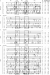
 ) and S (Silent;
  ) mutations is shown only when four or more mutated amino acids were observed. The proportions (%) of R mutations found in FR1, FR2 and FR3 (framework regions; cumulative length: 82 amino acids), and in CDR1 and CDR2 (complementarity determining regions; cumulative length: 22 amino acids) are shown for each sequence. *Clones considered to have arisen independently from antigen selection.

References
-
- Agematsu K, Hokibara S, Nagumo H, Komiyama A. CD27: a memory B-cell marker. Immunol Today. 2000;21:204–6. - PubMed
-
- Goodwin RG, Alderson MR, Smith CA, et al. Molecular and biological characterization of a ligand for CD27 defines a new family of cytokines with homology to tumor necrosis factor. Cell. 1993;73:447–56. - PubMed
-
- Kuppers R, Klein U, Hansmann ML, Rajewsky K. Cellular origin of human B-cell lymphomas. N Engl J Med. 1999;341:1520–9. - PubMed
Publication types
MeSH terms
Substances
LinkOut - more resources
Full Text Sources
Other Literature Sources
Research Materials

