. 2009 Sep;128(1 Suppl):e296-305.
doi: 10.1111/j.1365-2567.2008.02962.x. Epub 2008 Nov 7.

Expression of interferon-gamma and tumour necrosis factor-alpha messenger RNA does not correlate with protection in guinea pigs challenged with virulent Mycobacterium tuberculosis by the respiratory route

Affiliations

Expression of interferon-gamma and tumour necrosis factor-alpha messenger RNA does not correlate with protection in guinea pigs challenged with virulent Mycobacterium tuberculosis by the respiratory route

Amminikutty Jeevan et al. Immunology. 2009 Sep.

Abstract

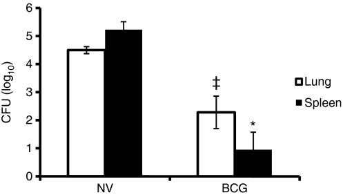
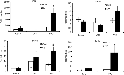
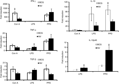
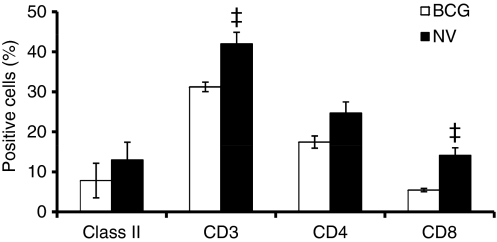
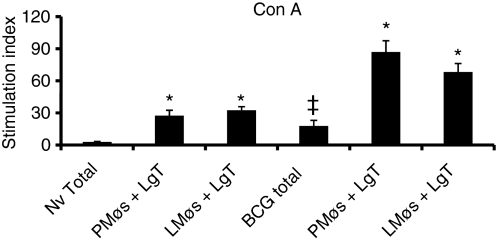
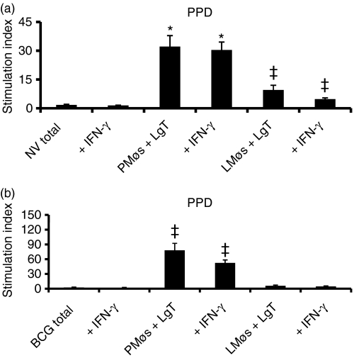
Cytokine messenger RNA (mRNA) expression was investigated in the spleen and lung digest cells of bacillus Calmette-Guérin (BCG)-vaccinated and non-vaccinated guinea pigs following low-dose, pulmonary exposure to virulent Mycobacterium tuberculosis. After purified protein derivative (PPD) stimulation, the levels of lung cell interferon-gamma (IFN-gamma), tumour necrosis factor-alpha (TNF-alpha) and spleen cell interleukin-12 (IL-12) p40 mRNAs were significantly increased in the non-vaccinated M. tuberculosis-infected guinea pigs compared to the BCG-vaccinated guinea pigs. In contrast, the expression of anti-inflammatory transforming growth factor-beta and IL-10 mRNAs was significantly enhanced in the spleens of BCG-vaccinated animals. Despite the presence of protective cytokine mRNA expression, the non-vaccinated guinea pigs had significantly higher lung and spleen bacterial burdens. In contrast, BCG-vaccinated guinea pigs controlled the bacterial multiplication in their lungs and spleens, indicating that both protective as well as anti-inflammatory cytokine responses are associated with a reduction in bacteria. In addition, lung digest cells from non-vaccinated guinea pigs contained a significantly higher percentage of neutrophils, CD3(+) and CD8(+) T cells, while the percentage of macrophages was increased in the BCG-vaccinated animals. Total and purified lung digest T cells co-cultured with lung macrophages (LMøs) proliferated poorly after PPD stimulation in both non-vaccinated and BCG-vaccinated animals while robust proliferation to PPD was observed when T cells were co-cultured with peritoneal macrophages (PMøs). Macrophages within the lung compartment appear to regulate the response of T cells irrespective of the vaccination status in guinea pigs. Taken together, our results suggest that type I cytokine mRNA expression is not associated with vaccine-induced protection in the low-dose guinea pig model of tuberculosis.


Figures

Figure 1
Figure 1
Effect of bacillus Calmette–Guérin (BCG) vaccination on lung and spleen colony-forming units (CFU). At 5 weeks after Mycobacterium tuberculosis infection, the spleen and the right lower lung lobe from non-vaccinated (NV) and BCG-vaccinated (BCG) guinea pigs were homogenized and appropriate dilutions were plated in duplicate onto Middlebrook 7H10 agar plates and incubated at 37° for 3 weeks. The number of CFU was counted and the results were calculated as mean log10 CFU per tissue. *P< 0·0007 and P< 0·01 when compared with non-vaccinated group as determined by Student’s t-test.
Figure 2
Figure 2
Cytokine messenger RNA (mRNA) expression in guinea pig lung digest cells. Total lung digest cells from non-vaccinated (NV) and bacillus Calmette–Guérin-vaccinated (BCG) guinea pigs 5 weeks following Mycobacterium tuberculosis infection were stimulated with Concanavalin A (Con A; 10 μg/ml), lipopolysaccharide (LPS; 1 μg/ml) or purified protein derivative (PPD; 25 μg/ml) for 24 hr. Interferon-γ (IFN-γ), tumour necrosis factor-α (TNF-α) mRNA, transforming growth factor-β (TGF-β) and interleukin-10 (IL-10) mRNA expression was quantified using real-time reverse transcription–polymerase chain reaction. Fold induction of mRNA was calculated from the threshold cycle values (Ct) normalized to HPRT Ct values and then to unstimulated cultures. The results are expressed as mean and the standard errors of means from five animals. The differences in the fold induction between cultures from non-vaccinated and BCG-vaccinated animals were determined by anova (P< 0·05).
Figure 3
Figure 3
Cytokine messenger RNA (mRNA) expression in guinea pig spleen cells. Total spleen cells from non-vaccinated (NV) and bacillus Calmette–Guérin-vaccinated (BCG) guinea pigs 5 weeks following Mycobacterium tuberculosis infection were stimulated with concanavalin A (Con A), lipopolysaccharide (LPS) or purified protein derivative (PPD) for 24 hr as described above. Interferon-γ (IFN-γ), tumour necrosis factor-α (TNF-α), transforming growth factor-β (TGF-β), interleukin-10 (IL-10) and IL-12p40 mRNA expression was quantified using real-time reverse transcription–polymerase chain reaction. Fold induction of mRNA was calculated as described in Fig. 2. The results are expressed as mean and the standard errors of means from five animals. The differences in the fold induction between the cultures from non-vaccinated and BCG-vaccinated groups were determined by anova (P< −0·05).
Figure 4
Figure 4
Phenotypic analysis of guinea pig lung digest cells. Lung cells obtained by enzymatic digestion from non-vaccinated (NV) and bacillus Calmette–Guérin-vaccinated (BCG) guinea pigs 5 weeks after infection with virulent Mycobacterium tuberculosis was characterized by flow cytometry. Proportions of major histocompatibility complex (MHC) class II+ cells, T cells and their subsets were determined by fluorescence-activated cell sorting analysis after staining the cells with monoclonal antibodies directed against the surface markers of MHC class II+ cells, T cells (CT5), CD4+ T cells (CT7), and CD8+ T cells (CT6). Results are expressed as % positive cells. P< 0·01 compared with BCG-vaccinated group as determined by Student’s t-test.
Figure 5
Figure 5
Proliferation of total lung digest cells and purified T cells to concanavalin A (Con A). Proliferation of total (2 × 10⁶/ml) lung digest cells (Total) and co-cultures of lung T (LgT) cells (3 × 10⁵/well) and peritoneal (PMøs) or lung (LMøs) macrophages (1 × 10⁵/well) from non-vaccinated (NV) and bacillus Calmette–Guérin-vaccinated (BCG) guinea pigs following Mycobacterium tuberculosis infection was assessed. Cells cultured for 4 days in the presence of Con A (10 μg/ml) were harvested after the addition of [3H]thymidine (1 μg/well) 6 hr earlier. The results are expressed as stimulation index calculated by dividing the counts per minute (c.p.m.) of stimulated cells by the c.p.m. of unstimulated cells. The results are the mean and the standard error of the mean from three experiments. There were five animals per group. *P< 0·001 in comparisons between total and co-cultures from non-vaccinated and BCG-vaccinated groups by anova. P< 0·05 when compared to the response in total cells from non-vaccinated guinea pigs.
Figure 6
Figure 6
Proliferation of total lung digest cells and purified T cells to purified protein derivative (PPD). Total lung cells (Total) or co-cultures of purified T (LgT) cells and peritoneal (PMøs) or lung (LMøs) macrophages from non-vaccinated (NV, a) and BCG-vaccinated (BCG, b) guinea pigs 5 weeks following Mycobacterium tuberculosis infection were stimulated with PPD (25 μg/ml) for 4 days and the [3H]thymidine uptake was assessed. The cell populations were also treated with recombinant guinea pig interferon-γ (IFN-γ; 200 ng/ml) at the beginning of the culture period. The results represent mean ± SEM from 5–10 animals. Results are expressed as stimulation index. *P< 0·001 and P< 0·05 when compared with total cells as determined by anova.

References

    1. Kaufmann S. Towards new leprosy and tuberculosis vaccines. Microbiol Sci. 1987;4:324–8. - PubMed
    1. Comstock GW. Epidemiology of tuberculosis. Am Rev Respir Dis. 1982;125:8–15. - PubMed
    1. Young LS, Inderlied CB, Berlin OG, Gottlieb MS. Mycobacterial infections in AIDS patients, with an emphasis on the Mycobacterium avium complex. Rev Infect Dis. 1986;8:1024–33. - PubMed
    1. Gandhi NR, Moll A, Sturm AW, et al. Extensively drug-resistant tuberculosis as a cause of death in patients co-infected with tuberculosis and HIV in a rural area of South Africa. Lancet. 2006;368:1575–80. - PubMed
    1. Smith D, Harding G, Chan J, Edwards M, Hank J, Muller D, Sobhi F. Potency of 10 BCG vaccines as evaluated by their influence on the bacillemic phase of experimental airborne tuberculosis in guinea pigs. J Biol Stand. 1979;7:179–97. - PubMed

Publication types

MeSH terms