

In vivo imaging demonstrates a time-line for new vessel formation in islet transplantation
- PMID: 19017287
- PMCID: PMC2837508
- DOI: 10.1111/j.1399-3046.2008.01088.x
In vivo imaging demonstrates a time-line for new vessel formation in islet transplantation
Abstract
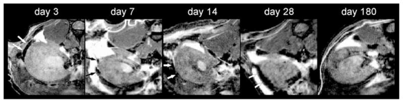
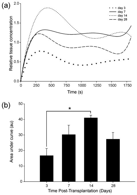
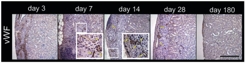
Vascularization of transplanted islets must be maintained to provide long-term graft function. In vivo assessment of new vessel formation in islet grafts has been poorly documented. The purpose of this study was to investigate whether neovascularization was detectable in vivo in a Feridex-labeled murine syngeneic subcapsular islet mass using DCE MRI over 180 days. Subcapsular transplants could be visualized at post-transplant days three, seven, 14, and 28 using T2-weighted MRI and at post-transplant day 180 by immunohistochemistry. Injection of the contrast agent gadolinium (Gd)-DTPA for DCE at three, seven, and 14 days showed increased signal in the transplant area consistent with new vessel formation. Areas under contrast enhancement curves suggested peak angiogenesis at 14 days. At 180 days, there was no observable change in signal intensity after contrast injection suggesting established vascularization or islet mass reduction. Immunohistochemistry confirmed MRI and DCE findings. These data suggest that islet angiogenesis occurs early after transplantation and is likely established after one month of transplantation. This study provides an in vivo time-line of neovascularization in subcapsular islet grafts. We anticipate that contrast extravasation captured by MRI may provide useful monitoring of graft angiogenesis if reproduced in a clinically relevant intraportal model.
Figures

References
-
- Shapiro AM, Lakey JR, Ryan EA, et al. Islet transplantation in seven patients with type 1 diabetes mellitus using a gluco-corticoid-free immunosuppressive regimen. N Engl J Med. 2000;343:230–238. - PubMed
-
- Ryan EA, Paty BW, Senior PA, et al. Five-year follow-up after clinical islet transplantation. Diabetes. 2005;54:2060–2069. - PubMed
-
- Davalli AM, Ogawa Y, Ricordi C, Scharp DW, Bonner -Weir S, Weir GC. A selective decrease in the beta cell mass of human islets transplanted into diabetic nude mice. Transplantation. 1995;59:817–820. - PubMed
-
- Carlsson PO, Palm F, Mattsson G. Low revascularization of experimentally transplanted human pancreatic islets. J Clin Endocrinol Metab. 2002;87:5418–5423. - PubMed
-
- Mattsson G, Jansson L, Carlsson PO. Decreased vascular density in mouse pancreatic islets after transplantation. Diabetes. 2002;51:1362–1366. - PubMed
Publication types
MeSH terms
Substances
Grants and funding
LinkOut - more resources
Full Text Sources
Other Literature Sources
Medical
