. 2009 Jan;50(1):78-89.
doi: 10.1093/pcp/pcn173. Epub 2008 Nov 18.

Multiple PLDs required for high salinity and water deficit tolerance in plants

Affiliations

Multiple PLDs required for high salinity and water deficit tolerance in plants

Bastiaan O R Bargmann et al. Plant Cell Physiol. 2009 Jan.

Abstract

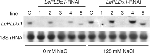
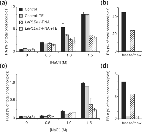
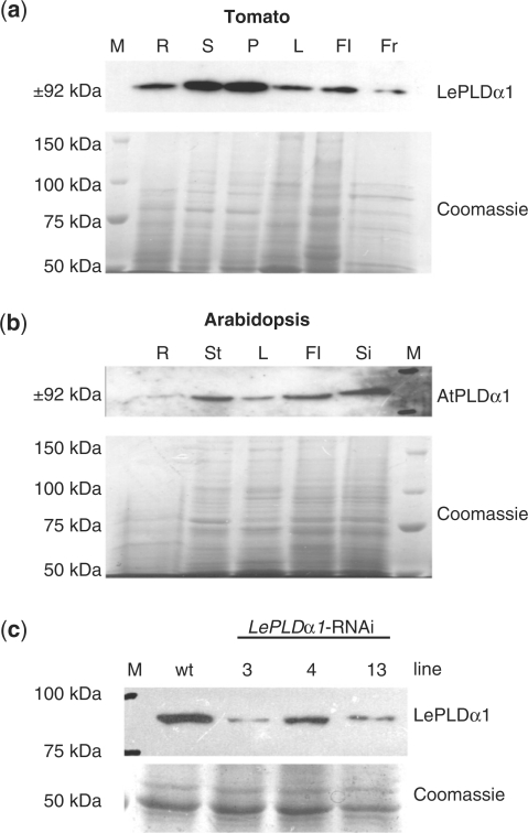
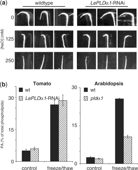
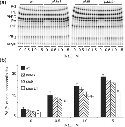
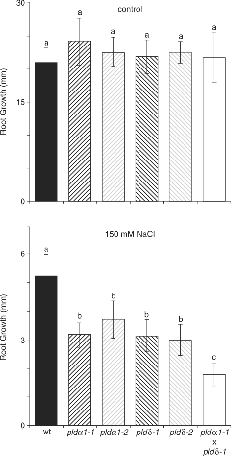
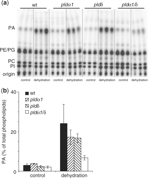
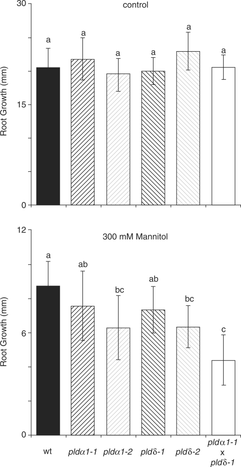
High salinity and drought have received much attention because they severely affect crop production worldwide. Analysis and comprehension of the plant's response to excessive salt and dehydration will aid in the development of stress-tolerant crop varieties. Signal transduction lies at the basis of the response to these stresses, and numerous signaling pathways have been implicated. Here, we provide further evidence for the involvement of phospholipase D (PLD) in the plant's response to high salinity and dehydration. A tomato (Lycopersicon esculentum) alpha-class PLD, LePLDalpha1, is transcriptionally up-regulated and activated in cell suspension cultures treated with salt. Gene silencing revealed that this PLD is indeed involved in the salt-induced phosphatidic acid production, but not exclusively. Genetically modified tomato plants with reduced LePLDalpha1 protein levels did not reveal altered salt tolerance. In Arabidopsis (Arabidopsis thaliana), both AtPLDalpha1 and AtPLDdelta were found to be activated in response to salt stress. Moreover, pldalpha1 and plddelta single and double knock-out mutants exhibited enhanced sensitivity to high salinity stress in a plate assay. Furthermore, we show that both PLDs are activated upon dehydration and the knock-out mutants are hypersensitive to hyperosmotic stress, displaying strongly reduced growth.


Figures

Fig. 1.
Fig. 1.
Silencing and salt-induced expression of LePLDα1 in tomato cell suspension cultures. RNA was extracted from independently transformed Msk8 cultures carrying an LePLDα1-RNAi construct (lines #1–5) or an empty control vector (C) that had been treated with 0 or 125 mM NaCl for 5 h. RNA was separated by gel electrophoresis, blotted and hybridized with a 32P-labeled LePLDα1 probe. An 18S rRNA probe was used as a loading control.
Fig. 2.
Fig. 2.
Salt-induced LePLDα1 activity in tomato cell suspension cultures. 32Pi-labeled control or LePLDα1-silenced (line 1) cell suspensions were left untreated, snap-frozen and thawed or treated with an equal volume of cell-free medium supplemented with increasing NaCl concentrations for 15 min, either without buffer or buffered with 50 mM Tris–HCl pH 7.5 and 10 mM EGTA (TE). Lipids were extracted, separated by TLC and analyzed by phosphoimaging. PA (a and b) and PBut (c and d) were quantified as a percentage of total radiolabeled lipids and are presented in a histogram (salt treatment n = 2, min and max values indicated; freeze/thaw n = 1).
Fig. 3.
Fig. 3.
Silencing LePLDα1 in tomato plants. (a) Proteins were extracted from roots (R), stems (S), petioles (P), leaves (L), flowers (Fl) and fruit (Fr) harvested from mature tomato plants. Proteins were separated by SDS–PAGE and blotted or stained with Coomassie brilliant blue as a loading control. A precision protein marker (M) was used to gauge the size of the detected band. (b) Protein blot analysis of AtPLDα1 protein levels was performed on proteins extracted from roots (R), inflorescence stems (St), leaves (L), flowers (Fl) and siliques (Si) of flowering Arabidopsis plants. (c) Protein blot analysis of LePLDα1 protein levels was performed on proteins extracted from 1-week-old wild-type (wt) and LePLDα1-silenced tomato seedlings.
Fig. 4.
Fig. 4.
Salt tolerance and lysis-induced PLD activity in LePLDα1-silenced tomato plants. (a) Tomato seeds from wild-type and LePLDα1-silenced plant lines were sown on agar plates and grown vertically in a growth chamber. After 1 week, seedlings were transferred to fresh plates supplemented with 0, 125 or 250 mM NaCl, rotated 180° and placed back in the growth chamber. Plates were scanned after 24 h. A representative silenced line is shown (line #3). (b) Leaf discs were excised from fully expanded leaves from wild-type and LePLDα1-silenced (line #13) tomato plants as well as wild-type and pldα1 knock-out Arabidopsis plants, and labeled overnight with 32Pi. Leaf discs were either left untreated or snap-frozen and thawed for 15 min. Lipids were extracted, separated by TLC and analyzed by phosphoimaging. PA was quantified as a percentage of total phospholipids and is presented in histograms ± SD (n = 3).
Fig. 5.
Fig. 5.
Salt-induced PLD activity in Arabidopsis T-DNA insertion lines. Leaf discs were excised from fully expanded leaves from control and pld knock-out plant lines and labeled overnight with 32Pi. Leaf discs were treated with increasing NaCl concentrations for 15 min. Lipids were extracted, separated by alkaline TLC (a) and analyzed by phosphoimaging (b). PA was quantified as a percentage of total radiolabeled lipids and is presented in a histogram ± SD (n = 3).
Fig. 6.
Fig. 6.
Reduced salt tolerance in Arabidopsis pld mutants. Seeds from wild-type, pldα1-1, pldα1-2, pldδ-1, pldδ-2 and pldα1-1/pldδ-1 knock-out mutant lines were sown on agar plates and grown vertically in a growth chamber. After 4 d, seedlings were transferred to fresh plates supplemented with 0 or 150 mM NaCl. Primary root growth was measured 4 d after transfer and is represented in histograms ± SD. Data were analyzed for significance by one-way ANOVA (Tukey post hoc, α = 0.001, n = 18–20).
Fig. 7.
Fig. 7.
Dehydration-induced PLD activity in Arabidopsis T-DNA insertion lines. Leaf discs were excised from fully expanded leaves from control and pld knock-out plant lines and labeled overnight with 32Pi. Leaf discs were treated by removing them from the labeling solution and placing them on filtration paper for 2 h. Lipids were extracted, separated by ethyl acetate TLC (a) and analyzed by phosphoimaging (b). PA was quantified as a percentage of total radiolabeled lipids and is presented in a histogram ± SD (n = 3).
Fig. 8.
Fig. 8.
Increased sensitivity to hyperosmotic stress in Arabidopsis pld mutants. Seeds from wild-type, pldα1-1, pldα1-2, pldδ-1, pldδ-2 and pldα1-1/pldδ-1 knock-out mutant lines were sown on agar plates and grown vertically in a growth chamber. After 4 d, seedlings were transferred to fresh plates supplemented with or without 300 mM mannitol. Primary root growth was measured 4 d after transfer and is represented in histograms ± SD. Data were analyzed for significance by one-way ANOVA (Tukey post hoc, α = 0.001, n = 15–16).

References

    1. Bargmann BOR, Munnik T. The role of phospholipase D in plant stress responses. Curr. Opin. Plant Biol. 2006;9:515–522. - PubMed
    1. Bargmann BOR, Laxalt AM, ter Riet B, Schouten E, van Leeuwen W, Dekker HL, et al. LePLDbeta1 activation and relocalization in suspension-cultured tomato cells treated with xylanase. Plant J. 2006;45:58–68. - PubMed
    1. Boudsocq M, Barbier-Brygoo H, Lauriere C. Identification of nine sucrose nonfermenting 1-related protein kinases 2 activated by hyperosmotic and saline stresses in Arabidopsis thaliana. J. Biol. Chem. 2004;279:41758–41766. - PubMed
    1. Boudsocq M, Laurière C. Osmotic signaling in plants: multiple pathways mediated by emerging kinase families. Plant Physiol. 2005;138:1185–1194. - PMC - PubMed
    1. Burza AM, Pekala I, Sikora J, Siedlecki P, Małagocki P, Bucholc M, et al. Nicotiana tabacum osmotic stress-activated kinase is regulated by phosphorylation on Ser-154 and Ser-158 in the kinase activation loop. J. Biol. Chem. 2006;281:34299–34311. - PubMed

Publication types

MeSH terms