. 2008 Nov 25;105(47):18366-71.
doi: 10.1073/pnas.0803437105. Epub 2008 Nov 18.

The Wnt modulator sFRP2 enhances mesenchymal stem cell engraftment, granulation tissue formation and myocardial repair

Affiliations

The Wnt modulator sFRP2 enhances mesenchymal stem cell engraftment, granulation tissue formation and myocardial repair

Maria P Alfaro et al. Proc Natl Acad Sci U S A. .

Abstract

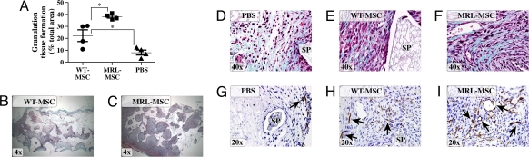
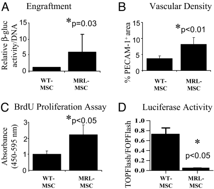
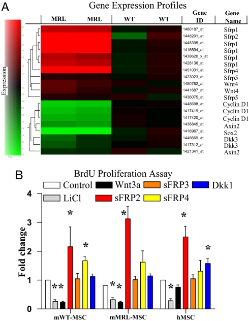
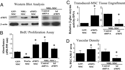
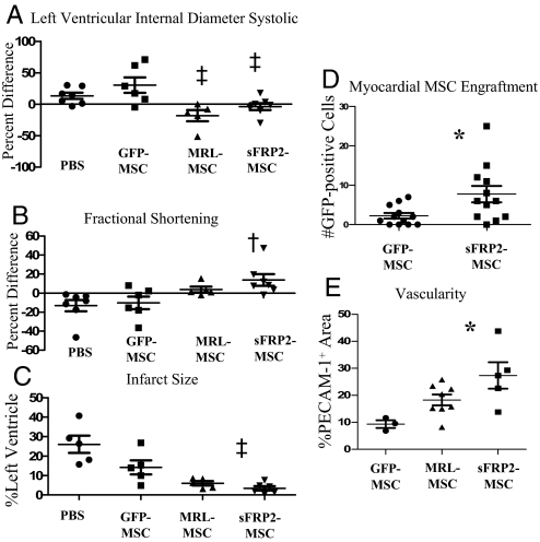
Cell-based therapies, using multipotent mesenchymal stem cells (MSCs) for organ regeneration, are being pursued for cardiac disease, orthopedic injuries and biomaterial fabrication. The molecular pathways that regulate MSC-mediated regeneration or enhance their therapeutic efficacy are, however, poorly understood. We compared MSCs isolated from MRL/MpJ mice, known to demonstrate enhanced regenerative capacity, to those from C57BL/6 (WT) mice. Compared with WT-MSCs, MRL-MSCs demonstrated increased proliferation, in vivo engraftment, experimental granulation tissue reconstitution, and tissue vascularity in a murine model of repair stimulation. The MRL-MSCs also reduced infarct size and improved function in a murine myocardial infarct model compared with WT-MSCs. Genomic and functional analysis indicated a downregulation of the canonical Wnt pathway in MRL-MSCs characterized by significant up-regulation of specific secreted frizzled-related proteins (sFRPs). Specific knockdown of sFRP2 by shRNA in MRL-MSCs decreased their proliferation and their engraftment in and the vascular density of MRL-MSC-generated experimental granulation tissue. These results led us to generate WT-MSCs overexpressing sFRP2 (sFRP2-MSCs) by retroviral transduction. sFRP2-MSCs maintained their ability for multilineage differentiation in vitro and, when implanted in vivo, recapitulated the MRL phenotype. Peri-infarct intramyocardial injection of sFRP2-MSCs resulted in enhanced engraftment, vascular density, reduced infarct size, and increased cardiac function after myocardial injury in mice. These findings implicate sFRP2 as a key molecule for the biogenesis of a superior regenerative phenotype in MSCs.


Conflict of interest statement

The authors declare no conflict of interest.

Figures

Fig. 1.
Fig. 1.
MRL-MSCs generated more advanced wound granulation tissue. (A) Graph of granulation tissue area in representative histologic sponge sections as a percentage of total sponge area. One-way ANOVA with Bonferrroni correction was used to compare data between WT vs. MRL or PBS, n = 4 in each group. Representative low power Trichrome images show decreased granulation tissue in WT- (B) vs. MRL- (C) MSC-loaded sponges. (D–F) High power Trichrome images show that MSC-loaded sponges (E and F) were more organized, highly cellular and with abundant type I collagen (blue) as compared with PBS control (D). (G–I) Representative immunostained sections using anti-PECAM-1 to designate vascular density from PBS-loaded (G), WT (H), or MRL (I) MSC-derived sponge granulation tissue. SP, sponge matrix, arrows point at positive stain, *, P < 0.05.
Fig. 2.
Fig. 2.
MRL-MSCs showed higher engraftment, vascularity. (A) β-gluc-specific activity (MSC marker) normalized to total cellular DNA content of paired WT- and MRL-MSC sponge granulation tissue samples were calculated and the average of the fold change for n = 7 animals were graphed to represent engrafted MSCs. (B) Vascular density graphed as percentage of immunopositive PECAM-1 area/total tissue area in histologic sections from granulation tissue. Data represents averages of multiple 40X fields from unpaired samples (n = 6). (C) In vitro proliferation of WT- and MRL-MSCs using BrdU ELISA; n = 5 experiments performed in triplicate. (D) Basal normalized luciferase activity; n = 2 experiments performed in duplicate. Unpaired Student's t test was used.
Fig. 3.
Fig. 3.
MRL-MSCs demonstrated a downregulation of canonical Wnt pathway by up-regulation of sFRPs. (A) Expression of known Wnt pathway genes (rows) in MSCs isolated from WT and MRL mice in duplicate experiments, from low (green) to high (red). Gene tree (to the left of the rows) corresponds to the degree of similarity of the pattern of expression for genes. (B) In vitro proliferation of murine and human MSCs relative to basal levels (e.g., media alone) in the presence of LiCl (10 mM) or various recombinant Wnt pathway factors: 50 ng/ml Wnt3a, 100 ng/ml sFRP2, 100 ng/ml sFRP3, 2 μg/ml sFRP4, or 100 ng/ml Dkk1. The data shown are the fold change relative to media alone within each given cell type. Data are from at least 3 experiments performed in triplicate. Unpaired Student's t test with Bonferroni's correction was used. *, P < 0.05.
Fig. 4.
Fig. 4.
sFRP2 promotes MSC proliferation, engraftment and vascular density of stem cell generated granulation tissue. (A) sFRP2-MSCs were sorted for GFP expression and analyzed by immunoblot for specific protein expression. WT-MSCs transduced with vector containing GFP alone (GFP-MSC) were also sorted in parallel. MRL-MSCs transduced with lentiviral control construct (Control shRNA) and MRL-MSCs with shRNA knockdown of sFRP2 (MRL-kd-sFRP2, clone 75B) were selected by puromycin resistance and analyzed by immunoblot for specific protein expression. (B) Cell proliferation assay of GFP-MSCs, MSCs expressing specific Wnt pathway components, Control shRNA, and MRL-kd-sFRP2 (clone 75B) n ≥ 3 independent experiments. (C) β-gluc specific activity normalized to total cellular DNA content of paired GFP-MSCs and sFRP2-MSC loaded granulation tissue and MRL-kd-sFRP2 paired to Control shRNA loaded sponges. n = 5 sponges of each condition. (D) Vascular density of granulation tissue derived from sFRP2-MSCs compared with control MSCs and MRL-kd-sFRP2 MSCs compared with Control shRNA graphed as percentage of immunopositive PECAM-1 area/total tissue area in 40× fields. One-way ANOVA with Bonferrroni correction was used to compare the proliferation data. Paired Student's t test was used for comparisons of engraftment and vascularity data. *, P ≤ 0.05.
Fig. 5.
Fig. 5.
sFRP2 aids in remodeling and improves function of injured hearts. Fractional shortening (A) and LVIDS (B) determined by echocardiography are plotted as percentage difference (Δ%) values (mean +/- SEM) between 7 and 30 days after infarct to reflect therapy-mediated impact on remodeling or function. Increased Δ% in FS measured in sFRP2-MSC-treated hearts compared with GFP-MSC-treated hearts demonstrates enhanced cardiac function. (C) Infarct size was significantly attenuated in hearts injected with sFRP2-MSCs compared with control GFP-MSCs. One-way ANOVA and Newman–Keuls multiple comparison test: P < 0.05 vs. PBS and GFP-MSC; P < 0.05 vs. GFP-MSC. (D) Engraftment of transplanted MSCs within myocardial sections 30 days after MI, immunohistochemistry for GFP. (E) PECAM-1-positive structures reflecting microvessel density within scar tissue were quantified. *, P < 0.05 using One-way ANOVA with Newman–Keuls post test. (Representative photomicrographs of infarct histology are shown in Fig. S5c.)

References

    1. Kinnaird T, et al. Local delivery of marrow-derived stromal cells augments collateral perfusion through paracrine mechanisms. Circ. 2004;109:1543–1549. - PubMed
    1. Falanga V, et al. Autologous BM-derived cultured MSCs delivered in a fibrin spray accelerate healing in murine and human cutaneous wounds. Tissue Eng. 2007;13:1299–1312. - PubMed
    1. Phinney DG, Prockop DJ. Concise review: Mesenchymal stem/multipotent stromal cells: The state of transdifferentiation and models of tissue repair-current views. Stem Cells. 2007;25:2896–2902. - PubMed
    1. Colter DC, Sekiya I, Prockop DJ. Identification of a subpopulation of rapidly self-renewing and multipotential adult stem cells in colonies of human marrow stromal cells. Proc Natl Acad Sci USA. 2001;98:7841–7845. - PMC - PubMed
    1. Jiang Y, et al. Pluripotency of MSCs derived from adult marrow. Nature. 2002;418:41–49. - PubMed

Publication types

LinkOut - more resources