. 2008 Nov 25;105(47):18513-8.
doi: 10.1073/pnas.0807967105. Epub 2008 Nov 18.

Anti-VEGF agents confer survival advantages to tumor-bearing mice by improving cancer-associated systemic syndrome

Affiliations

Anti-VEGF agents confer survival advantages to tumor-bearing mice by improving cancer-associated systemic syndrome

Yuan Xue et al. Proc Natl Acad Sci U S A. .

Abstract

The underlying mechanism by which anti-VEGF agents prolong cancer patient survival is poorly understood. We show that in a mouse tumor model, VEGF systemically impairs functions of multiple organs including those in the hematopoietic and endocrine systems, leading to early death. Anti-VEGF antibody, bevacizumab, and anti-VEGF receptor 2 (VEGFR-2), but not anti-VEGFR-1, reversed VEGF-induced cancer-associated systemic syndrome (CASS) and prevented death in tumor-bearing mice. Surprisingly, VEGFR2 blockage improved survival by rescuing mice from CASS without significantly compromising tumor growth, suggesting that "off-tumor" VEGF targets are more sensitive than the tumor vasculature to anti-VEGF drugs. Similarly, VEGF-induced CASS occurred in a spontaneous breast cancer mouse model overexpressing neu. Clinically, VEGF expression and CASS severity positively correlated in various human cancers. These findings define novel therapeutic targets of anti-VEGF agents and provide mechanistic insights into the action of this new class of clinically available anti-VEGF cancer drugs.


Conflict of interest statement

The authors declare no conflict of interest.

Figures
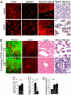
Fig. 1.
Fig. 1.
Vascular alterations in various organs. (A) Microvascular networks in liver, spleen, adrenal gland, and BM were revealed by immunohistochemical staining with anti-CD31. Arrows point to sinusoidal blood vessels. (B) Vascular networks in tumor, liver, and BM from the circulating levels of 0.8 ng/ml and 1.2 ng/ml VEGF in mice were compared. (C and D) Vascular areas were quantified by measuring CD31-positive signals and the mean values are presented (± SD). (E) Blood corticosterone levels were measured on day 14 after tumor implantation. Cx = cortex; M = medulla. (Scale bars in A and B, 50 μm.)
Fig. 2.
Fig. 2.
VEGF blocking and prolongation of survivals (A) At day 14 after treatment with MF1 and DC101, a representative mouse of each group was photographed. Arrows point to nose/mouth and paws. Asterisks mark the abdomens of mice. (B) Tumor volumes were measured at the indicated times to determine tumor growth rates. (D) The percentage of survival animals in each group is presented during a 15-day-treatment course. (E and F) After killing of animals on day 15 after treatment, livers and spleens were weighed and mean values are presented. (C) At the same time point, liver, spleen, adrenal gland, and BM of buffer-treated, MF1-treated, and DC101-treated mice (n = 8/group) were stained with H&E (top four sets of images). PA = portal area; RP = red pulp; WP = white pulp; Cx = cortex; and M = medulla. Vascular networks in tumors and livers were revealed by staining with a CD31 antibody (bottom two sets of images). (Scal bar, 50 μm.) (G) CD31 positive signals were quantified in tumor tissues. (H) VEGF tumors were allowed to grow into sizes of 0.8 cm3, followed by treatment with bevacizumab for 10 days. The mouth/nose and paws from a representative mouse of each group was photographed. (I-K) Tumor growth rates, liver weight, and spleen weight were measured. (L) The percentages of survival animals in bevacizumab- versus buffer-treated groups were presented during a 19-day experimental period.
Fig. 3.
Fig. 3.
CASS in a spontaneous mouse tumor model. Spontaneous mammary tumors developed in MMTVneu transgenic mice at 2-month age and mice were killed when they reached 4 months old. One group of mice (n = 6) received the anti-VEGFR-2 treatment at a dose of 800 μg/mouse. Paws (A), liver and spleen (C) were photographed. (B) Liver, spleen, and adrenal gland were evaluated by H&E staining (top three sets of images). The arrow indicates a hematopoietic islet in the liver tissue. Arrowheads indicate dilated sinusoidal blood vessels. Tissue sections of liver and adrenal gland were stained with anti-CD31 (bottom two sets of images). CV, central vein; RP, red pulp; WP, white pulp; Cx, cortex; M, medulla. (Scale bars, 50 μm.) Liver weight (D), spleen weight (E), and net body weight (F) were measured. Blood samples were collected and hemoglobin (G), hematocrit (H), and erythrocytes (I) were determined. (J) The serum levels of VEGF in various groups of mice were measured using a sensitive ELISA.
Fig. 4.
Fig. 4.
CASS in human cancer patients. Clinical samples were collected from RCC, bladder and prostate cancer patients. (A) Circulating levels of VEGF were measured by ELISA. (C) Histological micrographs of H&E and CD31 immunohistochemical staining of livers from RCC patients at a high (1.2 ng/ml) and a low (0.3 ng/ml) circulating VEGF level are presented. (Scale bars, 50 μm.) The development of hepatomegaly (B) and splenomegaly (D) were correlated with circulating VEGF levels. (E) Percentage of patients with ascites was correlated with the average circulating VEGF level. Levels of blood hemoglobin (F), ALT (G), AST (H), and albumin (I) were measured and correlated with circulating VEGF expression levels (J–M). HI, healthy individuals; PC, prostate cancer patients; BC, bladder cancer patients; RCC, renal cell carcinoma patients. Statistic analyses were indicated as in figures.

References

    1. Hurwitz H. Integrating the anti-VEGF-A humanized monoclonal antibody bevacizumab with chemotherapy in advanced colorectal cancer. Clin Colorectal Cancer. 2004;4(Suppl 2):S62–S68. - PubMed
    1. Motzer RJ, et al. Sunitinib versus interferon alfa in metastatic renal-cell carcinoma. N Engl J Med. 2007;356:115–124. - PubMed
    1. Ledford H. Drug markers questioned. Nature. 2008;452:510–511. - PubMed
    1. Jain RK. Normalization of tumor vasculature: An emerging concept in antiangiogenic therapy. Science. 2005;307:58–62. - PubMed
    1. Nathanson L, Hall TC. Introduction: Paraneoplastic syndromes. Semin Oncol. 1997;24:265–268. - PubMed

Publication types

MeSH terms

Substances