. 2008 Dec 2;105(48):18806-11.
doi: 10.1073/pnas.0807345105. Epub 2008 Nov 18.

An endothelin-1 switch specifies maxillomandibular identity

Affiliations

An endothelin-1 switch specifies maxillomandibular identity

Takahiro Sato et al. Proc Natl Acad Sci U S A. .

Abstract

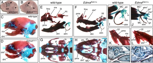
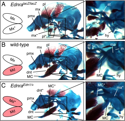
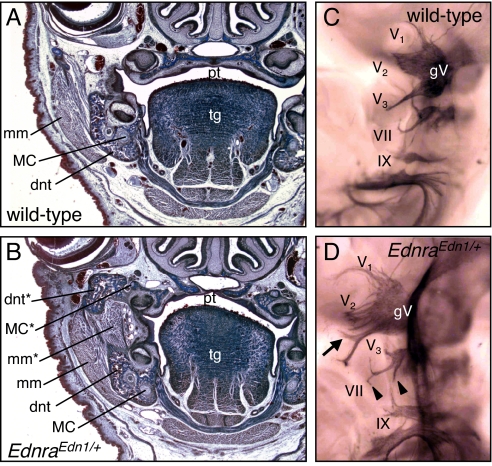
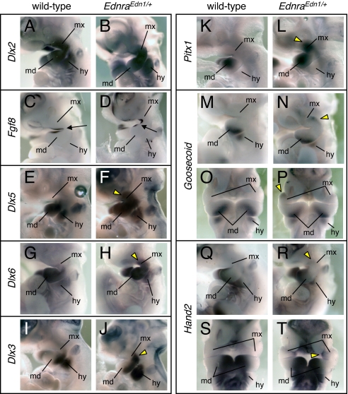
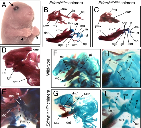
Articulated jaws are highly conserved structures characteristic of gnathostome evolution. Epithelial-mesenchymal interactions within the first pharyngeal arch (PA1) instruct cephalic neural crest cells (CNCCs) to form the different skeletal elements of the jaws. The endothelin-1 (Edn1)/endothelin receptor type-A (Ednra)-->Dlx5/6-->Hand2 signaling pathway is necessary for lower jaw formation. Here, we show that the Edn1 signaling is sufficient for the conversion of the maxillary arch to mandibular identity. Constitutive activation of Ednra induced the transformation of upper jaw, maxillary, structures into lower jaw, mandibular, structures with duplicated Meckel's cartilage and dermatocranial jaws constituted by 4 dentary bones. Misexpression of Hand2 in the Ednra domain caused a similar transformation. Skeletal transformations are accompanied by neuromuscular remodeling. Ednra is expressed by most CNCCs, but its constitutive activation affects predominantly PA1. We conclude that after migration CNCCs are not all equivalent, suggesting that their specification occurs in sequential steps. Also, we show that, within PA1, CNCCs are competent to form both mandibular and maxillary structures and that an Edn1 switch is responsible for the choice of either morphogenetic program.


Conflict of interest statement

The authors declare no conflict of interest.

Figures

Fig. 1.
Fig. 1.
Transformation of maxillary components in EdnraEdn1/+ mice. (A and B) Facial appearance of E18.5 wild-type (A) and EdnraEdn1/+ (B) mice. Vibrissae are absent in mutant mice that have open eyelids (arrows) and an anterior shift of the ear position (arrowheads). (C–L) Bone and cartilage elements of E18.5 wild-type (C, E, G, I, and K) and EdnraEdn1/+ (D, F, H, J, and L) skulls. (C and D) Lateral views of wild-type and EdnraEdn1/+ craniofacial skeletons. (E and F) The EdnraEdn1/+ mutant shows mirror-image duplication of the mandibular elements (dentary, MC, and malleus) at the expense of the maxillary elements (maxilla, jugal, squamosal, and incus). (G and H) Caudal views of wild-type and EdnraEdn1/+ craniofacial skeletons. The dentaries are removed to show the normal and transformed maxillae. Large parts of the palatine, pterygoid, lamina obturans, and ala temporalis are missing or severely deformed in the EdnraEdn1/+ mutant. (I and J) Ectotympanic and gonial bones are also duplicated in association with the ectopic MC in EdnraEdn1/+ mutants. (K and L) Caudal views of the premaxilla-maxilla junction. The tip of the transformed maxilla-dentary in the EdnraEdn1/+ mutant contains an ectopic incisor and fuses to the premaxilla. (M and N) Parasagittal sections of E18.5 wild-type (M) and EdnraEdn1/+ (N) mice stained by Mallory trichromic. The EdnraEdn1/+ mutant shows an ectopic incisor in addition to the orphotopic 1. Fusion between the premaxilla and transformed maxilla-dentary is observed in the mutant. at, ala temporalis; bs, basisphenoid; dnt, dentary; etm, ectotympanic; fmx, frontal process of maxilla; gn, gonial; hy, hyoid; in, incus; jg, jugal; lo, lamina obturans; ma, malleus; mx, maxilla; pl, palatine; pmx, premaxilla; ps, presphenoid; ptg, pterygoid; sq, squamosal; tg, tongue; UI, upper incisor; vbf, vibrissae follicle; vm, vomer; *, ectopic structure.
Fig. 2.
Fig. 2.
Comparison of craniofacial skeletons among E15.5 Ednra-null (EdnralacZ/lacZ) (A), wild-type (B) and Edn1-misexpressing (EdnraEdn1/+) (C) mice. Activated Edn1/Ednra signaling (colored in pink) correlates to the formation of MC and associated mandibular structures. at, ala temporalis; dnt, dentary; hy, hyoid; in, incus; ma, malleus; mx, maxilla; pl, palatine; pmx, premaxilla; *, ectopic structure.
Fig. 3.
Fig. 3.
Histological and whole-mount neuromuscular analysis of EdnraEdn1/+ mice. (A and B) Frontal sections of E18.5 wild-type (A) and EdnraEdn1/+ (B) mice stained by Mallory trichromic. Constitutive activation of Edn1/Ednra signaling results not only in the transformation of maxillary skeletal elements, but also in the appearance of new muscular components associated to the ectopic dentary, which can be interpreted as a duplicated masseter muscle. (C and D) Cranial nerves of E10.5 wild-type (C) and EdnraEdn1/+ (D) embryos stained by antineurofilament antibody. Ectopic branches of the mandibular (arrow) and facial (arrowheads) nerves are indicated. dnt, dentary; mm, masseter muscle; pl, palatine; tg, tongue; gV, trigeminal ganglion; V1, ophthalmic nerve; V2, maxillary nerve; V1, mandibular nerve; VII, facial nerve; IX, glossopharyngeal nerve; *, ectopic structure.
Fig. 4.
Fig. 4.
Gene expression analysis of the pharyngeal arches in EdnraEdn1/+ embryos. (A–T) Whole-mount in situ hybridization for Dlx2 (A and B), Fgf8 (C and D), Dlx5 (E and F), Dlx6 (G and H), Dlx3 (I and J), Pitx1 (K and L), Goosecoid (M–P), and Hand2 (Q–T) in E10.5 wild-type (A, C, E, G, I, K, M, O, Q, and S) and EdnraEdn1/+ (B, D, F, H, J, L, N, P, R, and T) embryos. Arrows indicate the junction of maxillary and mandibular arches. Yellow arrowheads indicate ectopic gene expression in the maxillary arch. hy, hyoid arch; md, mandibular arch; mx, maxillary arch.
Fig. 5.
Fig. 5.
Morphological transformation in EdnraHand2/+ ES cell-derived chimeric mice. (A) Representative surface appearance of E18.5 EdnraHand2/+ chimeric mice. Mutant mice demonstrate loss of vibrissae follicles, open eyelids (arrows), and polydactyly (arrowheads). (B–E) Craniofacial bone and cartilage elements of E18.5 EdnraNeo/+ ES cell-derived (B) and EdnraHand2/+ ES cell-derived (C–E) chimeric mice. Whereas EdnraNeo/+-chimeric mice show normal skeletal morphology (B), EdnraHand2/+-chimeric mice demonstrate transformation of the maxilla into dentary-like bone (C), which fuses to the premaxilla and contains an ectopic incisor at the tip (D). The condylar and angular processes of the dentary, gonial, ectotympanic, and squamosal bones were often malformed or absent (E). (F–I) Craniofacial skeletons of E15.5 wild-type (F and H) and EdnraHand2/+-chimeric (G and I) mice. Ectopic formation of MC-like, rod-shaped cartilage is present often with small and deformed ala temporalis in EdnraHand2/+-chimeric mice. The malleus, incus, stapes, and styloid process are deformed. agp, angular process; at, ala temporalis; cdp, condylar process; crp, coronoid process; dnt, dentary; etm, ectotympanic; fmx, frontal process of maxilla; gn, gonial; hy, hyoid; in, incus; jg, jugal; ma, malleus; mx, maxilla; pmx, premaxilla; sp, styloid process; sq, squamosal; st, stapes; UI, upper incisor; *, ectopic structure.

References

    1. Couly GF, Coltey PM, Le Douarin NM. The triple origin of skull in higher vertebrates: A study in quail-chick chimeras. Development. 1993;117:409–429. - PubMed
    1. Creuzet S, Couly G, Vincent C, Le Douarin NM. Negative effect of Hox gene expression on the development of the neural crest-derived facial skeleton. Development. 2002;129:4301–4313. - PubMed
    1. Köntges G, Lumsden A. Rhombencephalic neural crest segmentation is preserved throughout craniofacial ontogeny. Development. 1996;122:3229–3242. - PubMed
    1. Noden DM, Trainor PA. Relations and interactions between cranial mesoderm and neural crest populations. J Anat. 2005;207:575–601. - PMC - PubMed
    1. Trainor PA, Tam PP. Cranial paraxial mesoderm and neural crest cells of the mouse embryo: Co-distribution in the craniofacial mesenchyme but distinct segregation in branchial arches. Development. 1995;121:2569–2582. - PubMed

Publication types

MeSH terms

Substances