. 2008 Dec 1;181(11):7863-72.
doi: 10.4049/jimmunol.181.11.7863.

TREM-2 mediated signaling induces antigen uptake and retention in mature myeloid dendritic cells

Affiliations

TREM-2 mediated signaling induces antigen uptake and retention in mature myeloid dendritic cells

Suresh Radhakrishnan et al. J Immunol. .

Retraction in

Abstract

Myeloid dendritic cells (mDC) activated with a B7-DC-specific cross-linking IgM Ab (B7-DC XAb) take up and retain Ag and interact with T cell compartments to affect a number of biologic changes that together cause strong antitumor responses and blockade of inflammatory airway disease in animal models. The molecular events mediating the initial responses in mDC remain unclear. In this study we show that B7-DC XAb caused rapid phosphorylation of the adaptor protein DAP12 and intracellular kinases Syk and phospholipase C-gamma1. Pretreatment of mDC with the Syk inhibitor piceatannol blocked B7-DC XAb-induced Ag uptake with a concomitant loss of tumor protection in mice. Vaccination with tumor lysate-pulsed wild-type B7-DC XAb-activated mDC, but not TREM-2 knockout XAb-activated mDC, protected mice from lethal melanoma challenge. Multimolecular caps appeared within minutes of B7-DC XAb binding to either human or mouse mDC, and FRET analysis showed that class II, CD80, CD86, and TREM-2 are recruited in tight association on the cell surface. When TREM-2 expression was reduced in wild-type mDC using short hairpin RNA or by using mDC from TREM-2 knockout mice, in vitro DC failed to take up Ag after B7-DC XAb stimulation. These results directly link TREM-2 signaling with one change in the mDC phenotype that occurs in response to this unique Ab. The parallel signaling events observed in both human and mouse mDC support the hypothesis that B7-DC cross-linking may be useful as a therapeutic immune modulator in human patients.


Figures

Figure 1
Figure 1. Kinase and cytoskeleton reorganization inhibitors affect OVA-FITC uptake by matured human mDC
A, Human mDC were left untreated (black filled histogram) or were matured overnight with poly I:C. On day six, the mDC cultures were activated the isotype control antibody (grey unfilled histograms) or with B7-DC XAb (white unfilled histogram) and pulsed with OVA-FITC. Uptake of OVA-FITC was assessed 24 hours later by flow cytometry. B and C, Day 6 matured mDC were pre-incubated without or with the Src kinase inhibitor PP2 (0.3, 1, 3, or 10 μM), the PKC inhibitor Bim (50 nM), Rho A inhibitor Y-27632 (25 μM), PI3 kinase inhibitor LY294002 (10 μM), PLCγ inhibitor U73122 (10 μM), MEK inhibitor PD98059 (10 μM), p38 MAP kinase inhibitor SB203580 (1 μM), or Rac1 inhibitor NSC23766 (50 μM) prior to activation with isotype control antibody or B7-DC XAb. All cells were pulsed with OVA-FITC at the time of treatment and analyzed for OVA-FITC uptake 24 hours later by flow cytometry. Data are representative of 3 or more experiments.
Figure 2
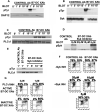
Figure 2. The DAP12-Syk-PLCγ is activated by B7-DC XAb and is required for regained antigen uptake in matured mDC
Matured human mDC were treated with the B7-DC XAb for varying times. Immunoprecipitates were prepared using antibodies specific for DAP12 (A), Syk (B), and PLCγ1 (C) and analyzed for activation by blotting for phosphotyrosine (pTyr). After stripping, the membranes were re-blotted with the specific antibodies used for immunoprecipitations. In (D) and (E), cells were pre-treated with the indicated concentrations or with 10 μM of the Syk inhibitor Piceatannol. In (F) and (G), cells were pretreated with Piceatannol or the active or inactive forms of PLCγ1 inhibitor (U73122/U73343) and antigen uptake was assessed by pulsing DC with FITC-conjugated ovalbumin and measuring intracellular fluorescence 16 hours later in CD11c+ cells by flow cytometry. Data are representative of 3 or more experiments.
Figure 3
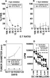
Figure 3. DCXAb-induced tumor protection in mice requires activation of Syk
Bone marrow derived mDC were pulsed with B16 melanoma tumor lysate and control antibody (○) or B7-DC XAb (•) in the absence (A) or presence of the Syk inhibitor Picetannol (B) prior to adoptive transfer into mice. Day 7 after transfer, splenocytes from mice were analyzed for the ability to kill the B16 targets in a chromium release assay (performed in triplicate using spleens pooled from 3 mice per group). C, mDC were pulsed with the tumor cell lysate and treated with control antibody (○), B7-DC XAb (•), or B7-DC XAb plus Piceatannol (□) prior to adoptive transfer into mice at the same time they were injected with B16 live tumor (5 mice per group). Mice were monitored for tumor growth (presented as the average size in mm plus or minus deviation from the mean). Mice with tumor size of 17×17mm were euthanized. D, mDC or mDC pretreated with Piceatannol were pulsed with OT-II specific peptide (ISQAVHAAHAEINEAGR) and control antibody or B7-DC XAb and incubated for 3 days in titrated doses with 3 × 10⁵ OT-II naïve T cells. The cultures were pulsed with 3H-thymidine 18 h prior to harvest. Error bars represent the standard deviation from the mean of triplicate samples.
Figure 4
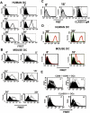
Figure 4. B7-DC XAb causes formation of multi-molecular caps on the cell surface of mouse and human mDC
A, Mouse mDC were tagged with fluorochrome-labeled antibody specific for MHC I-Ab (25−9−17-FITC) and CD80/CD86 (16.10A1-PE and GL-1-PE) 15 minutes prior to activation with B7-DC XAb or control IgM antibody for 15 minutes. PE, FITC (APC), DAPI, and merged confocal images are shown and are representative of the entire field. White bars indicate 20 microns. B, Same as in (A), except human cells were labeled with HLA-DR, DP, DQ-FITC (TU39), CD80 (2D10.4-PE) and CD86 (IT2.2-PE). Arrows indicate cap structures.
Figure 5
Figure 5. Reorganization of DCXAb cell surface results in close membrane clustering between Class II-CD80-CD86
The distribution of class II (labeled with APC-conjugated antibody) and CD80/CD86 molecules (labeled with PE-conjugated antibodies) was visualized over time by FRET following treatment of human (A) or mouse (B) mDC with B7-DC XAb (open histograms) or with isotype control antibody (filled histograms). C, Lack of induction of FRET of CD86-APC and CD80-PE in mouse mDC by the I-Ab specific IgM antibody 25−9−3 (open dark histograms) and isotype control antibody treated samples (filled histograms). Binding of the class II specific IgM antibody to the mouse mDC analyzed is also shown (far right panel). D, Inhibition of B7-DC XAb-induced FRET by B7-DC-specific IgG (green open histogram) compared to no IgG as control (red open histogram). Absence of FRET induced by isotype control antibody is also shown (filled histograms). E, Top: Class II-APC/CD11c-PE FRET in mDC from CD80−/−/CD86−/− mice. Left panel shows FRET signal at zero minutes after treatment with control antibody (filled histogram, MFI 80) or B7-DC XAb (open histogram, MFI 98). Right panel shows FRET signal 15 minutes after stimulation with isotype-matched control antibody (filled histogram, MFI 87) or B7-DC XAb (open histogram, MFI 190). Bottom: CD80-APC/CD86-PE FRET signal from class II−/− mDC. Left panel shows FRET signal at 0 minutes after stimulation with control antibody (filled histogram, MFI 13) or B7-DC XAb (open histogram, MFI 10). Right panel depicts FRET signal 15 minutes after stimulation with isotype-matched control antibody (filled histogram, MFI 12.5) or B7-DC XAb (open histogram, MFI 115). Data are representative of 3 or more experiments.
Figure 6
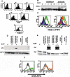
Figure 6. TREM-2 is recruited into B7-DC XAb-induced cap and is required for phosphorylation of DAP-12 and Syk
A, Class II-APC/TREM2-PE FRET was measured using six-day cultures of mouse mDC before (0’) or after stimulation with B7-DC XAb (open histograms) or isotype control antibody (filled histograms) for 5, 10, 15, or 30 min as labeled for five panels. Bottom panel shows expression of TREM-2 on unstimulated cells by staining with 237916-PE antibody (open histogram) compared to control (filled histogram). B, Class II complexes were immunoprecipitated from B7-DC XAb- or control antibody-treated mDC using biotin tagged I-Ab-specific KH74 antibody and analyzed for the presence of TREM-2 by Western blot. C, TREM-2 expression on myeloid dendritic cells transduced with virus encoding scrambled RNA (left panel) showing staining with TREM-2 antibody (green open histogram) or isotype control (filled histogram) in comparison to mDC transduced with virus encoding TREM-2 shRNA (right panel, orange histogram), scrambled RNA (right panel, green histogram), or uninfected DC probed with isotype control antibody (right panel, filled histogram). D and E, Tyrosine phosphorylation of Syk or DAP12 in mouse mDC transduced with shRNA specific for TREM-2 or a scrambled control sequence and activated with isotype control antibody or B7-DC XAb for 5 minutes. F, Uptake of OVA-APC by matured mDC. Left: mDC were transduced with the scrambled shRNA vector and treated with isotype control antibody (grey histogram) or with the B7-DC XAb (green histogram). Right: mDC were transduced with TREM-2 shRNA and treated with control antibody (grey histogram) or B7-DC XAb (orange histogram). Data are representative of 3 or more experiments.
Figure 7
Figure 7. TREM-2 is required for B7-DC XAb-induced activation of antigen uptake in mature mDC
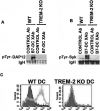
Tyrosine phosphorylation of DAP12 (A) or Syk (B) immunoprecipitated from myeloid dendritic cells derived from wildtype (WT) or TREM-2−/− (KO) mice after incubation with B7-DC XAb or isotype control. C, OVA-FITC uptake induced by the control antibody (filled histograms) or B7-DC XAb (open histograms) in matured mDC from wildtype (WT, left panel) or TREM-2−/− (KO, right panel) mice. Data are representative of 3 or more experiments.

References

    1. Mellman I, Steinman RM. Dendritic cells: specialized and regulated antigen processing machines. Cell. 2001;106:255–258. - PubMed
    1. Iwasaki A, Medzhitov R. Toll-like receptor control of the adaptive immune responses. Nat Immunol. 2004;5:987–995. - PubMed
    1. Takeda K, Akira S. TLR signaling pathways. Semin Immunol. 2004;16:3–9. - PubMed
    1. Banchereau J, Briere F, Caux C, Davoust J, Lebecque S, Liu YJ, Pulendran B, Palucka K. Immunobiology of dendritic cells. Annu Rev Immunol. 2000;18:767–811. - PubMed
    1. Banchereau J, Steinman RM. Dendritic cells and the control of immunity. Nature. 1998;392:245–252. - PubMed

Publication types

MeSH terms

Substances