. 2008 Dec 1;181(11):7948-57.
doi: 10.4049/jimmunol.181.11.7948.

Identification of human T cell antigens for the development of vaccines against Mycobacterium tuberculosis

Affiliations

Identification of human T cell antigens for the development of vaccines against Mycobacterium tuberculosis

Sylvie Bertholet et al. J Immunol. .

Abstract

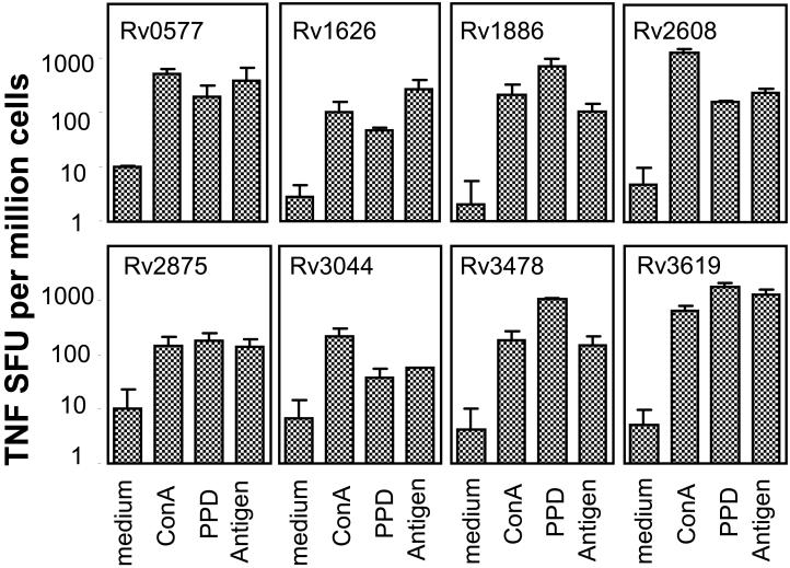
Development of a subunit vaccine for Mycobacterium tuberculosis (Mtb) depends on the identification of Ags that induce appropriate T cell responses. Using bioinformatics, we selected a panel of 94 Mtb genes based on criteria that included growth in macrophages, up- or down-regulation under hypoxic conditions, secretion, membrane association, or because they were members of the PE/PPE or EsX families. Recombinant proteins encoded by these genes were evaluated for IFN-gamma recall responses using PBMCs from healthy subjects previously exposed to Mtb. From this screen, dominant human T cell Ags were identified and 49 of these proteins, formulated in CpG, were evaluated as vaccine candidates in a mouse model of tuberculosis. Eighteen of the individual Ags conferred partial protection against challenge with virulent Mtb. A combination of three of these Ags further increased protection against Mtb to levels comparable to those achieved with bacillus Calmette-Guérin vaccination. Vaccine candidates that led to reduction in lung bacterial burden following challenge-induced pluripotent CD4 and CD8 T cells, including Th1 cell responses characterized by elevated levels of Ag-specific IgG2c, IFN-gamma, and TNF. Priority vaccine Ags elicited pluripotent CD4 and CD8 T responses in purified protein derivative-positive donor PBMCs. This study identified numerous novel human T cell Ags suitable to be included in subunit vaccines against tuberculosis.


Figures

Figure 1
Figure 1
Levels of IFN-γ released by antigen-stimulated human PBMC were measured in vitro. PPD+ and PPD- PBMC were incubated for 72 h in medium, PHA (10 μg/ml), Mtb lysate (10 μg/ml), or Mtb recombinant proteins (50 μg/ml). Antigens are grouped according to the selection criteria: m, membrane-associated; S, secreted proteins; P, PE/PPE; E, EsX; M, macrophage growth required; H, hypoxic response; D, database searches. (A) IFN-γ in supernatants was measured by sandwich ELISA. Mean (MeanAg - MeanMedium) ± SEM are shown for PPD+ (n=18) and PPD- (n=7) PBMC. (B) Percent of PPD+ positive responders.
Figure 1
Figure 1
Levels of IFN-γ released by antigen-stimulated human PBMC were measured in vitro. PPD+ and PPD- PBMC were incubated for 72 h in medium, PHA (10 μg/ml), Mtb lysate (10 μg/ml), or Mtb recombinant proteins (50 μg/ml). Antigens are grouped according to the selection criteria: m, membrane-associated; S, secreted proteins; P, PE/PPE; E, EsX; M, macrophage growth required; H, hypoxic response; D, database searches. (A) IFN-γ in supernatants was measured by sandwich ELISA. Mean (MeanAg - MeanMedium) ± SEM are shown for PPD+ (n=18) and PPD- (n=7) PBMC. (B) Percent of PPD+ positive responders.
Figure 2
Figure 2
Antibody responses to Mtb recombinant antigens were measured in sera isolated from mice vaccinated with Rv0577, Rv2875, Rv1886, Rv2608, Rv3478, Rv3619, Rv3620, Rv1813, and Rv3044 + CpG. Sera were tested for antigen-specific IgG1 and IgG2c antibody. (A) Mean of reciprocal dilution + SEM, and (B) IgG2c:IgG1 ratios are shown. *P < 0.05, Student’s t-Test. Data shown are representative of two independent experiments.
Figure 3
Figure 3
IFN-γ responses to Mtb recombinant antigens were determined in vaccinated mice. Splenocytes were prepared from animals immunized with Rv0577, Rv1626, Rv1813, Rv1886 (85B), Rv2875, Rv2608, Rv3619, Rv3620, Rv3044, and Rv3478 + CpG or saline, 3 wks after the last injection, and tested for IFN-γ in vitro recall responses. Cells were plated in duplicate at 2 × 10⁵ cells/well and cultured with medium, Con A (3 μg/ml), PPD (10 μg/ml), or each recombinant antigen (10 μg/ml) for 48 h. Frequencies of IFN-γ-secreting cells (spot-forming unit, SFU) were determined by ELISPOT. Mean + SD (n = 5 mice) shown are representative of two independent experiments.
Figure 4
Figure 4
TNF responses to Mtb recombinant antigens were determined in vaccinated mice. Splenocytes were prepared from animals immunized with Rv0577, Rv1626, Rv1813, Rv1886 (85B), Rv2875, Rv2608, Rv3619, Rv3620, Rv3044, and Rv3478 + CpG or saline, 3 wks after the last injection, and tested for TNF in vitro recall responses. Cells were plated in duplicate at 2 × 10⁵ cells/well and cultured with medium, Con A (3 μg/ml), PPD (10 μg/ml), or each recombinant antigen (10 μg/ml) for 48 h. Frequencies of TNF-secreting cells (SFU) were determined by ELISPOT. Mean + SD (n = 5 mice) shown are representative of two independent experiments.
Figure 5
Figure 5
Antigen-specific CD4 and CD8 T cell cytokine responses were analyzed in vaccinated mice. Splenocytes, isolated from animals immunized with Rv1813, Rv3620, Rv2608, Rv3619, Rv3478, or Rv1886 + CpG, or saline, were plated at 2 × 10⁶ cells/well and cultured with medium, or each recombinant antigen (20 μg/ml) for 12 h in the presence of GolgiStop. (A) CD4 T cells were identified by ICS based on CD3 and CD4 expression, and further gated as CD44high (antigen experienced) or CD44low (naïve). The percent of cells expressing two cytokines or more of IFN-γ, TNF, and IL-2 were determined. Means (n = 3) are shown. (B) CD8 antigen specific T cells were identified by ICS as CD3/CD44high/CD8 cells also positive for IFN-γ and/or TNF, and further analyzed for their content in GrB. Numbers of cytokine+ (IFN/TNF) and cytokine+GrB+ per million splenocytes were determined. Mean ± SD (n = 3 mice) shown are representative of two independent experiments.
Figure 6
Figure 6
The efficacy of a combination of three antigens adjuvanted with CpG was addressed. Mice were immunized s.c. three times, three wks apart with 25 μg CpG, 8 μg single antigen + CpG, 6 μg of each antigen + CpG (“3 Ags+CpG”), or once i.d. with 5×10⁴ BCG. Four weeks after the last boost, mice were challenged with Mtb H37Rv. (A) The number of viable bacteria (CFU Log10) in the lungs was determined 4 wks post-challenge. One way ANOVA followed by Dunnett’s Multiple Comparison Test was used for statistical analysis;* P < 0.05, ** P < 0.01. (B) Mice were bled one week after the last boost and sera were tested for antigen-specific IgG2c antibody. Mean of reciprocal dilution + SD (n = 3 mice) are shown. (C - D) Splenocytes were isolated from the vaccinated animals three wks after the last boost, plated in duplicate at 2 × 10⁵ cells/well, and cultured with medium, Con A (3 μg/ml), PPD (10 μg/ml), or each recombinant antigen (10 μg/ml) for 48 - 72 h. (C) IFN-γ levels measured by ELISA in 72 h supernatants. (D) Frequencies of TNF-secreting cells (SFU) determined by ELISPOT. Mean + SD (n = 3 mice) shown are representative of two independent experiments.
Figure 7
Figure 7
Human PPD+ CD4 T cells respond to recombinant Mtb antigen stimulation. PBMC from 6 healthy PPD+ subjects with diverse HLA types were incubated for 12 h in medium, PPD (10 μg/ml), or recombinant antigens (20 μg/ml) in the presence of anti CD28/CD49d and GolgiStop. T cells were identified by ICS based on CD3 expression, and further gated as CD4/CD45RO memory T cells. Percent of CD4 T cells expressing IFN-γ, TNF, or the two cytokines in response to a panel of Mtb antigens are shown.
Figure 8
Figure 8
Human PPD+ CD8 T cells respond to recombinant Mtb antigen stimulation. PBMC from 6 healthy PPD+ subjects with diverse HLA types were incubated for 12 h in medium, PPD (10 μg/ml), or recombinant antigens (20 μg/ml) in the presence of anti CD28/CD49d and GolgiStop. T cells were identified by ICS based on CD3 expression, and further gated as CD8/CD45RO memory T cells. Percent of CD8 T cells expressing IFN-γ, TNF, or the two cytokines in response to a panel of Mtb antigens are shown.

References

    1. WHO . Global Tuberculosis Control: Surveilance, Planning, Financing. World Health Organization; Geneva: 2007.
    1. Maartens G, Wilkinson RJ. Tuberculosis. Lancet. 2007;370:2030–2043. - PubMed
    1. Andersen P, Doherty TM. The success and failure of BCG - implications for a novel tuberculosis vaccine. Nat Rev Microbiol. 2005;3:656–662. - PubMed
    1. Doherty TM, Andersen P. Vaccines for tuberculosis: novel concepts and recent progress. Clin Microbiol Rev. 2005;18:687–702. - PMC - PubMed
    1. WHO . The WHO/IUTLD Global Project on Anti-Tuberculosis Resistance Surveillance. World Health Organization; Geneva: 2008. Anti-tuberculosis drug resistance in the world. Fourth global report.

Publication types

MeSH terms