. 2008 Dec 1;181(11):7977-84.
doi: 10.4049/jimmunol.181.11.7977.

Aging mice exhibit a functional defect in mucosal dendritic cell response against an intracellular pathogen

Affiliations

Aging mice exhibit a functional defect in mucosal dendritic cell response against an intracellular pathogen

Magali M Moretto et al. J Immunol. .

Abstract

Down-regulation of the immune response in aging individuals puts this population at a potential risk against infectious agents. In-depth studies conducted in humans and mouse models have demonstrated that with increasing age, the T cell immune response against pathogens is compromised and response to vaccinations is subdued. In the present study, using a mouse model, we demonstrate that older animals exhibit greater susceptibility to Encephalitozoon cuniculi infection, and their ability to evoke an Ag-specific T cell response at the gut mucosal site is reduced. The dampening of T cell immunity was due to the defective priming by the dendritic cells (DC) isolated from the mucosal tissues of aging animals. When primed with DC from younger mice, T cells from older animals were able to exhibit an optimal Ag-specific response. The functional defect in DC from older mice can be attributed to a large extent to reduced IL-15 message in these cells, which can be reversed by addition of exogenous IL-15 to the cultures. IL-15 treatment led to optimal expression of costimulatory molecules (CD80 and CD86) on the surface of older DC and restored their ability to prime a T cell response against the pathogen. To our knowledge, this is the first report which demonstrates the inability of the DC population from aging animals to prime a robust T cell response against an infectious agent. Moreover, the observation that IL-15 treatment can reverse this defect has far-reaching implications in developing strategies to increase vaccination protocols for aging populations.


Figures

Figure 1
Figure 1
Older mice cannot survive oral E. cuniculi infection. C57BL/6 mice of different age groups (4, 9 and 12 months) were orally infected with 2×10⁷ E. cuniculi spores (6 mice/group). Animals were monitored daily for mortality and morbidity until the end of the experiment. The experiment was repeated twice with similar results.
Figure 2
Figure 2
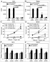
Defective mucosal immune response after E. cuniculi infection. C57BL/6 mice (8 week, 4, 9 and 12 month old) were infected per-orally with 2×10⁷ spores. Two weeks post-infection, mice were sacrificed (3 animals/group), MLN and spleen cell suspension were prepared. Proliferative response of MLN lymphocytes (A) and splenocytes (B) was measured by thymidine incorporation after 72h incubation with E. cuniculi spores. In a separate experiment, MLN (C) and spleen cell suspensions (D) were prepared and incubated with 51Cr labeled uninfected or infected macrophages at various effector to target ratios. After 4h incubation, the cytolytic activity was determined by radioisotope release into culture supernatants. MLN (E) and splenic (F) lymphocytes were isolated 14 days post E. cuniculi infection (3 mice/group). mRNA was prepared and assayed for IFNγ message by RT-PCR. Data are representative of two separate experiments.
Figure 3
Figure 3
MLN T cells from older mice fail to protect SCID recipients: MLN lymphocytes and splenocytes from 8 week old and 9 month old mice (4 mice/group) were isolated and adoptively transferred to SCID mice (5×10⁶ cells/mouse). Two days, post transfer, recipients were challenged per-orally with 2×10⁷ E. cuniculi spores. Morbidity and mortality were monitored daily until the end of the experiment. There were 6 animals per group and the study was performed twice with similar results.
Figure 4
Figure 4
(A) MLN DC from older mice are unable to prime young T cells in vitro. MLN DC from 8 week old and 9 month old mice were isolated (4 mice/group), plated at different concentrations and stimulated overnight with E. cuniculi spores. The next day, T cells isolated from naïve 8 week old mice (3 mice/group) were added to the culture. After 72h incubation, proliferative response was measured by thymidine incorporation. (B) T cells from young and older mice respond equally well to in vitro priming. MLN DC from 8 week old mice (4 mice) were isolated, plated at different concentrations and stimulated with E. cuniculi. After overnight incubation, T cells isolated from MLN of naïve 8 week old or 9 month old mice (3 mice/group) were added to the culture. After 72h incubation, proliferative response was measured by thymidine incorporation. (C) Defective IL-12 production by MLN DC from older mice. Culture supernatants were assayed for IL-12 production by ELISA. Results are representative of two separate experiments.
Figure 5
Figure 5
Splenic DC from older mice are able to prime splenocytes in vitro. Splenic DC from 8 week old and 9 month old mice were isolated, plated (20,000 DC/well) and stimulated with E. cuniculi spores. The next day, splenic T cells isolated from naïve 8 week old mice were added to the culture. (A) After 72h incubation, proliferative response was measured by thymidine incorporation. (B) Culture supernatants were assayed for IL-12 production by ELISA. Results are representative of two separate experiments.
Figure 6
Figure 6
Adoptive transfer of MLN DC form older mice fail to prime a CTL response in vivo. DC from MLN and spleen from 8 week and 9 month old mice were isolated, plated (5×10⁴ cells per well) and pulsed with irradiated E. cuniculi spores (2.5×10⁵ per well). After overnight incubation, cultures were harvested and cells were adoptively transferred to 9 month old mice (5×10⁵ per mouse) via iv route. Fourteen days later, recipient mice were sacrificed, MLN (A) and splenic (B) lymphocytes prepared and assayed for cytotoxicity. Experiment was performed twice with similar results and data are representative of one experiment.
Figure 7
Figure 7
IL-15 improves T cell priming by older DC and their costimulatory molecules expression. (A) MLN DC from older mice are unable to upregulate IL-15 message after E. cuniculi stimulation. MLN DC from 8 week-old and 9 month old mice were isolated and plated (20,000 DC/well). Cells were incubated 48 h with E. cuniculi spores and analyze for IL-15 mRNA expression normalized to β actin mRNA levels. Relative expression was measured by using the mean from each group and the formula RTL (relative transcript level) = 2−ΔCt × 1000. (B) IL-15 treatment restores ability of older DC to prime young T cells in vitro. MLN DC from 8 week old and 9 month old mice were isolated and plated (20,000 DC/well). DC were incubated overnight with E. cuniculi spores and exogenous IL-15 (100 ng/ml and 50 ng/ml). The next day, cell cultures were washed and T cells isolated from MLN of naïve 8 week old mice were added. After 72h incubation, proliferative response was measured by thymidine incorporation. (C) Treatment with IL-15 restores the ability of MLN DC from older mice to prime a CTL response. MLN DC from 8 week and 9 month old mice (10–15 mice/group) were isolated, plated (5×10⁴ DC/well) and stimulated overnight with E. cuniculi (2.5×10⁵ spores/well). The following day, purified T cells from naïve 8 week old mice (3 mice/group) were added to the culture and subsequently after 4 day incubation CTL assay performed. (D) IL-15 increases costimulatory molecules expression by older DC. MLN DC from 8 week and 9 month old mice were isolated, plated at 40,000 DC/well and incubated overnight with E. cuniculi spores. The next day, cells were harvested and labeled for MHC class II (i–iv), CD80 (v–vii), CD86 (ix–xii) and CD40 (xiii–xvi) (—8 week old DC + E. cuniculi, —8 week old DC + E. cuniculi + IL-15, formula image9 month old DC + E. cuniculi, formula image9 month old DC + E. cuniculi + IL-15). Results are presented as mean ± SD of triplicates and experiment was performed twice with similar results.

Similar articles

Cited by

References

    1. Zheng B, Han S, Takahashi Y, Kelsoe G. Immunosenescence and germinal center reaction. Immunol Rev. 1997;160:63–77. - PubMed
    1. Song H, Price PW, Cerny J. Age-related changes in antibody repertoire: contribution from T cells. Immunol Rev. 1997;160:55–62. - PubMed
    1. Garcia GG, Miller RA. Differential tyrosine phosphorylation of zeta chain dimers in mouse CD4 T lymphocytes: effect of age. Cell Immunol. 1997;175:51–57. - PubMed
    1. Haynes L, Linton PJ, Eaton SM, Tonkonogy SL, Swain SL. Interleukin 2, but not other common gamma chain-binding cytokines, can reverse the defect in generation of CD4 effector T cells from naive T cells of aged mice. J Exp Med. 1999;190:1013–1024. - PMC - PubMed
    1. Homann D, Teyton L, Oldstone MB. Differential regulation of antiviral T-cell immunity results in stable CD8+ but declining CD4+ T-cell memory. Nat Med. 2001;7:913–919. - PubMed

Publication types