. 2008 Dec 1;181(11):8027-35.
doi: 10.4049/jimmunol.181.11.8027.

Transgenic expression of cyclooxygenase-2 in hepatocytes accelerates endotoxin-induced acute liver failure

Affiliations

Transgenic expression of cyclooxygenase-2 in hepatocytes accelerates endotoxin-induced acute liver failure

Chang Han et al. J Immunol. .

Abstract

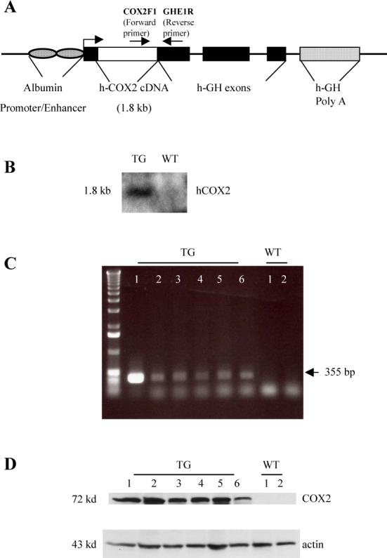
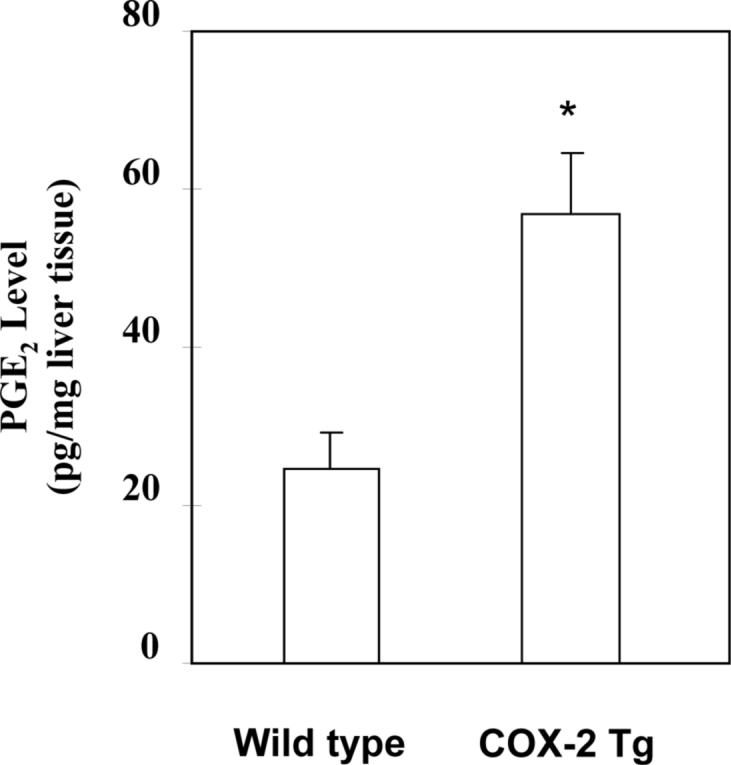
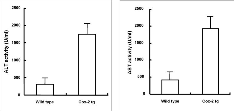
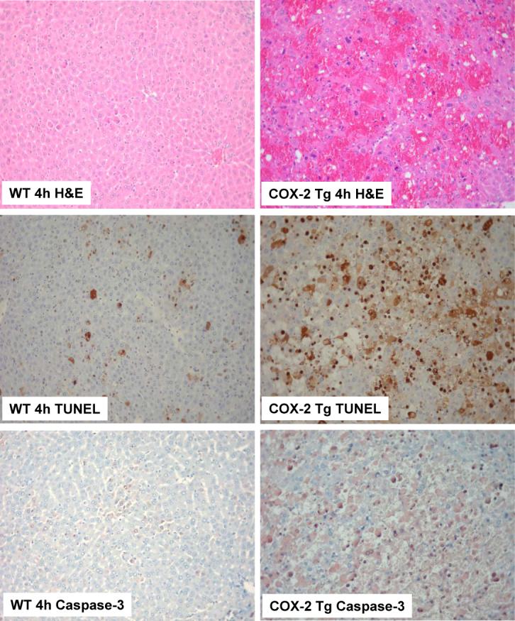
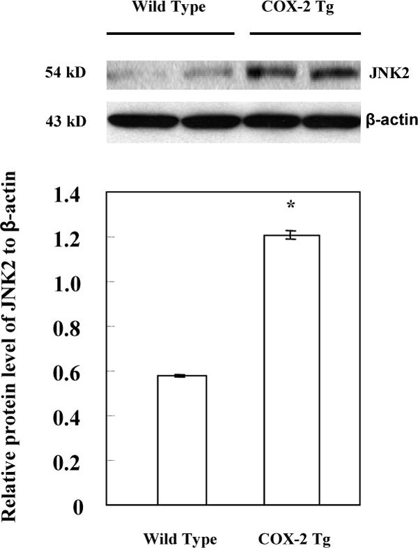
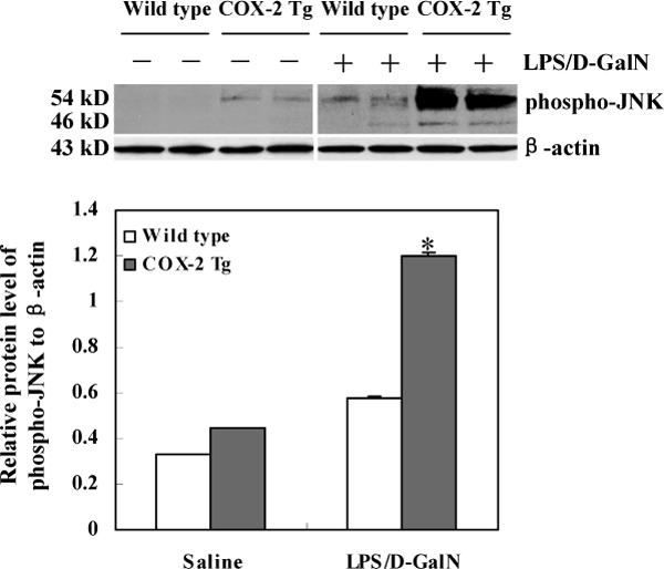
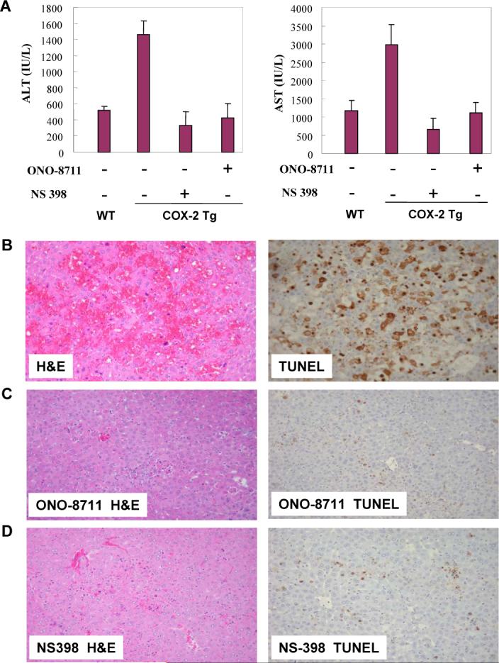
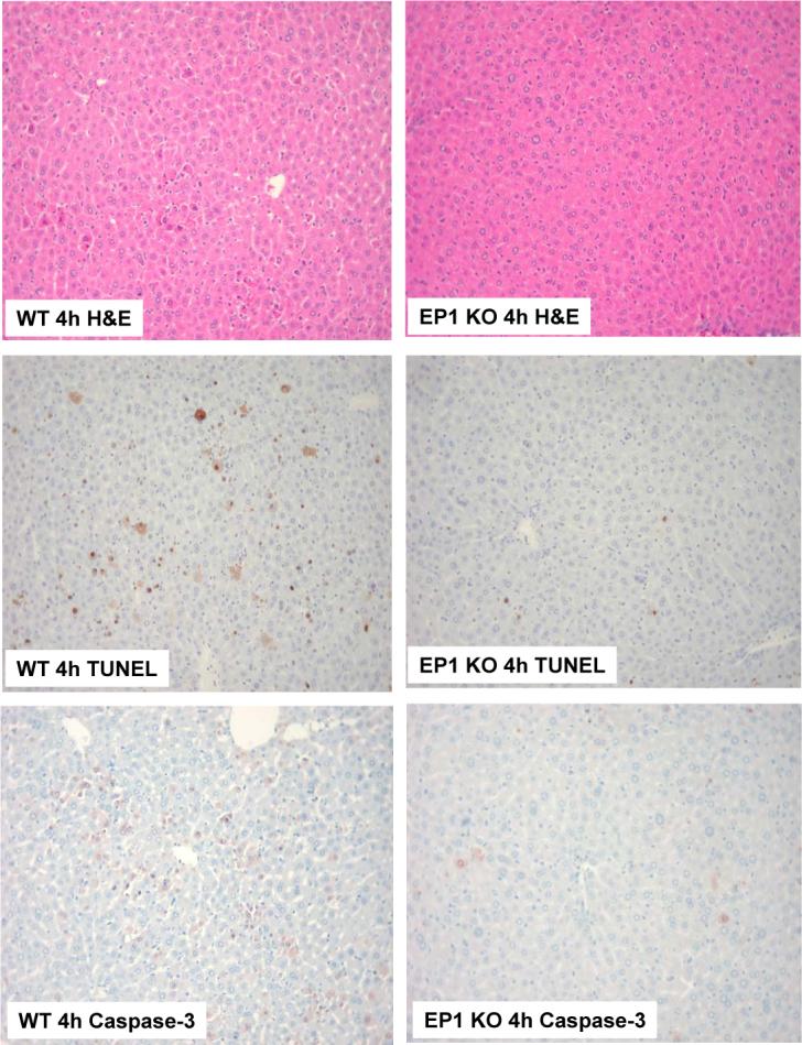
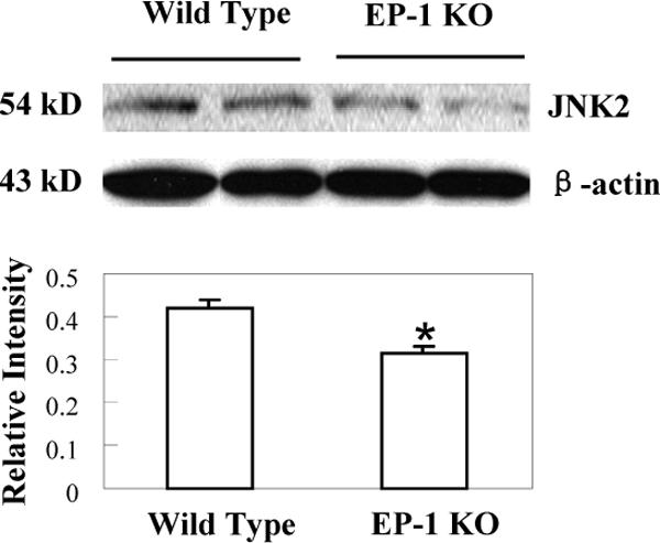
Bacterial LPS (endotoxin) is implicated in the pathogenesis of acute liver failure and several chronic inflammatory liver diseases. To evaluate the effect of hepatocyte cyclooxygenase (COX)-2 in LPS-induced liver injury, we generated transgenic mice with targeted expression of COX-2 in the liver by using the albumin promoter-enhancer driven vector and the animals produced were subjected to a standard experimental protocol of LPS-induced acute fulminant hepatic failure (i.p. injection of low dose of LPS in combination with d-galactosamine (d-GalN)). The COX-2 transgenic mice exhibited earlier mortality, higher serum aspartate aminotransferase and alanine aminotransferase levels and more prominent liver tissue damage (parenchymal hemorrhage, neutrophilic inflammation, hepatocyte apoptosis, and necrosis) than wild-type mice. Western blot analysis of the liver tissues showed that LPS/d-GalN treatment for 4 h induced much higher cleavage of poly(ADP-ribose) polymerase, caspase-3, and caspase-9 in COX-2 transgenic mice than in wild-type mice. Increased hepatic expression of JNK-2 in COX-2 transgenic mice suggest that up-regulation of JNK-2 may represent a potential mechanism for COX-2-mediated exacerbation of liver injury. Blocking the prostaglandin receptor, EP(1), prevented LPS/d-GalN-induced liver injury and hepatocyte apoptosis in COX-2 transgenic mice. Accordingly, the mice with genetic ablation of EP(1) showed less LPS/d-GalN-induced liver damage and less hepatocyte apoptosis with prolonged survival when compared with the wild-type mice. These findings demonstrate that COX-2 and its downstream prostaglandin receptor EP(1) signaling pathway accelerates LPS-induced liver injury. Therefore, blocking COX-2-EP(1) pathway may represent a potential approach for amelioration of LPS-induced liver injury.


Figures

Figure 1
Figure 1. Generation of COX-2 transgenic mice
The COX-2 transgene (complete human COX-2 cDNA cloned into the albumin promoter-driven vector) was microinjected into mouse zygotes to generate transgenic mice by established method. (A) Schematic representation of the human COX-2 transgene with albumin promoter. (B) Southern blot analysis of tail genomic DNA showing the presence of COX-2 transgene in transgenic (TG) but not in wild-type (WT) littermates. The DNA samples were digested with Bam H1; the blot was hybridized with [32P]-labeled human COX-2 cDNA probe. (C) PCR analysis of tail genomic DNA showing the presence of COX-2 transgene in transgenic (TG) mice but not in wild-type (WT) littermates. (D) Western blot showing successful expression of COX-2 protein in the liver tissues from COX-2 transgenic mice. Equal amounts of the mouse liver tissue proteins from transgenic mice (TG) and wild type (WT) littermates were subjected to SDS-PAGE and Western blotting using anti-human COX-2 antibody.
Figure 2
Figure 2. PGE2 levels in the liver tissues from wild type and COX-2 transgenic mice
Liver tissues were homogenized and extracted in 100 mg Amprep C18 minicolumn. The eluted samples were dried and the amounts of PGE2 were determined by specific enzymeimmune assay. The data are presented as mean±SD of pg/mg liver tissue (*ρ<0.01 compared to wild type mice, n=6).
Figure 3
Figure 3. Hepatic expression of COX-2 enhances serum transaminase levels induced by LPS
The COX-2 transgenic mice and the age/sex-matched wild type mice were administered intraperitoneally 30 ng/g body weight of LPS in combination with 800 μg/g body weight of D-galactosamine (D-GalN). The animals were sacrificed 4 hours after injection. Blood samples were collected and sera were separated for transaminase analysis. The COX-2 transgenic mice show significantly higher serum ALT and AST levels than the wild type mice after LPS/D-GalN treatment (p<0.01 compared to wild type mice treated with LPS/D-GalN) (similar transaminase levels were observed between COX-2 transgenic and wild type mice when the animals were not subjected to LPS/D-GalN treatment).
Figure 4
Figure 4. Hepatic expression of COX-2 enhances LPS-induced liver injury
The COX-2 transgenic mice and the age/sex-matched wild type mice were administered intraperitoneally 30 ng/g body weight of LPS in combination with 800 μg/g body weight of D-GalN. The animals were sacrificed 4 hours after injection and the liver tissues were harvested for histological evaluation. Formalin-fixed and paraffin-embedded sections (5 μm thick) were stained with hematoxylin and eosin (H&E), terminal deoxynucleotidy1-transferase-mediated deoxyuridine triphosphate-digoxigenin nick-end labeling (TUNEL), and caspase-3. (Upper panels) Histopathological characteristics of the liver tissues (H&E stain, 200X). The livers of COX-2 transgenic mice (right panel) exhibit more prominent hemorrhage necrosis, hepatocyte apoptosis and degeneration when compared to the livers of wild type mice (left panel). (Mid panels). TUNEL stain (200X) in liver tissues of LPS-treated mice. The number of TUNEL-positive hepatocytes in COX-2 transgenic mice is significantly higher than in wild type mice. (Lower panels) Caspase-3 immunostain (200X) in liver tissues. COX-2 transgenic mice show significantly higher numbers of caspase-3-positive apoptotic hepatocytes than wild type mice.
Figure 5
Figure 5. Liver tissue analysis for PARP, caspase-3 and caspase-9
The COX-2 transgenic mice, EP1 knockout mice and matched wild type mice were subjected to LPS and D-GalN injection (i.p.). The animals were sacrificed 4 hours after injection. The liver tissues were obtained and the cellular proteins were subjected to SDS-PAGE and Western blot analysis to determine the levels of PARP, caspase-3 and caspase-9 as described in the Methods. LPS/DGalN treatment for 4 hours induced much higher cleavage of PARP, caspase-3 and caspase-9 in COX-2 transgenic mice than in wild type or EP1 knockout mice.
Figure 6
Figure 6. Increased expression of JNK2 in COX-2 transgenic mice
The liver tissues from the COX-2 transgenic mice and their matched wild type mice were homogenized. The cellular proteins were subjected to SDS-PAGE and Western blot analysis to determine the protein level of JNK2. Western blot for β-actin was used as the loading control. Higher level of JNK2 was observed in the liver tissues from the COX-2 transgenic mice when compared to the wild type mice. The lower panel represents the ratio between JNK2 and β-actin by densitometry analysis (* p < 0.01).
Figure 7
Figure 7. Increased phosphorylation of JNK in COX-2 transgenic mice treated with LPS/D-GalN
The COX-2 transgenic mice and matched wild type mice were sacrificed 4 hours after LPS/D-GalN injection. The liver tissues were homogenized and the extracted proteins were subjected to SDS-PAGE and Western blot analysis using the antibody against phospho-JNK (Cell Signaling Technology, Danvers, MA). Western blot for β-actin was used as the loading control. The lower panel represents the ratio between phosphorylated p54-JNK2 and β-actin by densitometry analysis. *ρ<0.01 compared to wild type mice treated LPS/D-GalN or COX-2 Tg mice treated with saline.
Figure 8
Figure 8. The LPS/D-GalN-induced liver injury in COX-2 transgenic is attenuated by the EP1 receptor antagonist, ONO-8711 and by the COX-2 inhibitor, NS-398
The COX-2 transgenic mice received intraperitoneal injection of the EP1 receptor antagonist, ONO-8711 (2.5 μg/g body weight) or the COX-2 inhibitor, NS-398 (5 μg/g body weight), 45 minutes before administration of LPS/D-GalN. The animals were sacrificed 4 hours after LPS/D-GalN injection. Upon sacrifice the blood samples were collected for transaminase analysis, whereas the liver tissues were harvested for histopathological examination. (A) Serum ALT and AST levels in COX-2 transgenic mice with or without ONO-8711 or NS-398 pretreatment (wild type mice were included as control; all the mice received LPS/D-GalN injection). (B-D) Representative H&E and TUNEL stains (200X) of the liver tissues from COX-2 transgenic mice pretreated with ONO-8711 (C), NS-398 (D) or without pretreatment (B) (all the mice received LPS/D-GalN injection). The TUNEL-positive hepatocytes in the mice pretreated with ONO-8711 (5.59±0.01%) or NS-398 (13.75±0.07%) is significantly lower than in mice without pretreatment (47.78±0.73%, p < 0.01).
Figure 9
Figure 9. Genetic ablation of EP1 receptor prevents LPS-induced liver injury
EP1 knockout mice and matched wild type mice were subjected to LPS and D-GalN injection (i.p.). The animals were sacrificed 4 hours after injection and the liver tissues were harvested for histological evaluation. Formalin-fixed and paraffin-embedded sections (5 μm thick) were stained with H&E, TUNEL, and caspase-3. (Upper panels) H&E stain of the liver tissues from LPS-treated mice (200X). (Mid panels) TUNEL stain (200X) of the liver tissues. The number of TUNEL-positive hepatocytes in EP1 knockout mice is less than in wild type mice. (Lower panels) Caspase-3 immunostain (200X) of the liver tissues. The EP1 knockout mice show less caspase-3-positive hepatocytes than wild type mice.
Figure 10
Figure 10. Decreased expression of JNK2 in EP1 knockout mice treated with LPS/D-GalN
EP1 knockout mice and matched wild type mice were sacrificed 4 hours after LPS and D-GalN injection. The liver tissues were homogenized and the extracted proteins were subjected to SDS-PAGE and Western blot analysis to determine the protein level of JNK2. Western blot for β-actin was used as the loading control. Reduced JNK2 protein was observed in the EP1 knockout livers when compared to the wild type controls. The lower panel represents the ratio between JNK2 and β-actin by densitometry analysis (* p < 0.01 compared to wild type mice).

References

    1. Hotchkiss RS, Karl IE. The pathophysiology and treatment of sepsis. N Engl J Med. 2003;348:138–150. - PubMed
    1. Williams R. Classification, etiology, and considerations of outcome in acute liver failure. Semin Liver Dis. 1996;16:343–348. - PubMed
    1. Ostapowicz G, Lee WM. Acute hepatic failure: a Western perspective. J Gastroenterol Hepatol. 2000;15:480–488. - PubMed
    1. Kang YJ, Wingerd BA, Arakawa T, Smith WL. Cyclooxygenase-2 gene transcription in a macrophage model of inflammation. J Immunol. 2006;177:8111–8122. - PubMed
    1. Callejas NA, Bosca L, Williams CS, Du BR, Martin-Sanz P. Regulation of cyclooxygenase 2 expression in hepatocytes by CCAAT/enhancer-binding proteins. Gastroenterology. 2000;119:493–501. - PubMed

Publication types

MeSH terms