. 2008 Dec 1;181(11):8044-52.
doi: 10.4049/jimmunol.181.11.8044.

Multidrug resistance-associated transporter 2 regulates mucosal inflammation by facilitating the synthesis of hepoxilin A3

Affiliations

Multidrug resistance-associated transporter 2 regulates mucosal inflammation by facilitating the synthesis of hepoxilin A3

Michael Pazos et al. J Immunol. .

Abstract

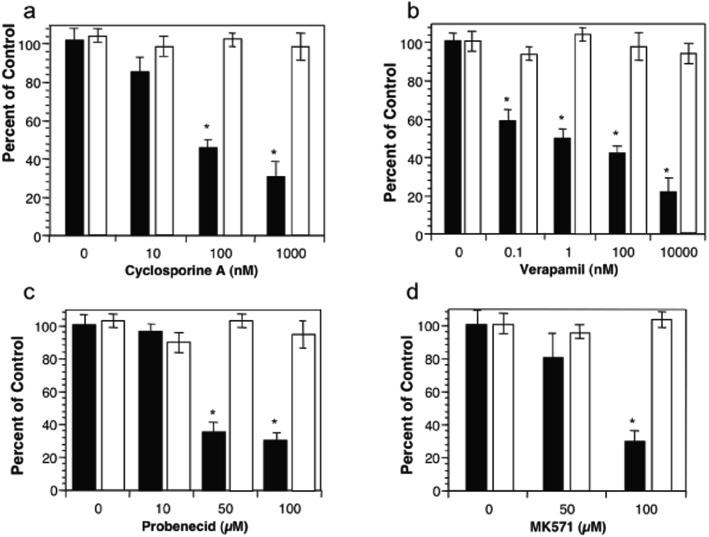
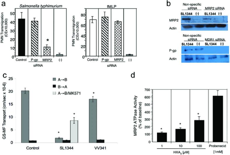
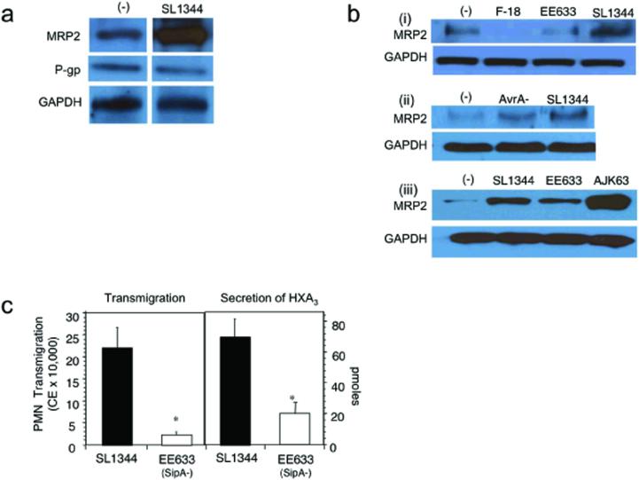
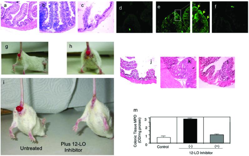
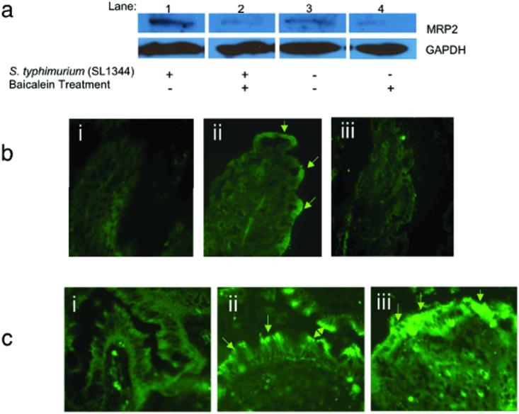
Neutrophil transmigration across mucosal surfaces contributes to dysfunction of epithelial barrier properties, a characteristic underlying many mucosal inflammatory diseases. Thus, insight into the directional movement of neutrophils across epithelial barriers will provide important information relating to the mechanisms of such inflammatory disorders. The eicosanoid hepoxilin A(3), an endogenous product of 12-lipoxygenase activity, is secreted from the apical surface of the epithelial barrier and establishes a chemotactic gradient to guide neutrophils from the submucosa across epithelia to the luminal site of an inflammatory stimulus, the final step in neutrophil recruitment. Currently, little is known regarding how hepoxilin A(3) is secreted from the intestinal epithelium during an inflammatory insult. In this study, we reveal that hepoxilin A(3) is a substrate for the apical efflux ATP-binding protein transporter multidrug resistance-associated protein 2 (MRP2). Moreover, using multiple in vitro and in vivo models, we show that induction of intestinal inflammation profoundly up-regulates apical expression of MRP2, and that interfering with hepoxilin A(3) synthesis and/or inhibition of MRP2 function results in a marked reduction in inflammation and severity of disease. Lastly, examination of inflamed intestinal epithelia in human biopsies revealed up-regulation of MRP2. Thus, blocking hepoxilin A(3) synthesis and/or inhibiting MRP2 may lead to the development of new therapeutic strategies for the treatment of epithelial-associated inflammatory conditions.


Figures

Figure 1
Figure 1
The effect of P-gp (MDR1) and MRP2 inhibitors on the ability of S. typhimurium to induce PMN transepithelial migration. T84 cell monolayers were treated for 1 hr with the P-gp inhibitors (cyclosporine A (10-1000 nM; (a)) or verapamil (0.1-10000 nM; (b)) or the MRP2 inhibitors (probenecid (10-100 μM; (c)) or MK571 (50-100 μM; (d)). The pre-treated monolayers were then infected at the apical surface with wild-type S. typhimurium SL1344 and judged for their ability to induce PMN transepithelial migration (see Methods). Closed bars (black) represent S. typhimurium induced PMN transepithelial migration while the open bars (white) represent PMN migration to imposed gradients of fMLP. Data are expressed as the mean and SD of triplicate samples. This experiment is representative of more than three performed. * P < 0.01
Figure 2
Figure 2
Demonstration of the functional importance of the apical ABC transporter MRP2 during PMN transepithelial migration and as a substrate for HXA3. a) Polarized intestinal cell monolayers silenced for the expression of MRP2 or P-gp (MDR1) were assessed for the ability to promote S. typhimurium-induced PMN transepithelial migration. Data are represented as mean ± SD of triplicate samples and are representative of three experiments; * P < 0.01. b) Immunoblot analysis of the modulation in protein expression of MRP2 and P-gp (MDR1) based on siRNA, as described in the Methods section. Results are representative of three independent experiments. c) MRP2 functional activity was assessed by measuring GS-MF efflux from polarized monolayers of T84 infected in the absence and presence of S. typhimurium. T84 cell monolayers were infected apically for 1 h with either SL1344 (wild type S. typhimurium) or VV341 (an isogenic derivative of SL1344 which lacks the hilA gene and is invasion deficient). Control represents baseline transport in the presence of physiologic buffer (HBSS). A→B represents transport in the apical to basolateral direction; B→A represents transport in the basolateral to apical direction; MK571 is an MRP2 inhibitor (100 μM containing 2% DMSO). Data are expressed as mean ± SD of triplicate samples and are representative of at least three independent experiments; * P < 0.01. d) Inorganic phosphate (Pi) yielded by hydrolysis of ATP used by the substrate HXA3 is represented as percent of the baseline response. Pi was measured as (nmol/mg/protein/min) and baseline values were determined to be 1.6 ± 0.15. Bars represent the mean ± SD of triplicate determinations representative of at least three independent experiments; * P < 0.05.
Figure 3
Figure 3
MRP2 is up-regulated by an inflammatory pathway activated by S. typhimurium that involves a SipA dependent-mechanism. a) T84 cells were infected apically with SL1344 for 1 h, then lysed, and enriched for the TX-100 insoluble membrane fraction as described under Methods. Incubation with the physiologic buffer HBSS served as the baseline control for MRP2 expression. 25 μg of protein was separated on a 10% polyacrylamide PAGE gel and immunoblotted for MRP2. b) To determine specificity of the response (i) T84 cell monolayers were apically infected with SL1344, the isogenic SipA mutant strain (EE633), and a normal intestinal human flora E. coli isolate, E. coli F-18, or left uninfected in the presence of HBSS ((−) control). (ii) T84 cells were infected apically for 1 h in the presence of either SL1344 or its isogenic AvrA (S. typhimurium effector protein) mutant strain. (−) represents the baseline buffer control. (iii) T84 cell monolayers were apically infected for 1 h in the presence of SL1344, EE633, or the sipA complemented strain AJK63. This strain is derived from EE633 containing the pAK68C plasmid that encodes sipA. (−) represents the baseline buffer control. For each immunoblot, following bacterial infection the cells were lysed and then enriched for the TX-100 insoluble membrane fraction. 25 μg of protein was separated on 10% polyacrylamide PAGE gels and immunoblotted for MRP2. The data represent a single experiment and are repetitive of at least three experiments performed. c) SipA affects the release of HXA3 from model intestinal epithelia. Monolayers of T84 epithelial cells were infected for 1 h with SL1344 or EE633 S. typhimurium strain. PMN transepithelial cell migration (left) and HXA3 secretion (right) were measured. HXA3 was identified according to Method 1 as described in Methods and Materials. Data are presented as the mean ± SD of assays performed in triplicate and are representative of three independent experiments; *P < 0.01.
Figure 4
Figure 4
MRP2 expression is modulated during active states of intestinal inflammation. a-c) Histopathology of the proximal large colon of mice following a 48 h infection with physiologic buffer (HBSS; a), SL1344 (b), and EE633 (c). Sections are stained with hemotoxylin and eosin (H and E) and represent a magnification of 20X. d-f) Immunohistochemistry of the expression pattern of MRP2 in the proximal large colon performed on the same set of mice described for a-c. Panels d, e, f represent buffer control, SL1344, and EE633 infected mice, respectively; 10X magnification. The enhanced boxed image in panel e illustrates the apical expression of MRP2 as denoted by the green arrows. g and h) Gross examination of the anal region in mice induced to exhibit characteristics of IBD using the CD4+ CD45RBhi T cell adoptive transfer model. Following induction of disease the mice generally exhibit significant rectal prolapse. i) Gross examination of the diseased mice (CD4+ CD45RBhi T cell adoptive transfer model) following a therapeutic intervention strategy using the 12/15-LOX inhibitor baicalein. j-l) H and E stained proximal colonic sections of mice not induced for IBD (j), mice induced for IBD (k), and mice induced for disease and then therapeutically treated with baicalein (l); 20X magnification. m) PMN infiltration into the proximal colon quantified by tissue myeloperoxidase (MPO). Data are expressed as the mean ± SD and represent groups of five mice per data set and the experiment was performed three times with similar results.
Figure 5
Figure 5
12/15-LOX activity and the expression of MRP2 are linked events in response to active states of intestinal inflammation. a) Immunoblot analysis of MRP2 expression during apical infection of SL1344 in the absence and presence of the 12-LOX inhibitor baicalein, as described in Methods. For each sample 25 μg of protein was isolated from the membrane fraction and run on a 10% polyacrylamide PAGE gel and immunoblotted for MRP2. The data represent a single experiment and are repetitive of at least three experiments performed. b) Immunohistochemistry depicting the localization of MRP2 to the apical epithelial surface of the proximal colon in mice treated in the absence and presence of 12/15-LOX inhibition using the CD4+ CD45RBhi T cell adoptive transfer model of IBD: (i) represents the healthy control which did not undergo the adoptive transfer of the CD4+ CD45RBhi T cells; (ii) represents mice induced for IBD; and; (iii) represents mice induced for IBD and then therapeutically treated with baicalein. 10X magnification. c) Human intestinal biopsy specimens stained for MRP2: (i) healthy colonic section; (ii) colonic section from a patient with active Crohn's disease, (iii); colonic section from a patient with active ulcerative colitis (20X magnification). Data represent a single experiment that is representative of eight patient colonic biopsy specimens examined (four each for Crohn's and ulcerative colitis patients).

Similar articles

Cited by

References

    1. McCormick BA, Hofman PM, Kim J, Carnes DK, Miller SI, Madara JL. Surface attachment of Salmonella typhimurium to intestinal epithelia imprints the subepithelial matrix with gradients chemotactic for neutrophils. J Cell Biol. 1995;131:1599–1608. - PMC - PubMed
    1. Mrsny RJ, Gewirtz AT, Siccardi D, Savidge T, Hurley BP, Madara JL, McCormick BA. Identification of hepoxilin A3 in inflammatory events: a required role in neutrophil migration across intestinal epithelia. Proc Natl Acad Sci U S A. 2004;101:7421–7426. - PMC - PubMed
    1. Pace-Asciak CR, Reynaud D, Demin P, Nigam S. The hepoxilins. A review. Adv Exp Med Biol. 1999;447:123–132. - PubMed
    1. Nigam S, Zafiriou MP, Deva R, Ciccoli R, Roux-Van der Merwe R. Structure, biochemistry and biology of hepoxilins: an update. Febs J. 2007;274:3503–3512. - PubMed
    1. Nigam S, Patabhiraman S, Ciccoli R, Ishdorj G, Schwarz K, Petrucev B, Kuhn H, Haeggstrom JZ. The rat leukocyte-type 12-lipoxygenase exhibits an intrinsic hepoxilin A3 synthase activity. J Biol Chem. 2004;279:29023–29030. - PubMed

Publication types

MeSH terms

Substances