. 2008 Dec 1;181(11):8153-61.
doi: 10.4049/jimmunol.181.11.8153.

Restoration of adenosine deaminase-deficient human thymocyte development in vitro by inhibition of deoxynucleoside kinases

Affiliations

Restoration of adenosine deaminase-deficient human thymocyte development in vitro by inhibition of deoxynucleoside kinases

Michelle L Joachims et al. J Immunol. .

Abstract

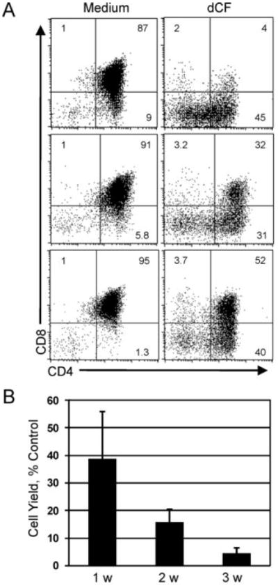
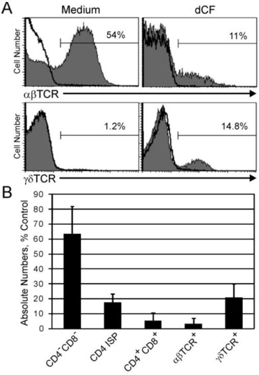
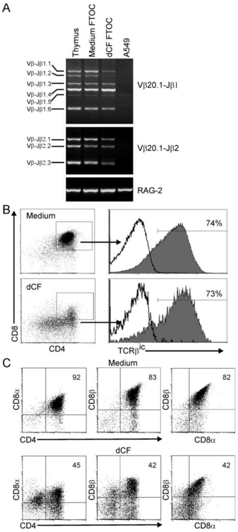
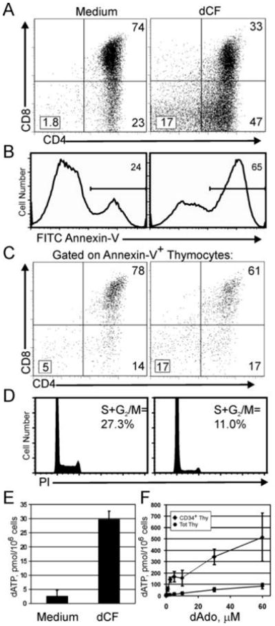
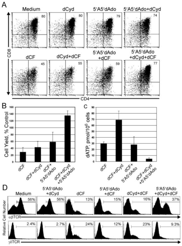
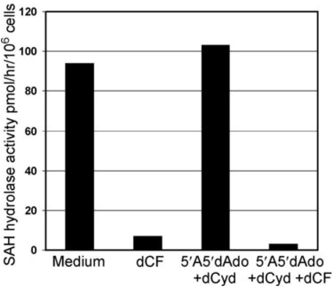
Mutations in the gene encoding adenosine deaminase (ADA), a purine salvage enzyme, lead to immunodeficiency in humans. Although ADA deficiency has been analyzed in cell culture and murine models, information is lacking concerning its impact on the development of human thymocytes. We have used chimeric human/mouse fetal thymic organ culture to study ADA-deficient human thymocyte development in an "in vivo-like" environment where toxic metabolites accumulate in situ. Inhibition of ADA during human thymocyte development resulted in a severe reduction in cellular expansion as well as impaired differentiation, largely affecting mature thymocyte populations. Thymocyte differentiation was not blocked at a discrete stage; rather, the paucity of mature thymocytes was due to the induction of apoptosis as evidenced by activation of caspases and was accompanied by the accumulation of intracellular dATP. Inhibition of adenosine kinase and deoxycytidine kinase prevented the accumulation of dATP and restored thymocyte differentiation and proliferation. Our work reveals that multiple deoxynucleoside kinases are involved in the phosphorylation of deoxyadenosine when ADA is absent, and suggests an alternate therapeutic strategy for treatment of ADA-deficient patients.


Figures

Figure 1
Figure 1
Human thymocyte yield and differentiation are severely impaired in ADA-deficient hu/moFTOC. A. CD34+DN cells from human thymus were cultured in hu/moFTOC in Yssel’s complete medium ± 5 μM dCF for 3 wk, then harvested, stained with antibodies to CD4 and CD8. Three individual experiments are shown. B. Cell yield from dCF-treated cultures expressed as a percentage of control cultures harvested at wk 1, 2 and 3 of culture (mean ± SD, n ≥ 6). Live cells/lobe were determined by trypan blue staining and counting.
Figure 2
Figure 2
Mature thymocytes are most affected in ADA-deficient hu/moFTOC. A. Cells from hu/moFTOC ± 5 μM dCF were harvested at 3 wk, stained with antibodies to αβTCR and γδTCR, and analyzed by flow cytometry (shaded histograms). Isotype control antibody staining is indicated by the black line histograms. B. Absolute numbers of human thymocyte subpopulations in dCF-treated hu/moFTOC at 3 wk expressed as a percentage of control cultures (mean ± SD, n ≥ 3).
Figure 3
Figure 3
Human thymocyte development in the absence of ADA is not blocked at a particular stage. A. Cells from hu/moFTOC ± 5 μM dCF were harvested at 3 wk and genomic DNA prepared. TCR gene rearrangements from human Vβ20.1 to either the Jβ1 or the Jβ2 region were analyzed by PCR and agarose gel electrophoresis. Genomic DNA from total human thymocytes (Thymus) or human fibroblast cells (A549) were used as positive and negative controls, respectively. B and C. Cells from cultures were harvested as in A, stained with antibodies to CD4, CD8α, CD8β (C) and analyzed using flow cytometry. Following the surface stains, the cells were stained for TCRβic (B) as described in Materials and Methods. The dot plots shown are gated on all live cells, and the histograms for TCRβic expression are shown gated on the DP cells from each condition. Isotype control staining is indicated by the black line histograms.
Figure 4
Figure 4
Induction of apoptosis, inhibition of cell proliferation and accumulation of dATP in cells from ADA-inhibited hu/moFTOC. A-D: Hu/moFTOCs were incubated for 11 days ± 5 μM dCF; then cells were harvested and stained. One representative experiment out of 3 is shown. A. Surface staining with anti-CD4 and anti-CD8 of total thymocytes. B. Annexin-V staining on total thymocytes from A. C.CD4/CD8 phenotype of Annexin-V+ cells in A. D. Cell cycle analysis of thymocytes from 11d culture stained with propidium iodide as described in Materials and Methods. E. Cells from 11d hu/moFTOCs were processed for dATP analysis as described in Materials and Methods. dATP is expressed in pmol/million cells (mean ± SD, n=4). F. Total and CD34+ thymocytes were incubated with 5 μM dCF ± dAdo for 20 h in suspension culture; then cells were harvested and analyzed for dATP as in E (mean ± SD, n=4).
Figure 5
Figure 5
Rescue of ADA-deficient phenotype and dATP accumulation in hu/moFTOC by inhibition of deoxynucleoside kinases. A. Human CD34+ cells were cultured in hu/moFTOC in medium alone, 5 μM 5′A5′dAdo, 50 μM dCyd, or 50 μM dCyd + 5 μM 5′A5′dAdo ± 5 μM dCF for 10 d, then harvested, counted, and stained with antibodies to CD4 and CD8. B. Cell yield from experiments as in A, expressed as a percentage of each control condition (mean ± SD, n=4). C. Analysis of dATP accumulation for experiments as in A. dATP values are expressed as pmol/million cells (mean ± SD, n=4). D. Hu/moFTOCs incubated with the indicated combinations of 5 μM dCF, 5 μM 5′A5′dAdo and 50 μM dCyd were harvested after 3 wk, stained with antibodies to either αβTCR or γδTCR and analyzed by flow cytometry.
Figure 6
Figure 6
SAH hydrolase activity is inhibited in rescued ADA-deficient hu/moFTOC. SAH hydrolase activity was measured as described in Materials and Methods in cell extracts of thymocytes harvested from hu/moFTOC cultured for 12 d. Results are expressed as pmol/hr/106 cells. The data shown are representative of two separate experiments.

References

    1. Giblett ER, Anderson JE, Cohen F, Pollara B, Meuwissen HJ. Adenosine-deaminase deficiency in two patients with severely impaired cellular immunity. Lancet. 1972;2:1067–1069. - PubMed
    1. Hershfield MS, Mitchell BS. Immunodeficiency diseases caused by adenosine deaminase deficiency and purine nucleoside phosphorylase deficiency. In: Scriver CR, Sly WS, Childs B, Beaudet AL, Valle D, Kinzler K, Vogelstein B, editors. The metabolic and molecular bases of inherited disease. Vol. 2. McGraw-Hill, Inc.; New York: 2001. p. 2585.
    1. Blackburn MR, Kellems RE. Adenosine deaminase deficiency: metabolic basis of immune deficiency and pulmonary inflammation. Adv Immunol. 2005;86:1–41. - PubMed
    1. Linch DC, Levinsky RJ, Rodeck CH, Maclennan KA, Simmonds HA. Prenatal diagnosis of three cases of severe combined immunodeficiency: severe T cell deficiency during the first half of gestation in fetuses with adenosine deaminase deficiency. Clin Exp Immunol. 1984;56:223–232. - PMC - PubMed
    1. Donofrio J, Coleman MS, Hutton JJ. Overproduction of adenine deoxynucleosides and deoxynucleotides in adenosine deaminase deficiency with severe combined immunodeficiency disease. J Clin Invest. 1978;62:884–887. - PMC - PubMed

Publication types

MeSH terms

Substances