

Preservation of neuronal number despite age-related cortical brain atrophy in elderly subjects without Alzheimer disease
- PMID: 19018241
- PMCID: PMC2734185
- DOI: 10.1097/NEN.0b013e31818fc72f
Preservation of neuronal number despite age-related cortical brain atrophy in elderly subjects without Alzheimer disease
Abstract
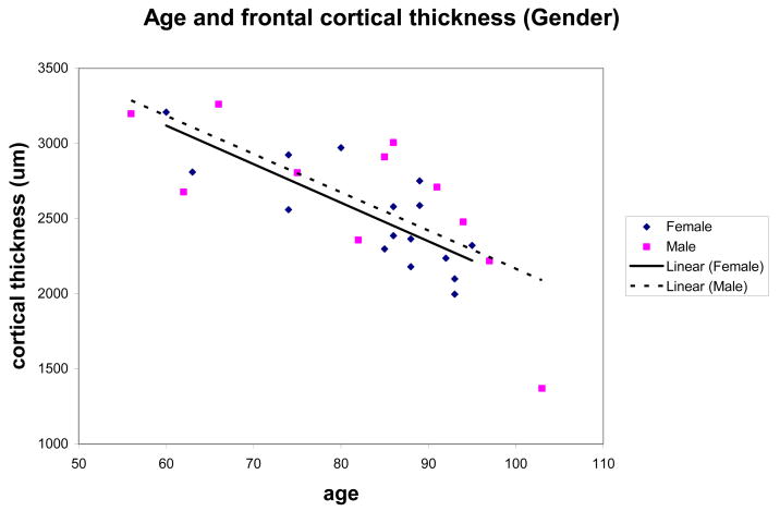
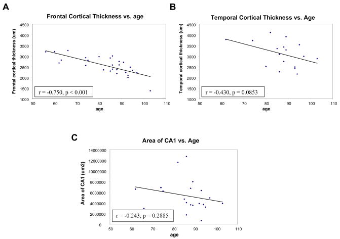
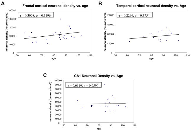
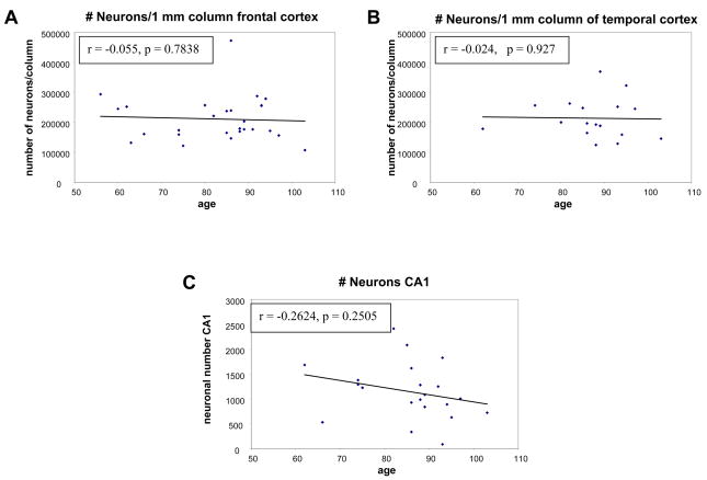
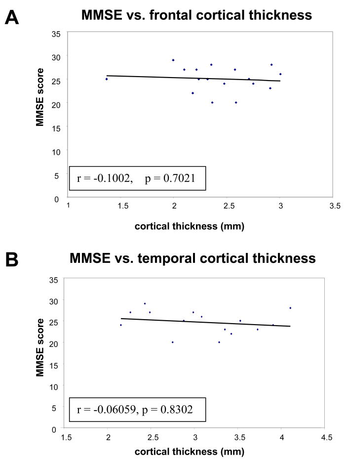
Cerebral volume loss has long been associated with normal aging, but whether this is due to aging itself or to age-related diseases, including incipient Alzheimer disease, is uncertain. To understand the changes that occur in the aging brain, we examined the cerebral cortex of 27 normal individuals ranging in age from 56 to 103 years. None fulfilled the criteria for the neuropathologic diagnosis of Alzheimer disease or other neurodegenerative disease. Seventeen of the elderly participants had cognitive testing an average of 6.7 months prior to death. We used quantitative approaches to analyze cortical thickness, neuronal number, and density. Frontal and temporal neocortical regions had clear evidence of cortical thinning with age, but total neuronal numbers in frontal and temporal neocortical regions remained relatively constant during a 50-year age range. These data suggest that loss of neuronal and dendritic architecture, rather than loss of neurons, underlies neocortical volume loss with increasing age in the absence of Alzheimer disease.
Figures
References
-
- Scahill RI, Frost C, Jenkins R, Whitwell JL, Rossor MN, Fox NC. A longitudinal study of brain volume changes in normal aging using serial registered magnetic resonance imaging. Arch Neurol. 2003;60:989–94. - PubMed
-
- Fotenos AF, Snyder AZ, Girton LE, Morris JC, Buckner RL. Normative estimates of cross-sectional and longitudinal brain volume decline in aging and AD. Neurology. 2005;64:1032–39. - PubMed
-
- De Toledo-Morrell L, Goncharova I, Dickerson B, Wilson RS, Bennett DA. From healthy aging to early Alzheimer’s disease: In vivo detection of entorhinal cortex atrophy. Ann N Y Acad Sci. 2000;911:240–53. - PubMed
Publication types
MeSH terms
Grants and funding
- R03 AG024633/AG/NIA NIH HHS/United States
- R01 AG008487/AG/NIA NIH HHS/United States
- P50 AG005134/AG/NIA NIH HHS/United States
- T32 AG023480/AG/NIA NIH HHS/United States
- R01-AG021133/AG/NIA NIH HHS/United States
- AG004390/AG/NIA NIH HHS/United States
- R01 AG021133/AG/NIA NIH HHS/United States
- AG08487/AG/NIA NIH HHS/United States
- P30 AG062421/AG/NIA NIH HHS/United States
- R21-AG023661/AG/NIA NIH HHS/United States
- R21 AG023661/AG/NIA NIH HHS/United States
- P01 AG004390/AG/NIA NIH HHS/United States
- R03-AG024633/AG/NIA NIH HHS/United States
- AG05134/AG/NIA NIH HHS/United States
LinkOut - more resources
Full Text Sources
Medical
