. 2008 Nov 18;6(11):e284.
doi: 10.1371/journal.pbio.0060284.

Drosophila neurotrophins reveal a common mechanism for nervous system formation

Affiliations

Drosophila neurotrophins reveal a common mechanism for nervous system formation

Bangfu Zhu et al. PLoS Biol. .

Abstract

Neurotrophic interactions occur in Drosophila, but to date, no neurotrophic factor had been found. Neurotrophins are the main vertebrate secreted signalling molecules that link nervous system structure and function: they regulate neuronal survival, targeting, synaptic plasticity, memory and cognition. We have identified a neurotrophic factor in flies, Drosophila Neurotrophin (DNT1), structurally related to all known neurotrophins and highly conserved in insects. By investigating with genetics the consequences of removing DNT1 or adding it in excess, we show that DNT1 maintains neuronal survival, as more neurons die in DNT1 mutants and expression of DNT1 rescues naturally occurring cell death, and it enables targeting by motor neurons. We show that Spätzle and a further fly neurotrophin superfamily member, DNT2, also have neurotrophic functions in flies. Our findings imply that most likely a neurotrophin was present in the common ancestor of all bilateral organisms, giving rise to invertebrate and vertebrate neurotrophins through gene or whole-genome duplications. This work provides a missing link between aspects of neuronal function in flies and vertebrates, and it opens the opportunity to use Drosophila to investigate further aspects of neurotrophin function and to model related diseases.


Conflict of interest statement

Competing interests. The authors have declared that no competing interests exist.

Figures

Figure 1
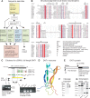
Figure 1. Identification of a Drosophila Neurotrophin
(A) Bioinformatic searches used to identify DNT1. (B) Sequence-structure alignment of the Cysknot domains of DNT1 and representative neurotrophins: Arrows: β-strands; spirals: α-helices; identical residues are shown in white over red (6 Cys and Gln conserved in all NTs); conservative substitutions in the hydrophic core in red. (C) Evidence for the existence of cDNA3 spanning the pro- and Cysknot domains. DNT1 locus showing exons (green), introns (gaps), and UTRs (red), Cysknot domain (shaded). P1, P2: predicted promoters. Yellow box indicates the region deleted in the DNT141 and DNT155 mutant alleles. cDNA3 was amplified by PCR on a larval/pupal cDNA library using primers to the 5′ untranslated end of CG18318 or from the ATG and to the 3′ untranslated end of CG18318. RT-PCR was carried out on RNA using bridging primers: 5′ primers were located at the 3′ of cDNA1 from CG32244, and 3′ primers were located at the 3′ end of the Cysknot domain. Amplification by RT-PCR with these primers is only possible if an mRNA spanning both CG32244 and CG32242 exists. (D) Model of the DNT1 protomer: blue is N-terminus, red is C-terminus, predicted disulphide bonds shown as ball-and-stick model. (E) DNT1 protein. SP: signal peptide; p.c: predicted cleavage site at position 498; Cysknot (black). (F) Cleavage products (arrows) of DNT1(cDNA3-V5) detected with anti-V5 in S2 cells. (G) The DNT1 Cysknot (CK) or Cysknot3′tail (CK3′tail) form dimers (arrows) when expressed in S2 cells: western blots showing tagged SP-CK-V5 and SP-CK3′tail-V5 detected with anti-V5 antibodies run under reducing (R) and nonreducing (Non-R) conditions. The dimers dissociate into monomers in reducing conditions.
Figure 2
Figure 2. DNT1 Is Highly Conserved in Insects
Alignment of the DNT1 Cysknot domain plus some flanking sequences to orthologs from insects with sequenced genomes, including 12 Drosophila species, three mosquito species (Anopheles and Culex), honey bee (Apis), jewel wasp (Nasonia), and human body louse (Pediculus). Identical residues are shown in white over red; conservative substitutions in red. DNT1 is very highly conserved throughout the whole protein sequence in all Drosophila species (full sequence not shown, but note sequences flanking the Cysknot) and very highly conserved within the Cysknot in all insects, but outside Drosophila, the sequences outside the Cysknot diverge.
Figure 3
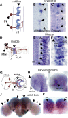
Figure 3. DNT1 Is Expressed in Target Cells
In situ hybridisations to DNT1 transcripts: (B) probe that detects all isoforms and (C, E, F, and H–K) DNT1-Cysknot–specific probe. (A, D, and G) Target tissues expressing DNT1 (blue), targeting neurons (brown). DNT1 expression in: (B and C) the embryonic midline (ml) (arrows); (E and F) the embryonic muscle; (F) higher magnification, showing muscles 6, 7, 12, and 13, arrows (A–F), anterior is up; (H and I) the larval lamina (la, arrows); (J and K) the adult brain: (J) dorsal view, DNT1 is expressed in multiple locations (arrows) including the optic lobes (ol) and central brain; (K) ventral view of central brain, transcripts are present in the cell bodies surrounding the calyx (ca) neuropile (arrows) of the mushroom bodies, site of learning and memory. (K) shows the same specimen as (J). IN, interneuron; me, medulla; MN, motor neuron.
Figure 4
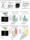
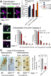
Figure 4. DNT1 Promotes Neuronal Survival in the CNS
(A) Embryos are stained with Caspase-3, and six to seven trunk segments of the nerve cord (VNC) are scanned at the confocal microscope (see Text S1), excluding the head and the posterior end. After acquisition, apoptotic cells within a region of interest (ROI) comprising the scanned VNC and excluding the epidermis are quantified automatically. GOF, gain of function; LOF, loss of function; wt, wild type. (B and D) Examples of Caspase-3–stained VNCs. GAL4 targeted expression to: all neurons (elavGAL4) and midline (simGAL4). Illustration of GOF and RNAi constructs used. (C and E) Results from the automatic quantification of α-Caspase-3–positive cells using DeadEasy software of (stage 13/14 for GOF and stage 17 for LOF). RNAi targeted to the pro-domain or the Cysknot in embryos heterozygote for DNT141 or Df(3L)ED4342. RNAi targeted to the pro-domain knocks-down all DNT1 transcripts, thus all these RNAi constructs reduce cDNA3 levels. Black asterisks are comparisons to wild type, red asterisks to controls. Triple asterisks (***) indicate p < 0.001, double asterisks (**) indicate p < 0.01, and a single asterisk (*) indicates p < 0.05. Numbers over graphs are sample sizes: n = number of embryos. For p-values and statistics tests, see Text S1. CK, DNT1-Cystine-knot; CK3+, Cysknot3-tail; FL, full-length cDNA3-GFP; pro, pro-domain cDNA1; wt, wild type embryos.
Figure 5
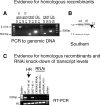
Figure 5. Evidence for Homologous Recombination and RNAi
(A and B) Evidence that DNT141 and DNT155 are homologous recombinant alleles: (A) PCR evidence that DNT141 (41) and DNT155 (55) are null alleles as the coding region of DNT1 has been replaced for that of the white gene: 5′ arm: product using primers to white and tie; 3′ arm: product using primers to white and CG13720; DNT: 5′ fragment of coding region. CK, Cysknot domain; wt, wild-type control (yw stock). (B) Southern blot evidence that in DNT141, the wild-type EcoRI 5.6-kb band shifts to 7.2 kb due to the insertion of white. (C) Verification of lack of transcripts in DNT1 homologous recombinant mutants and knock-down upon targeted RNAi. RT-PCR showing lack of transcripts in DNT141 null mutant embryos and normal levels of transcripts in DNT141/+ heterozygous and wild-type embryos (controls). Transcript levels are reduced upon targeted DNT1 RNAi to the midline using simGAL4 for all three RNAi constructs.
Figure 6
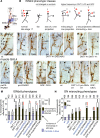
Figure 6. Apoptosis and Loss of Interneurons and Motor Neurons upon DNT1 Loss of Function
Embryonic VNC stained with Caspase-3, and the interneurons and motor neuron markers HB9 and Eve. Quantification of colocalising cells is done manually by looking at each individual section. (A) Colocalisation (arrows, single 0.5-μm section confocal images) of Caspase-3 (green) and HB9 (magenta) (stage 16–17). Graph shows that the percentage of segments with more than one apoptotic neuron increases in DNT1 mutants, n = number of segments. wt, wild type. (B) Colocalisation of Eve (magenta) and Caspase-3 (green), quantification on the right. INs are EL interneurons, MNs are U/CQ motor neurons, n = number of embryos. (C) Loss of Eve-positive cells, arrows point at missing cells in DNT141 mutant embryos. On the right, quantification of Eve-positive cells per segment, numbers over bars: n = number of segments. Triple asterisks (***) indicate p < 0.001, double asterisks (**) indicate p < 0.01, and a single asterisk (*) indicates p < 0.05. For p-values and statistics tests, see Text S1.
Figure 7
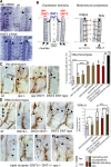
Figure 7. DNT1 Enables Motor-Axon Targeting
(A) Projections of motor neurons to the embryonic muscles. (B) Altered DNT1 function results in an increase in ISNb/d misrouting phenotypes. In wild type, there are three stereotypic projections (I, II, and III). Two phenotypes were observed in all genotypes with comparable frequency: “fan” of multiple thin projections originating from II, and “loss” of one or more projections. Misrouting phenotypes, including concomitant effects in two or more projections (e.g., misrouting in two projections, or misrouting in one plus loss of another), are found with higher frequency in experimental genotypes (some examples drawn). (C–H) ISNb/d motor neuron targeting at muscles 7, 6, 13, and 12 visualised with FasII antibodies (brown) in stage 17: (C) wild-type embryos; (D) DNT141/Df(3L)ED4342 transheterozygote mutants; (E) DNT141 mutants; (F and G) upon targeted RNAi to the muscle: (F) 24BGAL4 > pWCysknotRNAi;DfED4342 and (G) 24BGAL4 > pro-RNAi;DfED4342; (H) upon expression of Cysknot3′tail at the muscle (24BGAL4 > UASCysknot3′tail). GOF, gain of function; LOF, loss of function; mr, misrouting; wt, wild type. (I–L) SN projections: (I) wild type; (J) DNT141/Df(3L)ED4342; (K) 24BGAL4 > pro-RNAi;DfED4342; (L) 24BGAL4 > UASCysknot3-tail. Arrowheads point to projections or misroutings, asterisks to missing projections. Dorsal is up, anterior to the left. (M and N) Quantification of ISNb/d and SN phenotypes: misrouting and effects in two or more projections are shown in brown and for controls in blue. Triple asterisks (***) indicate p < 0.001, double asterisks (**) indicate p < 0.01, and a single asterisk (*) indicates p < 0.05. Black asterisks are comparisons to wild type, red asterisks to controls. Numbers over graphs indicate number of hemisegments. For statistics tests and p-values, see Text S1.
Figure 8
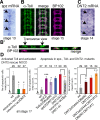
Figure 8. Spz and DNT2 Have Neurotrophic Function
(A) In situ hybridisation in a stage 13 embryo showing spz transcripts at the CNS midline. (B) Distribution of anti-Toll protein (green) in all CNS neuropile axons, as seen by colocalisation (merge) with the axonal marker BP102 (magenta) at stage 16. Toll is also present in some midline cells. (B′) Transverse sections through the neuropile to show that Toll coincides with the distribution of BP102. (C) In situ hybridisation in a stage 14 embryo showing DNT2 transcripts at the CNS midline. (D) Automatic quantification of Caspase-3 in loss-of-function and gain-of-function conditions for spz, Toll, and DNT2 using DeadEasy software. Expression of activated Toll and DNT2 (UASDNT2-Cysknot) in all neurons (with ElavGAL4) rescue NOCD: stage 17 embryos. Apoptosis in spz2, Tollr3/Dfro80b, and DNT2e03444 mutants: stage 17 embryos. The incidence of apoptosis does not increase in spz2DNT141 double mutants compared to either of the single mutants. Rescue: expression of activated Spz (UASspz-Cysknot) in all neurons with elavGAL4 in DNT141 null mutant embryos only partially rescues cell death. Apoptosis increases in DNT1DNT2 double mutants compared to the single mutants. Triple asterisks (***) indicate p < 0.001, double asterisks (**) indicate p < 0.01, and a single asterisk (*) indicates p < 0.05. Numbers over bars are n = number of embryos. For p-values and statistics tests, see Text S1. wt, wild type.
Figure 9
Figure 9. Complementary and Synergistic Functions of DNTs in Targeting
(A) Distribution of spz mRNA in two lateral bands below the epidermis, at the location of the segment boundary muscle (SBM) and lateral transverse (LT) muscles. Below, distribution of DNT2 transcripts in two domains: over muscles 6, 7, 12, and 13 overlapping with DNT1 expression (arrowheads) and a second domain overlapping with spz expression (arrow). DNT2 is also expressed in other muscles (not shown). ml, midline. (B) Diagrams illustrating the complementary expression domains of DNT1, DNT2, and Spz. On the far right, the motor neuron projections from ISNb/d are shown on the left half, and the projections from SN are shown on the right half for clarity. ISNb/d project to muscles 6, 7, 12, and 13 that express DNT1 and DNT2; SNa project to SBM and LT muscles that express spz and DNT2. (C and D) Quantifications of axonal phenotypes are colour coded by genotype: controls in blue; single mutants in gold; double mutants in red; and triple mutants in dark brown. (C) Axonal phenotypes of the SNa motor axons, quantification on the right. There is a significantly higher percentage of hemisegments with axonal misrouting defects in the SNa projections in spz2 , spz2DNT141 double, and DNT2eo3444DNT141 spz2 triple mutants than in DNT141 mutants. No significant increase in axonal misroutings was found for ISNb/d projections in spz2 mutants. (D) ISNb/d phenotypes in DNT2 eo3444/Df6092, DNT2eo3444DNT141 double-, and DNT2eo3444DNT141 spz2 triple-mutant embryos. Both the frequency and severity of misrouting phenotypes (arrowheads) increase in the double and triple mutants, including loss of all projections (asterisks). Two hemisegments are shown for the triple mutant. (E and F) Dramatic misroutings in DNT2eo3444DNT141 spz2 triple-mutant embryos in (E) the transverse nerve (TN) (which, however, do not increase in frequency) and (F) ISN (penetrance in triple mutants 12.7% vs. 0%–1.8% in single and double mutants, respectively). Triple asterisks (***) indicate p < 0.001, double asterisks (**) indicate p < 0.01, and a single asterisk (*) indicates p < 0.05. Numbers over bars are n = number of hemisegments. (C–F) all stage 17. All axonal images show projections in one hemisegment, except for far right in (D and F) that show two. For p-values and statistic tests, see Text S1.
Figure 10
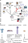
Figure 10. A Common Origin for the NT Superfamily Underlies Nervous System Structure and Function
(A) NT superfamily members identified in protostomes (Drosophila) and deuterostomes (vertebrates, Amphioxus, sea urchin, and acorn worm) imply that NTs were present in Urbilateria, their common ancestor. A protostomian NT gene would have duplicated to give rise to DNT1, DNT2, and spz in insects or perhaps earlier. A chordate NT duplicated twice to give rise to the four vertebrate NTs, and the NGF ortholog duplicated again in fish to result in NT6/7. Identified Trk and p75 receptors are also shown; Trk-like receptors lack some extracellular domains. NT receptors and signalling mechanisms may have diversified through evolution. Annelids, flatworms, nematodes, and tunicates are not shown, see Figure S1. (B) Hypothesis that the NTs are required in all centralised nervous systems to link structure and function. NTs are also present at least in acorn worm with a nerve net, a diffuse nervous system, where NT may play a subset of functions, suggesting that NTs could also be present in lower animals (e.g., Cnidarians). Drosophila can be used as a model system for NT-related studies.

References

    1. Levi-Montalcini R. The nerve growth factor 35 years later. Science. 1987;237:1154–1162. - PubMed
    1. Blum R, Konnerth A. Neurotrophin-mediated rapid signaling in the central nervous system: mechanisms and functions. Physiology. 2005;20:70–78. - PubMed
    1. Lu B, Pang PT, Woo NH. The yin and yang of neurotrophin action. Nat Rev Neurosci. 2005;6:603–614. - PubMed
    1. Davies AM. Regulation of neuronal survival and death by extracellular signals during development. EMBO J. 2003;22:2537–2545. - PMC - PubMed
    1. Zweifel L. Functions and mechanisms of retrograde neurotrophin signalling. Nat Rev Neurosci. 2005;6:615–625. - PubMed

Publication types

MeSH terms