. 2008 Nov;9(6):741-51.
doi: 10.1111/j.1364-3703.2008.00498.x.

Identification of differentially expressed root genes upon rhizomania disease

Affiliations

Identification of differentially expressed root genes upon rhizomania disease

Laure Schmidlin et al. Mol Plant Pathol. 2008 Nov.

Abstract

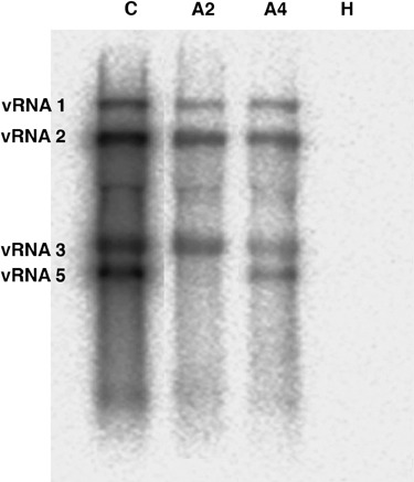
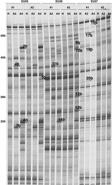
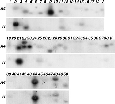
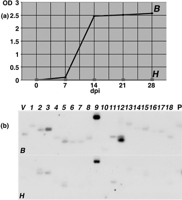
Rhizomania is one of the most devastating sugar beet diseases. It is caused by Beet necrotic yellow vein virus (BNYVV), which induces abnormal rootlet proliferation. To understand better the physiological and molecular basis of the disorder, transcriptome analysis was performed by restriction fragment differential display polymerase chain reaction (RFDD-PCR), which provided differential gene expression profiles between non-infected and infected sugar beet roots. Two distinct viral isolates were used to detect specific or general virus-induced genes. Differentially expressed genes were selected and identified by sequence analysis, followed by reverse Northern and reverse transcriptase PCR experiments. These latter analyses of different plants (Beta vulgaris and Beta macrocarpa) infected under distinct standardized conditions revealed specific and variable expressions. Candidate genes were linked to cell development, metabolism, defence signalling and oxidative stress. In addition, the expression of already characterized genes linked to defence response (pathogenesis-related protein genes), auxin signalling and cell elongation was also studied to further examine some aspects of the disease. Differential expression was retrieved in both B. vulgaris and B. macrocarpa. However, some candidate genes were found to be deregulated in only one plant species, suggesting differential response to BNYVV or specific responses to the BNYVV vector.


Figures

Figure 1
Figure 1
BNYVV RNA detection within total RNAs from Beta vulgaris roots infected by B‐type (A2) or P‐type (A4) BNYVV viral isolates. Total RNAs from non‐infected roots (H) were also tested. Viral RNA 1, 2, 3 and 5 were detected by Northern blotting with specific riboprobes. RNA 4 probe was omitted to allow the visualization of the RNA 5 species. Positions of viral RNAs were determined with the detection of viral RNAs within total RNAs from Chenopodium quinoa leaf lesions induced by a P‐type BNYVV isolate (C). Positions of viral species are indicated on the left.
Figure 2
Figure 2
Autoradiogram obtained from RFDD‐PCR analysis showing the amplification products of RNAs from Beta vulgaris healthy roots (H) used as reference, and from roots infected by a B‐type BNYVV isolate (A2) or a P‐type BNYVV isolate (A4). Amplification reactions were made in duplicates (nos. 1 and 2) using the labelled 0‐extension primer in combination with one of the three displayPROBE primers (EU25, EU26 and EU27) tested in this example. Differentially expressed bands (detected here in infected samples) are indicated by dots and FDD name reported in Table 1. Names in italic correspond to BNYVV amplified sequences. Numbers on the left refer to the size of PCR fragments (base pairs).
Figure 3
Figure 3
Expression patterns of genes selected after the initial characterization. Reverse Northern analysis consisted of replicate membranes harbouring PCR products of cDNA clones hybridized with 32P‐labelled cDNA probes derived from Beta vulgaris healthy (H panels) or infected (A4 panels) root mRNAs. The identifier of each cDNA indicated on the top is also reported in Table 1. A viral sequence (V) was used as a positive control for hybridizations.
Figure 4
Figure 4
(a) BNYVV detection in Beta vulgaris roots infected by a B‐type BNYVV isolate (B) using standardized conditions (bioassay, SESVanderHave laboratory) or in control roots (H). Viral contents were monitored by ELISA analysis for BNYVV CP protein detection. Results are expressed as an optical density value (OD). (b) Differential expression of RFDD‐PCR candidate genes after B. vulgaris standardized infections. Reverse Northern hybridizations were performed with 32P‐labelled cDNA probes derived from B. vulgaris healthy (H) or infected (B) root RNAs extracted 14 days after treatment. The identifier of each cDNA indicated on the top is also reported in Table 1. A viral sequence (V) and a B. vulgaris beta‐1,3‐glucanase (accession no. BQ591809) PR‐2 sequence (P) were used as controls.
Figure 5
Figure 5
(a) BNYVV localization in Beta macrocarpa roots after plant systemic infection. Viral contents of B. macrocarpa roots harvested 13 days after infection (I) or control roots (H) were monitored by immunoprinting and permitted the localization of infected tissues (i) and non‐infected tissues (h) selected for subsequent total RNA extraction. Background on control roots (H) corresponds to the processing of chemiluminescent reagent by cellular peroxidases. (b) Differential expression of RFDD‐PCR genes after B. macrocarpa systemic infection. Reverse Northern hybridizations were performed with cDNA probes derived from B. macrocarpa healthy (h) or infected (i) root RNAs. Identifiers of cDNA clones are indicated on the top. A viral sequence (V) was used as a positive control, as well as a B. vulgaris beta‐1,3‐glucanase PR‐2 (P) sequence (accession no. BQ591809) and a ubiquitin (U) sequence (accession no. BQ583989).
Figure 6
Figure 6
Expression patterns of RFDD‐PCR candidate genes and other genes of interest in Beta vulgaris and Beta macrocarpa roots upon response to early BNYVV infection. Analyses were conducted on healthy (H) or infected (B) B. vulgaris root total RNAs extracted after 7 days of bioassay treatment and on healthy (h) or infected (i) B. macrocarpa root total RNAs extracted after 13 days of plant systemic infection. Semi‐quantitative RT‐PCR was standardized with the L11 ribosomal protein gene (accession no. BE590348) and the BNYVV RNA 3 sequence was used as a viral control. Signals were quantified with ImageJ 1.38w software and transcript accumulation was expressed as a ratio between infected value and non‐infected value (ratio I/H or i/h). Identifiers (no.) of RFDD‐PCR cDNAs are those indicated in Table 1.

Similar articles

Cited by

References

    1. Angot, A. , Vergunst, A. , Genin, S. and Peeters, N. (2007) Exploitation of eukaryotic ubiquitin signaling pathways by effectors translocated by bacterial type III and type IV secretion systems. PLoS Pathog. 3, e3. - PMC - PubMed
    1. Asai, T. , Stone, J.M. , Heard, J.E. , Kovtun, Y. , Yorgey, P. , Sheen, J. and Ausubel, F. (2000) Fumonisin B1‐induced cell death in arabidopsis protoplasts requires jasmonate‐, ethylene‐, and salicylate‐dependent signaling pathways. Plant Cell, 12, 1823–1836. - PMC - PubMed
    1. Burketova, L. , Stillerova, K. and Feltlova, M. (2003) Immunohistological localization of chitinase and beta‐1,3‐glucanase in rhizomania‐diseased and benzothiadiazole treated sugar beet roots. Physiol. Mol. Plant Pathol. 63, 47–54.
    1. Cassab, G.I. (1998) Plant cell wall proteins. Annu. Rev. Plant Physiol. Plant Mol. Biol. 49, 281–309. - PubMed
    1. Chen, Z. , Agnew, J.L. , Cohen, J.D. , He, P. , Shan, L. , Sheen, J. and Kunkel, B.N. (2007) Pseudomonas syringae type III effector AvrRpt2 alters Arabidopsis thaliana auxin physiology. Proc. Natl. Acad. Sci. USA, 104, 20131–20136. - PMC - PubMed

Publication types

MeSH terms

LinkOut - more resources