. 2009 Jul;127(3):408-17.
doi: 10.1111/j.1365-2567.2008.02984.x. Epub 2008 Nov 14.

Evidence for a human leucocyte antigen-DM-induced structural change in human leucocyte antigen-DObeta

Affiliations

Evidence for a human leucocyte antigen-DM-induced structural change in human leucocyte antigen-DObeta

Francis Deshaies et al. Immunology. 2009 Jul.

Abstract

Human leucocyte antigen (HLA)-DO is a non-classical major histocompatibility complex class II molecule which modulates the function of HLA-DM and the loading of antigenic peptides on molecules such as HLA-DR. The bulk of HLA-DO associates with HLA-DM and this interaction is critical for HLA-DO egress from the endoplasmic reticulum. HLA-DM assists the early steps of HLA-DO maturation presumably through the stabilization of the interactions between the N-terminal regions of the alpha and beta chains. To evaluate a possible role for HLA-DM in influencing the conformation of HLA-DO, we made use of a monoclonal antibody, Mags.DO5, that was raised against HLA-DO/DM complexes. Using transfected cells expressing mismatched heterodimers between HLA-DR and -DO chains, we found that the epitope for Mags.DO5 is located on the DObeta chain and that Mags.DO5 reactivity was increased upon cotransfection with HLA-DM. Our results suggest that HLA-DM influences the folding of HLA-DO in the endoplasmic reticulum. A mutant HLA-DO showing reduced capacity for endoplasmic reticulum egress was better recognized by Mags.DO5 in the presence of HLA-DM. On the other hand, an HLA-DO mutant capable of endoplasmic reticulum egress on its own was efficiently recognized by Mags.DO5, irrespective of the presence of HLA-DM. Taken together, our results suggest that HLA-DM acts as a private chaperone, directly assisting the folding of HLA-DO to promote egress from the endoplasmic reticulum.


Figures
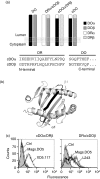
Figure 1
Figure 1
Mags.DO5 epitope is located on the β chain of DO (a) Schematic representation of the various DO constructs used in this study. The predicted amino acid sequence of the N-terminal region of mature cDO chains is shown. (b) The position of the DR α (black) and β (dark grey) regions grafted on cDO chains are highlighted on a top view of human leucocyte antigen-DR. α1 and β1 indicate the position of the N terminus of each domain. (c) Flow cytometry analysis of HeLa cells stably expressing mixed cDOα/DRβ or DRα/cDOβ pairs and stained for cell surface expression using DO-specific Mags.DO5 (bold line), DRβ-specific XD5.117 (thin line; left panel) or DRα-specific L243 (thin line; right panel). Filled histograms represent control staining using HeLa cells incubated only with the Alexa-488-coupled secondary GAM antibody.
Figure 2
Figure 2
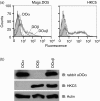
Mags.DO5 monoclonal antibody (mAb) is conformational (a) HEK293T cells were transiently transfected with either DOα alone (light grey), DOβ alone (dark) or DOα+DOβ (grey) complementary DNAs and stained with Mags.DO5 and HKC5 after permeabilization. Cells were analysed by flow cytometry. Filled histograms represent control cells stained with the GAM-Alexa-488 secondary antibody. This experiment was done a minimum of 10 times with similar results. (b) Cell lysates were prepared and analysed on immunoblots for the expression of DOα (upper panel), DOβ (middle panel) and actin (lower panel).
Figure 3
Figure 3
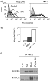
Coexpression of DM increases Mags.DO5 reactivity (a) HEK293T cells were transfected with either DO (thick line) or DO +DM (thin line) cDNAs, fixed, permeabilized and stained after 48 hr with Mags.DO5 (left panel) and HKC5 (right panel) monoclonal antibodies (mAbs). (b) HEK293T cells were transiently transfected with either DOβ alone, DOαDOβ or DOαDOβ+ DM complementary DNAs (cDNAs) and stained with Mags.DO5 and HKC5 before flow cytometry analysis. The y-axis represents the ratio between the mean fluorescence values obtained for the two mAbs. Similar results were obtained in three other independent experiments. (c) HEK293T cells were transiently transfected with either DOα alone, DOβ alone, DOαDOβ or DOαDOβ+DM cDNAs. After 48 hr, cells were lysed and immunoprecipitation was performed using the DOβ-specific HKC5 mAb. Samples were analysed on immunoblots by probing with HKC5 or the DOα-specific rabbit antiserum.
Figure 4
Figure 4
DM-induced conformation change in DO occurs in endoplasmic reticulum (ER) (a) HEK293T cells were transfected with DO alone, DO and DM wild-type (wt) or DO and DMY. After 48 hr, cell surface DO was stained using Mags.DO5. The mean fluorescence values were 119 and 194 for DM and DMY, respectively (not shown). (b) Cells were permeabilized and stained with HKC5 (left panel) or Mags.DO5 (right panel). (c) Mean fluorescence values obtained in (b) were plotted as the Mags.DO5 over HKC5 ratio. This experiment is representative of three independent experiments.
Figure 5
Figure 5
DM affects the conformation of a transport-incompetent DO mutant. (a) HeLa (left panels) and HeLa DM (right panels) cells were transduced with AdDO (thin line) or AdDO VVE (thick line) adenoviral constructs. Cells were fixed, permeabilized and stained with HKC5 (upper panels) or Mags.DO5 (lower panels). (b) Cells were lysed and half of the post-nuclear supernatants was treated with EndoH. Samples were analysed on immunoblots using first the DOα-specific rabbit antiserum and then the control actin-specific probe. Similar results were obtained in an independent experiment.
Figure 6
Figure 6
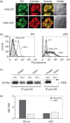
Transport-competent cDO molecules reveal the Mags.DO5 epitope independent of DM (a) Immunofluorescence microscopy analysis of cDO subcellular localization. HeLa DM-negative cells were stably transfected with either wild-type (wt) DO (a–d) or cDO chimera (e–h), permeabilized and incubated with Mag.DO5 and rabbit anti-calnexin followed by Alexa 488-labelled goat anti-mouse antibodies (a,e), biotinylated goat anti-rabbit antibody and Texas-red conjugated streptavidin (b,f). (c) and (g) show the merge of wt DO or cDO images with those obtained for calnexin. (d) and (h) show the cells in visible light. (b) Flow cytometry analysis of HeLa cells stably transfected with DO (left panel) or cDO (right panel). Cells were permeabilized and stained with Mags.DO5 or HKC5. (c) HEK293T cells were transfected with cDO and DM, lysed in Chaps or Triton X-100 (Triton) and DO was immunoprecipitated with the DOα-specific rabbit antiserum. Samples were analysed on immunoblots using the DMβ-specific rabbit antiserum. Control Raji cells were lysed in the same conditions and immunoprecipitation was performed for DO or for DR using XD5. (d) HEK293T cells were transiently transfected with DO or cDO in the absence or presence of DM. After 48 hr, cells were permeabilized and stained with Mags.DO5 and HKC5. Mean fluorescence values obtained for DM+ and DM cells were plotted as a ratio. These ratios are representative of at least two other experiments.
Figure 7
Figure 7
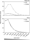
Coexpression of DM reveals the Mags.DO5 epitope on DO molecules. HEK293T cells transiently expressing DO or DO +DM were permeabilized 48 hr post-transfection and stained with serial dilutions of the Mags.DO5 (top panel) or HKC5 (lower panel) monoclonal antibodies. These experiments were repeated three times with similar results.

References

    1. Cresswell P. Assembly, transport, and function of MHC class-II molecules. Annu Rev Immunol. 1994;12:259–93. - PubMed
    1. Bakke O, Dobberstein B. MHC class II-associated invariant chain contains a sorting signal for endosomal compartments. Cell. 1990;63:707–16. - PubMed
    1. Lotteau V, Teyton L, Peleraux A, et al. Intracellular transport of class II MHC molecules directed by invariant chain. Nature. 1990;348:600–5. - PubMed
    1. Roche PA, Cresswell P. Invariant chain association with HLA-DR molecules inhibits immunogenic peptide binding. Nature. 1990;345:615–8. - PubMed
    1. Roche PA, Cresswell P. Proteolysis of the class II-associated invariant chain generates a peptide binding site in intracellular HLA-DR molecules. Proc Natl Acad Sci USA. 1991;88:3150–4. - PMC - PubMed

Publication types