. 2009 Jul;127(3):326-37.
doi: 10.1111/j.1365-2567.2008.02979.x. Epub 2008 Nov 14.

Selective induction of the Notch ligand Jagged-1 in macrophages by soluble egg antigen from Schistosoma mansoni involves ERK signalling

Affiliations

Selective induction of the Notch ligand Jagged-1 in macrophages by soluble egg antigen from Schistosoma mansoni involves ERK signalling

Felicia Goh et al. Immunology. 2009 Jul.

Abstract

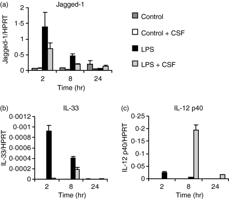
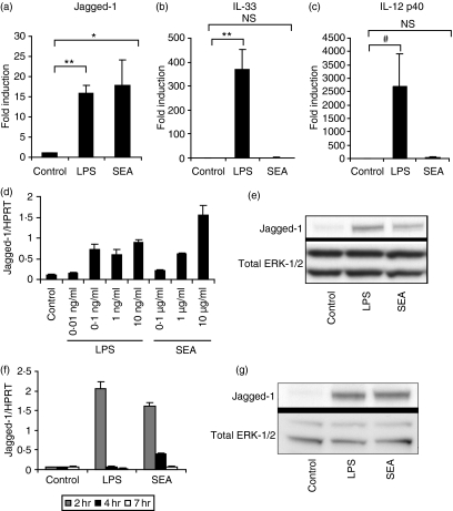
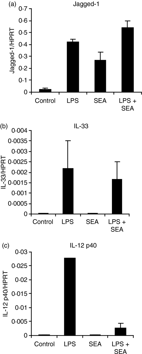
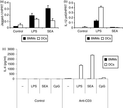
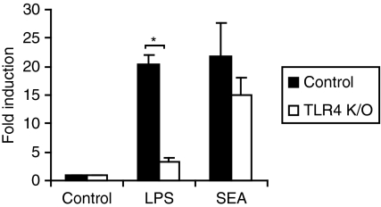
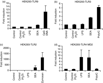
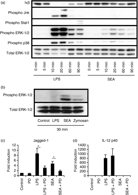
Soluble egg antigen (SEA) from the helminth Schistosoma mansoni promotes T helper type 2 (Th2) responses by modulating antigen-presenting cell function. The Jagged/Notch pathway has recently been implicated in driving Th2 development. We show here that SEA rapidly up-regulated mRNA and protein expression of the Notch ligand Jagged-1 in both murine bone marrow-derived macrophages (BMMs) and human monocyte-derived macrophages (HMDMs). Another potential Th2-promoting factor, interleukin (IL)-33, was not transcriptionally induced by SEA in BMMs. Up-regulation of Jagged-1 mRNA by SEA was also apparent in conventional dendritic cells (DCs), although the effect was less striking than in BMMs. Conversely, SEA-pulsed DCs, but not BMMs, promoted IL-4 production upon T-cell activation, suggesting that Jagged-1 induction alone is insufficient for instructing Th2 development. A comparison of the responses initiated in BMMs by SEA and the bacterial endotoxin lipopolysaccharide (LPS) revealed common activation of extracellular signal-regulated kinase-1/2 (ERK-1/2) and p38 phosphorylation, as well as induction of Jagged-1 mRNA. However, only LPS triggered IkappaB degradation, phosphorylation of c-Jun N-terminal kinase (Jnk) and signal transducer and activator of transcription 1 (Stat1) Tyr701, and IL-33 and IL-12p40 mRNA up-regulation. Inducible gene expression was modified by the presence of the macrophage growth factor colony-stimulating factor (CSF)-1, which inhibited Jagged-1 induction by SEA and LPS, but enhanced LPS-induced IL-12p40 expression. Unlike LPS, SEA robustly activated signalling in HEK293 cells expressing either Toll-like receptor 2 (TLR2) or TLR4/MD2. Pharmacological inhibition of the ERK-1/2 pathway impaired SEA- and LPS-inducible Jagged-1 expression in BMMs. Taken together, our data suggest that Jagged-1 is an ERK-dependent target of TLR signalling that has a macrophage-specific function in the response to SEA.


Figures

Figure 1
Figure 1
Colony-stimulating factor (CSF)-1 suppresses lipopolysaccharide (LPS)-inducible Jagged-1 and interleukin (IL)-33 mRNA expression, but increases IL-12p40 expression in murine bone marrow macrophages (BMMs). BMMs were plated overnight in the presence or absence of CSF-1 and then treated for 2, 8 and 24 hr with 10 ng/ml LPS or medium as a control. Gene expression (mean of triplicates + standard error of the mean) was quantified by real-time polymerase chain reaction (PCR) for Jagged-1 (a), IL-33 (b) and IL-12p40 (c). The results shown are representative of six similar experiments. HPRT, hypoxanthine phosphoribosyltransferase.
Figure 2
Figure 2
Soluble egg antigen (SEA) selectively up-regulates Jagged-1 mRNA and protein expression in mouse and human macrophages. Bone marrow macrophages (BMMs) [plated overnight without colony-stimulating factor (CSF)-1] were treated for 2 hr with 10 ng/ml lipopolysaccharide (LPS), 10 μg/ml SEA or medium as a control (a, b, c), or treated for 2 hr with various concentrations of LPS or SEA (d). Human monocyte-derived macrophages (HMDMs) were treated for 2, 4 and 7 hr (f). Gene expression was calculated by real-time polymerase chain reaction (PCR) for Jagged-1 (a, d, f), interleukin (IL)-33 (b) and IL-12p40 (c). For quantification of protein expression, BMMs (e) or HMDMs (g) were plated overnight in the absence of CSF-1 and treated for 4 hr with 10 ng/ml LPS, 10 μg/ml SEA or medium as a control. Total Jagged-1 and extracellular signal-regulated kinase-1/2 (ERK-1/2) protein were visualized by immunoblotting. Results for the mouse experiments are from six independent experiments, displayed as mean + SEM (a, b, c), or are representative of three or more similar experiments (d, e). Results for the human experiments (f, g) are representative of two similar experiments using different donors. *, P < 0·05; **, P < 0·01; #, P = 0·058; NS, not significant for either LPS or SEA versus control treatments.
Figure 3
Figure 3
Combinatorial effects of soluble egg antigen (SEA) and lipopolysaccharide (LPS) on regulated gene expression in bone marrow macrophages (BMMs). BMMs were plated overnight in the absence of colony-stimulating factor (CSF)-1, and then treated for 2 hr with 10 ng/ml LPS, 10 μg/ml SEA, both, or medium as a control. Gene expression (mean of triplicates + standard error of the mean) was calculated by real-time polymerase chain reaction (PCR) for Jagged-1 (a), interleukin (IL)-33 (b) and IL-12p40 (c). The results shown are indicative of seven similar experiments.
Figure 4
Figure 4
Jagged-1 induction in antigen-presenting cells (APCs) does not correlate with permissiveness for T helper type 2 (Th2) responses (a and b). Bone marrow macrophages (BMMs) [plated overnight without colony-stimulating factor (CSF)-1] or dendritic cells (DCs) were treated for 2 hr with 10 ng/ml lipopolysaccharide (LPS), 10 μg/ml soluble egg antigen (SEA) or medium as a control. Gene expression was calculated by real-time polymerase chain reaction (PCR) for Jagged-1 (a) and interleukin (IL)-12p40 (b). (c) BMMs or DCs were stimulated overnight with medium, 10 ng/ml LPS, 10 μg/ml SEA or 0·1 μm CpG DNA, and then co-cultured with purified mouse splenocytes for 7 days, followed by anti-CD3 stimulation in combination with a second treatment of medium, LPS, SEA or CpG DNA for 3 days. IL-4 levels in cell culture supernatants were determined by enzyme-linked immunosorbent assay (ELISA). Data are representative of three or more similar experiments.
Figure 6
Figure 6
Induction of Jagged-1 mRNA by soluble egg antigen (SEA) is not dependent upon Toll-like receptor 4 (TLR4). TLR4−/− (K/O) and wild-type bone marrow macrophages (BMMs) were treated for 2 hr with 10 ng/ml lipopolysaccharide (LPS), 10 μg/ml SEA or medium control. Jagged-1 mRNA levels were determined by real-time polymerase chain reaction (PCR) and data from six independent experiments are displayed as fold induction (relative to unstimulated cells) + standard error of the mean. *, P < 0·00001 compared with wild-type cells.
Figure 5
Figure 5
Soluble egg antigen (SEA) signals in HEK293 cells expressing Toll-like receptor 2 (TLR2) or TLR4/MD2, but not TLR9. HEK293 cells expressing TLR9 (a), TLR3 (b), TLR2 (c) or TLR4/MD2 (d) were treated for 24 hr with 10 μg/ml SEA, TLR-specific positive controls (10 ng/ml LPS, 0·1 μg/ml CpG DNA (2006), 30 μg/ml PolyIC and 20 μg/ml zymosan) and vehicle controls. Interleukin (IL)-8 production in culture supernatants was determined by enzyme-linked immunosorbent assay (ELISA). Results from three independent experiments (except for HEK293 cells expressing TLR3, which represents four independent experiments) are displayed as fold induction (relative to unstimulated cells) + standard error of the mean.
Figure 7
Figure 7
Soluble egg antigen (SEA) selectively regulates Toll-like receptor (TLR) signalling pathways in bone marrow macrophages (BMMs). (a) BMMs were treated for 0, 5, 10, 20, 60 and 90 min with 10 ng/ml lipopolysaccharide (LPS) or 10 μg/ml SEA. Levels of IκB-α, phospho c-Jun N-terminal kinase (Jnk) (Thr183/Tyr185), phospho signal transducer and activator of transcription 1 (Stat1) (Tyr701), phospho extracellular signal-regulated kinase-1/2 (ERK-1/2) (Thr202/Tyr204), phospho p38 (Thr180/Tyr182) and total ERK-1/2 were visualized by immunoblotting. The results displayed are representative of three independent experiments. (b) HEK293-TLR2 cells were stimulated with 10 ng/ml LPS, 10 μg/ml SEA, 20 μg/ml zymosan or medium control for 30 min and ERK-1/2 levels visualized by immunoblotting. (c, d) BMMs were pre-incubated for 15 min with and without 30 μm PD-98059, followed by stimulation for 2 hr with 10 ng/ml LPS, 10 μg/ml SEA, or medium control. Jagged-1 (c) and interleukin (IL)-12p40 (d) mRNA levels were determined by real-time polymerase chain reaction (PCR), and data from seven independent experiments are displayed as fold induction (relative to unstimulated cells) + standard error of the mean. *, P < 0·05 compared with non-PD-98059-treated samples.

Similar articles

Cited by

References

    1. Kidd P. Th1/Th2 balance: the hypothesis, its limitations, and implications for health and disease. Altern Med Rev. 2003;8:223–46. - PubMed
    1. Miyatake S, Arai N, Arai K. Chromatin remodeling and T helper subset differentiation. IUBMB Life. 2000;49:473–8. - PubMed
    1. Xu DM, Chan WL, Leung BP, et al. Selective expression and functions of interleukin 18 receptor on T helper (Th) type 1 but not Th2 cells. J Exp Med. 1998;188:1485–92. - PMC - PubMed
    1. Townsend MJ, Fallon PG, Matthews DJ, Jolin HE, McKenzie AN. T1/ST2-deficient mice demonstrate the importance of T1/ST2 in developing primary T helper cell type 2 responses. J Exp Med. 2000;191:1069–76. - PMC - PubMed
    1. Schmitz J, Owyang A, Oldham E, et al. IL-33, an interleukin-1-like cytokine that signals via the IL-1 receptor-related protein ST2 and induces T helper type 2-associated cytokines. Immunity. 2005;23:479–90. - PubMed

Publication types

MeSH terms