. 2008 Dec;70(6):1441-52.
doi: 10.1111/j.1365-2958.2008.06492.x. Epub 2008 Oct 30.

The ribosomal stalk is required for ribosome binding, depurination of the rRNA and cytotoxicity of ricin A chain in Saccharomyces cerevisiae

Affiliations

The ribosomal stalk is required for ribosome binding, depurination of the rRNA and cytotoxicity of ricin A chain in Saccharomyces cerevisiae

Jia-Chi Chiou et al. Mol Microbiol. 2008 Dec.

Abstract

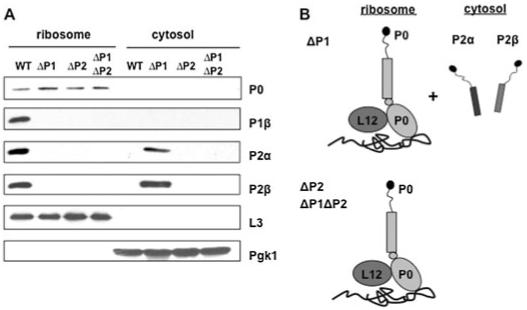
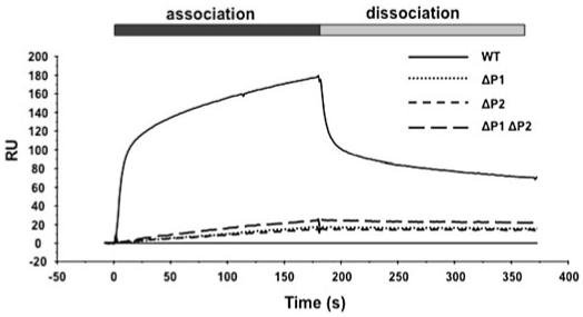
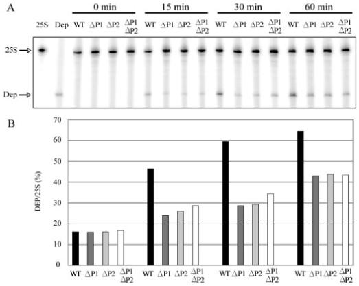
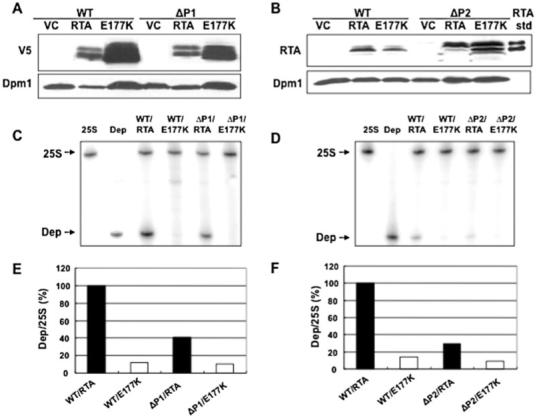
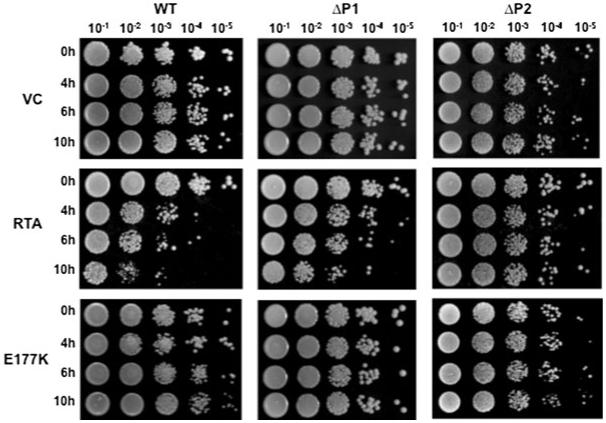
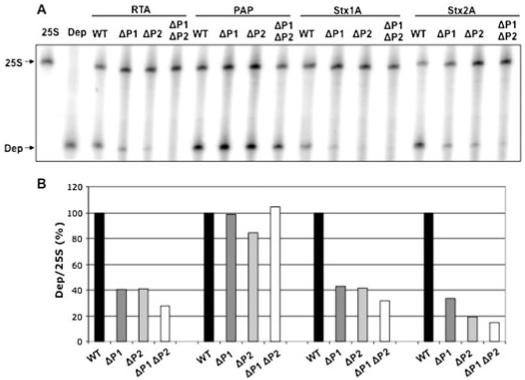
Ribosome inactivating proteins (RIPs) like ricin, pokeweed antiviral protein (PAP) and Shiga-like toxins 1 and 2 (Stx1 and Stx2) share the same substrate, the alpha-sarcin/ricin loop, but differ in their specificities towards prokaryotic and eukaryotic ribosomes. Ricin depurinates the eukaryotic ribosomes more efficiently than the prokaryotic ribosomes, while PAP can depurinate both types of ribosomes. Accumulating evidence suggests that different docking sites on the ribosome might be used by different RIPs, providing a basis for understanding the mechanism underlying their kingdom specificity. Our previous results demonstrated that PAP binds to the ribosomal protein L3 to depurinate the alpha-sarcin/ricin loop and binding of PAP to L3 was critical for its cytotoxicity. Here, we used surface plasmon resonance to demonstrate that ricin toxin A chain (RTA) binds to the P1 and P2 proteins of the ribosomal stalk in Saccharomyces cerevisiae. Ribosomes from the P protein mutants were depurinated less than the wild-type ribosomes when treated with RTA in vitro. Ribosome depurination was reduced when RTA was expressed in the DeltaP1 and DeltaP2 mutants in vivo and these mutants were more resistant to the cytotoxicity of RTA than the wild-type cells. We further show that while RTA, Stx1 and Stx2 have similar requirements for ribosome depurination, PAP has different requirements, providing evidence that the interaction of RIPs with different ribosomal proteins is responsible for their ribosome specificity.


Figures

Fig. 1
Fig. 1
Immunoblot analysis of the ribosomal stalk proteins. A. Yeast cells were fractionated into ribosomal and cytosolic fractions. Equal amount of total protein (10 μg) was analysed on a 12% SDS-PAGE and probed with antibodies against P0, P1β,P2α and P2β. Anti-L3 and anti-αPgk1p (phosphoglycerate kinase) were used as the loading controls for the ribosome and the cytosol fractions respectively. The immunoblot analysis was repeated three times by using different ribosome preparations. B. Schematic representation of the stalk structure in the ΔP1, ΔP2 and ΔP1ΔP2 mutants.
Fig. 2
Fig. 2
Interaction of RTA with ribosomes in the stalk mutants using the Biacore 3000. The N-terminal His-tagged RTA was coupled to the NTA chip as the ligand and the salt-washed monomeric ribosomes isolated from the P protein mutants or the isogenic wild-type strain were used as the analyte. As a control, the N-terminal His-tagged EGFP was immobilized on the reference chip. Ribosomes at 5 nM were passed over the target and the reference surface. The signal obtained from the reference surface was subtracted from the target surface containing the His-tagged RTA to correct for non-specific binding.
Fig. 3
Fig. 3
In vitro depurination of yeast rRNA by RTA. A. Yeast ribosomes (20 pmol) were incubated with 10 ng of RTA at 30°C for 0, 15, 30 and 60 min. The rRNA was extracted and used in the dual-primer extension assay as described in the Experimental procedures. The reaction products were separated on a 7 M urea/6% polyacrylamide gel. The first lane corresponds to the primer extension product with the 25S rRNA primer (25S) alone; the second lane corresponds to the extension product with the primer used to detect depurination (Dep) alone. B. The extent of ribosome depurination was quantified using a PhosphorImager. The depurination levels were determined by the intensities of depurination bands divided by the intensities of the 25S bands. The experiment was repeated at least three times with different ribosome preparations.
Fig. 4
Fig. 4
In vivo expression of RTA and depurination of rRNA. Immunoblot analysis was carried out at 10 h post induction. Each strain was transformed with vector (VC), preRTA (RTA) or an inactive RTA mutant (E177K). Membrane fractions were isolated (Li et al., 2007). A. RTA expression was detected using a monoclonal antibody against the V5 epitope. B. RTA expression was detected using a polyclonal antibody against RTA. Anti-Dpm1 was used as the loading control. Purified RTA (20 ng) from castor bean was loaded as the RTA standard. C and D. The total RNA isolated from the same cells was used in the dual-primer extension assay. E and F. The depurination levels were quantified from three different experiments as described in Fig. 3 and expressed as a percentage of the depurination in the wild-type cells, which was shown as 100%. The analysis was repeated three times with different transformants.
Fig. 5
Fig. 5
Viability of yeast cells expressing RTA and E177K. The yeast cells were transformed with a plasmid carrying RTA under the GAL1 promoter. Cells expressing E177K or harbouring the vector (VC) were used as controls. The cells were first grown in SD medium supplemented with 2% glucose and then transferred to SD medium supplemented with 2% galactose. At indicated times after induction, a series of 10-fold dilutions were plated on media containing 2% glucose and grown at 30°C for 2 days. The analysis was repeated three times with different transformants.
Fig. 6
Fig. 6
In vitro depurination of yeast ribosomes by different RIPs. A. Yeast ribosomes (20 pmol) were incubated with 10 ng of RTA, PAP or 26 ng of Stx1 and Stx2 in the presence of 30 μM DTT at 30°C for 30 min. The rRNA was extracted and used in the dual-primer extension assay. B. The depurination levels were quantified as described in Fig. 3. The level of depurination in the ΔP1, ΔP2 and ΔP1ΔP2 mutants was calculated as per cent of the wild type, which was shown as 100%. The analysis was repeated three times by using different ribosome preparations.

Similar articles

Cited by

References

    1. Algire MA, Maag D, Savio P, Acker MG, Tarun SZ, Jr, Sachs AB, et al. Development and characterization of a reconstituted yeast translation initiation system. RNA. 2002;8:382–397. - PMC - PubMed
    1. Ayub MJ, Smulski CR, Ma KW, Levin MJ, Shaw PC, Wong KB. The C-terminal end of P proteins mediates ribosome inactivation by trichosanthin but does not affect the pokeweed antiviral protein activity. Biochem Biophys Res Commun. 2008;369:314–319. - PubMed
    1. Ban N, Nissen P, Hansen J, Moore PB, Steitz TA. The complete atomic structure of the large ribosomal subunit at 2.4 A resolution. Science. 2000;289:905–920. - PubMed
    1. Baykal U, Tumer NE. The C-terminus of pokeweed antiviral protein has distinct roles in transport to the cytosol, ribosome depurination and cytotoxicity. Plant J. 2007;49:995–1007. - PubMed
    1. Chan DS, Chu LO, Lee KM, Too PH, Ma KW, Sze KH, et al. Interaction between trichosanthin, a ribosome-inactivating protein, and the ribosomal stalkprotein P2 by chemical shift perturbation and mutagenesis analyses. Nucleic Acids Res. 2007;35:1660–1672. - PMC - PubMed

Publication types